1. Introduction
The KAATSU training involves exercise with moderate blood flow restriction, which is known to increase muscle strength and mass (Takarada et al., 2000; Nakajima et al., 2011), and aerobic capacity (Abe et al., 2010). The poten-tial underlying mechanism of KAATSU training is an in-crease in metabolic stress that facilitates systemic hormone production and activation of fast-twitch muscle fibers (Pearson and Hussain, 2015). Because low-intensity KAATSU training produces a sufficient training effect in cardiac patients (Nakajima et al., 2010), it is used in the rehabilitation of cardiac patients who cannot exercise at a high intensity due to risks for developing cardiac or respi-ratory complications (Atkins et al., 1976).
Exercise intensity during cardiac rehabilitation is nor-mally set below anaerobic threshold (AT) (JCS Joint Working Group, 2014). Previous studies, however, which examined the effects of blood flow restriction on muscle activation, heart rate (HR), and ratings of perceived exer-tion (RPE), set exercise intensity based on peak power output (Corvino et al., 2017; Thomas et al., 2018) or VO-2max without KAATSU (Ozaki et al., 2010; Kim et al., 2015) during aerobic exercise. How the application of KAATSU modifies muscle activation, HR, and RPE while exercising just below AT remains unclear. Thus, the pur-pose of this case study was to determine for the first time the effects of KAATSU on muscle activation, HR, and RPE while exercising at an intensity just below AT in a healthy young male.
The effects of moderate blood flow restriction induced
by KAATSU on muscle activation, heart rate, and rate of
perceived exertion during low-intensity aerobic exercise:
A pilot study
Yuta Mizushima
1), Azusa Uematsu
2), Hayato Ishizaka
1), Shigeru Toyoda
3), Takashi Mizushima
1), Teruo
Inoue
3), Yoshiaki Sato
4), Tibor Hortobágyi
5), Toshiaki Nakajima
3, 6)Int. J. KAATSU Training Res. 2020; 16: 1-4
Abstract
[Objective] We examined the effects of moderate blood flow restriction due to KAATSU on rectus femoris activation, heart rate (HR), and ratings of perceived exertion (RPE) while cycling at an in-tensity just below anaerobic threshold (AT).
[Methods] A healthy young male (age = 23 y) performed symptom-limited graded exercise testing on a cycle ergometer with KAATSU (180 SKU) or without KAATSU on 2 separate days. We record-ed surface electromyogram (EMG) from the right rectus femoris, HR, RPE for breathlessness and effort in lower limbs.
[Results] The participant reached AT with lower physical and perceived effort with KAATSU than without KAATSU. Muscle activation was similar during exercise between with and without KAATSU just below AT.
[Conclusion] Exercise just below AT with KAATSU vs. without KAATSU elicited lower physical and perceived effort without difference in muscle activation. Therefore, low-intensity aerobic exercise with KAATSU might be an effective, low-burden, and safe method for the rehabilitation of patients with cardiovascular disease.
Key words: Cycle exercise, Electromyogram, KAATSU, Anaerobic threshold, Low-intensity exercise.
Corresponding author: Azusa Uematsu, Ph.D., Department of Health and Sport Sciences, Premedical Sciences, Dokkyo Medical University, 880 Kitako-bayashi, Mibu, Tochigi, Japan 321-0293
E-mail: auematsu@ dokkyomed.ac.jp, Tel: +81-282-87-2119, Fax: +87-282-86-7265 See end of article for authors’ affiliations
CASE REPORT
1) Department of Rehabilitation, Dokkyo Medical University, Tochigi, Japan
2) Department of Health and Sport Sciences, Premedical Sciences, Dokkyo Medical University, Tochigi, Japan 3) Department of Cardiovascular Medicine, School of Medicine, Dokkyo Medical University, Tochigi, Japan 4) Center for KAATSU Research at Harvard Medical School, Massachusetts, USA
5) University Medical Center Groningen, University of Groningen, Groningen, The Netherlands 6) Department of Medical KAATSU Training, Dokkyo Medical University, Tochigi, Japan
2 The effects of moderate blood flow restriction induced by KAATSU on muscle activation, heart rate, and rate of perceived exertion during low-intensity aerobic exercise: A pilot study
2. Material and Methods
2.1 Participant
A healthy young male (age = 23 years, height = 173.0 cm, weight = 60.0 kg) participated in this study who was informed of the procedures and gave written informed consent before starting the study. The study protocol was conducted according to Declaration of Helsinki and ap-proved by The University Ethics Committee (approval number: 27074).
2.2 Experimental Protocol
The participant visited a laboratory on two days, sepa-rated by 1 week. He performed a symptom-limited graded exercise test using a cycle ergometer (Strength Ergo 8, Fukuda Denshi, Tokyo, Japan) without KAATSU on Day 1 and with KAATSU on Day 2. The experiment started by recording electromyographic (EMG) activity of the right rectus femoris during a maximal voluntary contrac-tion (MVC). The graded exercise test started with a 4 minute-long warm up and was followed by 1-minute-long stages in 20 W increments while keeping cycling cadence constant at 50 revolution per minute.
2.3 Moderate blood flow restriction induced by KAAT-SU
The compact KAATSU system (KAATSU Nano, KAATSU Global, Huntington Beach, CA, USA) was used to artificially restrict blood flow in the thigh. We placed the 60-mm-wide pneumatic cuff around the proximal end of each thigh while the participant was seated on the seat of the bicycle ergometer. Moderate blood flow restriction induced by KAATSU does not interfere with muscle func-tion and produces a training effect (Iida et al., 2007). Thus, the pressure in the cuff was set at 180 standard KAATSU unit (SKU) for moderate blood flow restriction of the lower limbs. We applied the pressure for the entire duration of the graded exercise test.
Data Collection
We recorded surface EMG activity from the right rectus femoris with active surface electrodes (2 mm width, 10 mm length, 10 mm between electrodes, SS-2096, Nihon Kohden, Tokyo, Japan). The skin was wiped clean with al-cohol-soaked cotton to reduce skin impedance. The earth electrode was placed over the skin of the right anterior su-perior iliac spine. The EMG signal was transmitted (ZB-581G, Nihon Kohden, Tokyo, Japan) to a receiver (ZR-550H, Nihon Kohden, Tokyo, Japan) which was connected to a multi telemeter system (WEB-5500, Ni-hon Kohden, Tokyo Japan). To monitor the angular posi-tion of the right knee joint during exercise, a goniometer (SG 150, Biometrics, Newport, UK) was affixed to the lateral side of the right knee with double sided tape. The EMG and goniometer signals were sampled at 2 kHz us-ing a multi telemeter system. The EMG signal was band-pass filtered (15-500 Hz) and the goniometer signal was
low-pass filtered (6 Hz) using a signal processing software (Spike 2, Cambridge Electronics Devices, Cambridge, UK).
Respiratory data were obtained using a breath-by-breath gas analyzer (AE-310, Minato Medical Science, Osaka, Ja-pan). HR was recorded with a stress test system (ML-9000, Fukuda Denshi, Tokyo, Japan). We collected RPE data relative to the sensation of breathlessness and effort in the knee extensors as exercise intensity increased, using a Borg scale of 6 to 20 (Borg 1982).
Data Analysis
We determined knee joint extension phase during cycle exercise using the goniometer signal. The EMG signal was full-wave rectified and averaged for the knee extension phase of 3 cycles at each intensity. EMG amplitude was normalized as %MVC to compare between Days 1 and 2. We analyzed the peak and average EMG in the knee joint extension phase.
Based on ventilatory equivalent (VE) and VO2 data, AT was determined as an increased point of VE/VO2. Then, we defined the data from 1 minute just before at AT as “just below AT”.
We compared data with and without KAATSU and in-tensity-matched to just below AT with KAATSU (KAAT-SU -AT intensity) of without KAAT(KAAT-SU.
3. Results
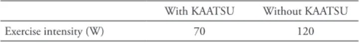
Table 1 shows exercise intensity just below AT with and without KAATSU. Exercise intensity just below AT was 41.2% lower in with KAATSU vs. without KAATSU. The exercise intensity just below AT with and without KAAT-SU was 36.1% and 62.9%, respectively, relative to the in-tensity of peak power output without KAATSU (194 W).
Table 2 shows EMG of the right rectus femoris, HR, and RPE just below AT with and without KAATSU and KAATSU -AT intensity of without KAATSU. Peak and average EMG amplitude in knee extension phase was sim-ilar between just below AT with and without KAATSU. Both EMG outcomes just below AT with KAATSU was substantially higher compared with KAATSU -AT intensi-ty of without KAATSU. HR, RPE for sensation of breath-lessness and effort in lower limbs were the higher just be-low AT without KAATSU but not different between just below AT with KAATSU and KAATSU -AT intensity of without KAATSU.
Table 1. Exercise intensity just below AT with and without KAATSU
With KAATSU Without KAATSU Exercise intensity (W) 70 120 AT, anaerobic threshold
3 Yuta Mizushima, Azusa Uematsu, Hayato Ishizaka., et al
4. Discussion
We examined the effects of moderate blood flow restric-tion induced by KAATSU on muscle activarestric-tion, HR, and RPE while cycling just below AT in a healthy young male. The main findings were that: 1) the right rectus femoris activation did not differ just below AT, but was substan-tially higher during KAATSU compared without KAAT-SU when exercise intensity was matched to just below AT with KAATSU and 2) the participant reached to AT with low physical and subjective burden during aerobic exercise using KAATSU compared with exercise without KAAT-SU.
A previous study reported that moderate blood flow re-striction induced by KAATSU increases muscle activation during low-intensity resistance training in healthy young adults (Yasuda et al., 2009; Yasuda et al., 2014; Leonneke et al., 2015) and also in cardiovascular patients (Ishizaka et al., 2019). In the aerobic exercise study, Kim et al. (2015) reported that muscle activation of vastus lateralis of low-intensity with KAATSU was lower than high-in-tensity without KAATSU (40% and 75% of peak power output without KAATSU, respectively). In the present case study, exercise intensity just below AT with and with-out KAATSU was 36.1% and 61.9%, respectively, of peak power out without KAATSU. Lucia et al. (1999) revealed that muscle activation increases drastically around 80-90% of peak power output during incremental exercise in elite young male cyclists. Therefore, it is possible that the KAATSU-induced increase of muscle activation was great-er than exgreat-ercise intensity-dependent increase of muscle ac-tivation so that muscle acac-tivation just below AT with KAATSU did not differ from that of without KAATSU, but substantially higher than KAATSU-AT intensity of without KAATSU.
Ozaki et al (2010) reported that blood flow restriction increases HR at rest and during low-intensity exercise, but Mendonca et al. (2014) showed that HR was slightly higher during blood flow restriction than without blood flow restriction. They also reported that blood flow restric-tion increased VO2 during walking, and VO2 tended to increase but HR reached a ceiling with exercise progres-sion (Mendonca et al., 2014). In the present case study, the participant reached AT with lower intensity, HR, and RPE with moderate blood flow restriction induced by
KAATSU compared with without KAATSU, suggesting that physical and subjective burden was lower at AT with KAATSU but future studies using a large sample size will be needed to reduce the high variability in HR and RPE (Mendonca et al., 2014).
5. Conclusion
This study shows that moderate blood flow restriction induced by KAATSU decreases the exercise intensity, HR, and RPE to reach a metabolic state just below AT while the knee extensor muscle activation was similar during cy-cling with and without KAATSU. Therefore, we suggest that low-intensity aerobic exercise with KAATSU has the potential to help the rehabilitation of patients with cardiac conditions. The benefit of exercising with KAATSU in cardiac patients could include improved muscle function at a low rate of perceived exertion and physical effort.
Acknowledgements
This study was supported in part by JSPS KAKENHI Grant Number 19H03981 (to T.N.) and 20K11166 (to A.U.).
References
1) Takarada Y, Takazawa H, Sato Y, Takebayashi S, Tanaka Y, Ishii N
(2000) Effects of resistance exercise combined with moderate vas-cular occlusion on musvas-cular function in humans. J Appl Physiol 88: 2097-2106.
2) Nakajima T, Yasuda T, Sato Y, Morita T, Yamasoba T (2011) Effects
of exercise and anti-aging. Anti-Aging Med 9: 92-102.
3) Abe T, Fujita S, Nakajima T, Sakamaki M, Ozaki H, Ogasawara R,
Sugaya M, Kudo M, Kurano M, Yasuda T, Sato Y, Ohshima H, Mu-kai C, Ishii N (2010) Effects of low-intensity cycle training with re-stricted leg blood flow on thigh muscle volume and VO2max in young
men. J Sports Sci Med 9: 452-458.
4) Pearson SJ, Hussain SR (2015) A review on the mechanisms of
blood-flow restriction resistance training-induced muscle hypertro-phy. Sports Med 45: 187-200.
5) Nakajima T, Kurano M, Sakagami F, Iida H, Fukushima K, Fukuda T,
Takano H, Madarame H, Yasuda T, Nagata T, Sato Y, Yamasoba T, Morita T (2010) Effects of low-intensity KAATSU resistance training on skeletal muscle size/strength and endurance capacity in patients with ischemic heart disease. Int J KAATSU Train Res 6: 1-7.
6) Atkins JM, Matthews OA, Blomqvist CG, Mullins CB (1976)
Inci-dence of arrhythmias induced by isometric and dynamic exercise. Br Heart J 38: 465-471.
7) JCS Joint Working Group (2014) Guidelines for rehabilitation in pa-Table 2. EMG of rectus femoris in knee extension phase, HR, and RPE just below AT with and without KAATSU
and intensity-matched to just below AT with KAATSU of without KAATSU (KAATSU-AT without KAATSU). With KAATSU Without KAATSU KAATSU-AT without KAATSU
Peak EMG (%MVC) 34.5 31.8 25.4
Average EMG (%MVC) 8.8 7.7 5.8
HR (bpm) 102 131 106
RPE for breathlessness 7 10 8
PPE for effort in lower limbs 8 12 8
4 The effects of moderate blood flow restriction induced by KAATSU on muscle activation, heart rate, and rate of perceived exertion during low-intensity aerobic exercise: A pilot study
tients with cardiovascular disease (JCS 2012). Circ J 78: 2022-2093.
8) Corvino RB, Rossiter HB, Loch T, Martins JC, Caputo F (2017)
Physi-ological responses to interval endurance exercise at different levels of blood flow restriction. Eur J Appl Physiol 117(1): 39-52.
9) Thomas HJ, Scott BR, Peiffer JJ (2018) Acute physiological responses
to low-intensity blood flow restriction cycling. J Sci Med Sport 21(9): 969-974.
10) Ozaki H, Brechue WF, Sakamaki M, Yasuda T, Nishikawa M, Aoki
N, Ogita F, Abe T (2010) Metabolic and cardiovascular responses to upright cycle exercise with leg blood flow reduction. J Sports Sci Med 9(2): 224-230.
11) Kim D, Loenneke JP, Thiebaud RS, Abe T, Bemben MG (2015) The
acute muscular effects of cycling with and without different degrees of blood flow restriction. Acta Physiol Hung 102(4): 428-441. 12) Iida H, Kurano M, Takano H, Kubota N, Morita T, Meguro K, Sato Y,
Abe T, Yamazaki Y, Uno K, Takenaka K, Hirose K, Nakajima T (2007) Hemodynamic and neurohumoral responses to the restric-tion of femoral blood flow by KAATSU in healthy subjects. Eur J Appl Physiol 100(3): 275-285.
13) Borg GA (1982) Psychophysical bases of perceived exertioin. Med
Sci Sports Exerc 14: 377.
14) Yasuda T, Brechue WF, Fujita T, Shirakawa J, Sato Y, Abe T (2009)
Muscle activation during low-intensity muscle contractions with re-stricted blood flow. J Shorts Sci 27: 479-489.
15) Yasuda T, Fukushima K, Fukuda T, Iida H, Imuta H, Sato Y,
Ya-masoba T, Nakajima T (2014) Effect of low-intensity, elastic band exercise combined with blood flow restriction on muscle activation. Scand J Med Sci Sports 24: 55-61.
16) Loenneke JP, Kim D, Fahs CA, Thiebaud RS, Abe T, Larson RD,
Bemben DA, Bemben MG (2015) Effects of exercise with and
with-out different degree of blood flow restriction on torque and muscle activation. Muscle Nerve 51: 713-721.
17) Ishizaka H, Uematsu A, Mizushima Y, Nozawa N, Katayanagi S,
Matsumoto K, Nishikawa K, Takahashi R, Arakawa T, Sawaguchi T, Yasuda T, Yamaguchi S, Ogawa H, Shibasaki I, Toyoda S, Horto-bágyi T, Fukuda H, Inoue T, Mizushima T, Nakajima T (2019) Blood flow restriction increases the neural activation of the knee extensors during very low-intensity leg extension exercise in cardiovascular patients: A pilot study. J Clin Med 8: 1252.
18) Lucía A, Sanchéz O, Carvajal A, Chicharro JL (1999) Analysis of
the aerobic-anaerobic transition in elite cyclists during incremental exercise with the use of electromyography. Br J Sports Med 33: 178-185.
19) Mendonca GV, Vaz JR, Teixeira MS, Grácio T, Pezarat-Correia P
(2014) Metabolic cost of locomotion during treadmill walking with blood flow restriction. Clin Physiol Funct Imaging 34: 308-316.
Authors’ affiliations
Mizushima Y, Ishizaka H, Mizushima T. Department of Rehabilitation,
Dokkyo Medical University, Tochigi, Japan
Uematsu A. Department of Health and Sport Sciences, Premedical
Sci-ences, Dokkyo Medical University, Tochigi, Japan
Toyoda S, Nakajima T, Inoue T. Department of Cardiovascular
Medi-cine, School of MediMedi-cine, Dokkyo Medical University, Tochigi, Japan
Sato Y. Center for KAATSU Research of Harvard Medical School,
Mas-sachusetts, USA
Hortobágyi T. University Medical Center Groningen, University of
Groningen, Groningen, The Netherlands
Nakajima T. Department of Medical KAATSU Training, Dokkyo Medical