RESEARCH ARTICLE
Effectiveness, safety, and factors associated
with the clinical success of endoscopic biliary
drainage for patients with hepatocellular
carcinoma: a retrospective multicenter study
Akihiro Matsumi
1, Hironari Kato
1*, Toru Ueki
2, Etsuji Ishida
3, Masahiro Takatani
4, Masakuni Fujii
5, Masaki Wato
6,
Tatsuya Toyokawa
7, Ryo Harada
8, Hirofumi Tsugeno
9, Minoru Matsubara
10, Hiroshi Matsushita
11and Hiroyuki Okada
1Abstract
Background: Only a few reports have assessed the effectiveness of endoscopic biliary drainage (EBD) in hepatocel‑ lular carcinoma (HCC) patients with obstructive jaundice and liver dysfunction.
Methods: This was a retrospective study based on the clinical databases from the Okayama University Hospital and 10 affiliated hospitals. All patients received EBD for jaundice or liver dysfunction. The indication for EBD was aggrava‑ tion of jaundice or liver dysfunction with intrahepatic bile duct (IHBD) dilation. The technical and clinical success rate, complications, factors associated with clinical failure, and survival duration were evaluated.
Results: A total of 107 patients were enrolled in this study. Technical success was achieved in 105 of 107 patients (98.1%). Clinical success was achieved in 85 of 105 patients (81%). Complications related to endoscopic retrograde cholangiography (ERC) occurred in 3 (2.8%) patients. Child–Pugh class C (odds ratio 3.90, 95% confidence interval [CI] 1.47–10.4, p = 0.0046) was the only factor associated with clinical failure, irrespective of successful drainage. The median survival duration was significantly longer in patients with clinical success than in those without clinical success (5.0 months vs. 0.93 months; hazard ratio [HR] 3.2, 95% CI 1.87–5.37). HCC Stage I/II/III (HR 0.57, CI 0.34–0.95,
p = 0.032), absence of portal thrombosis (HR 0.52, CI 0.32–0.85, p = 0.0099), and clinical success (HR 0.39, CI 0.21–0.70, p = 0.0018) were significant factors associated with a long survival.
Conclusions: EBD for obstructive jaundice and liver dysfunction in patients with HCC can be performed safely with a high technical success rate. Clinical success can improve the survival duration, even in patients expected to have a poor prognosis.
Trial registration: Retrospectively registered.
Keywords: Endoscopic retrograde cholangiopancreatography, Jaundice, Hepatocellular carcinoma, Liver dysfunction
© The Author(s) 2021. Open Access This article is licensed under a Creative Commons Attribution 4.0 International License, which permits use, sharing, adaptation, distribution and reproduction in any medium or format, as long as you give appropriate credit to the original author(s) and the source, provide a link to the Creative Commons licence, and indicate if changes were made. The images or other third party material in this article are included in the article’s Creative Commons licence, unless indicated otherwise in a credit line to the material. If material is not included in the article’s Creative Commons licence and your intended use is not permitted by statutory regulation or exceeds the permitted use, you will need to obtain permission directly from the copyright holder. To view a copy of this licence, visit http://creat iveco mmons .org/licen ses/by/4.0/. The Creative Commons Public Domain Dedication waiver (http://creat iveco mmons .org/publi cdoma in/zero/1.0/) applies to the data made available in this article, unless otherwise stated in a credit line to the data.
Background
Hepatocellular carcinoma (HCC) is the most common cancer worldwide and the most frequent cause of death by cancer. With advances made in the HCC diagnosis and treatment techniques in recent years, the rates of an
Open Access
*Correspondence: kato‑[email protected]‑u.ac.jp
1 Department of Gastroenterology and Hepatology, Okayama University
Graduate School of Medicine, Dentistry, and Pharmaceutical Sciences, 2‑5‑1 Shikata‑cho, Kita‑ku, Okayama 700‑8558, Japan
early tumor diagnosis and long-term survival have been increasing [1–8].
Jaundice is encountered in 5%-44% of patients with HCC at the time of the initial diagnosis and frequently occurs during the later stages of disease [2]. Jaundice and liver dysfunction are usually caused by diffuse tumor infiltration into the liver parenchyma, tumor invasion, and/or progressive terminal liver failure resulting from advanced underlying cirrhosis. Obstructive jaundice caused by tumor invasion or hemobilia is rare in HCC, with an incidence of 0.5%-13% [4], and HCC treat-ments, such as percutaneous tumor ablation (PTA), arte-rial embolization, and radiation therapy, rarely cause obstructive jaundice [9]. However, it is closely connected with patients’ mortality as well as jaundice caused by liver dysfunction. Although endoscopic biliary drain-age (EBD) is considered useful and effective in patients with obstructive jaundice due to other malignant dis-eases such as bile duct cancer and pancreatic cancer, only a few studies have investigated the effectiveness of EBD in HCC patients with obstructive jaundice and liver dys-function or obstructive cholangitis, and most of them have only included a small number of patients [3].
We carried out a retrospective analysis of the outcomes of EBD for obstructive jaundice or liver dysfunction with or without cholangitis caused by HCC in a large number of patients and clarified the factors associated with treat-ment success and prognosis extension.
Methods
This retrospective study was based on the clinical data-bases of the Okayama University Hospital and 10 affili-ated hospitals. Data were extracted from the patients who underwent endoscopic stent placement for obstruc-tive jaundice caused by HCC from April 2013 to October 2018. Biliary strictures considered to be associated with therapy for HCC, such as PTA, trans-arterial emboliza-tion, and radiation therapy, were omitted. Informed con-sent was obtained via an opt-out option on the website. The Institutional Review Board of Okayama University approved this study.
Reference for endoscopic treatment
The indication for endoscopic stent placement was aggra-vation of jaundice or liver dysfunction with intrahepatic bile duct (IHBD) dilation. Some patients had cholangi-tis in addition to jaundice or liver dysfunction. Jaundice was defined as a serum total bilirubin concentration over 3 mg/dl, and liver dysfunction was defined as hepatobil-iary enzyme elevation compared to the baseline of each patient. IHBD dilatation was defined when an axial scan by computed tomography, magnetic resonance imaging,
or ultrasound sonography showed the lumen of the intra-hepatic bile duct to be more than 2 mm in diameter.
In accordance with Tokyo Guidelines 2018 [10], chol-angitis was defined as the presence of leukocytosis, elevation of C-reactive protein (CRP), or a fever (temper-ature ≥ 38 °C), in addition to the findings indicating chol-estasis and/or the IHBD dilatation previously mentioned.
Endoscopic procedures
Endoscopic retrograde cholangiography (ERC) was performed using a duodenoscope (TJF260V, JF260V; Olympus Optical Co., Tokyo, Japan) in all cases. After confirming the biliary stricture caused by HCC with ERC, a plastic stent (PS) or endoscopic nasobiliary drain-age (ENBD) tube was placed. The stent diameter (5–7 Fr) varied depending on the diameter of the bile duct. The number of PS or ENBD tubes was chosen according to the number of branches divided by the stricture. In cases with several divided branches, a maximum of three stents was placed into the major branches: left hepatic duct, anterior branch of the right hepatic duct, and posterior branch of the right hepatic duct. ENBD was sometimes used for initial drainage for patients with hemobilia due to bleeding of HCC. If ENBD was effective and hemo-stasis was confirmed, the ENBD tube was replaced with a PS to avoid cholestasis with a clot, even in cases with-out biliary stricture. Metallic stents were not used due to reports indicating that the survival rate was significantly extended in patients with HCC undergoing endoscopic biliary draining with PSs compared to those treated with metallic stents [7]. In cases in which endoscopic proce-dures were deemed difficult, percutaneous transhepatic biliary drainage (PTBD) was employed. If clinical success could not be achieved despite successful stent placement, then the type of stent, e.g. straight type or Zimmon type, was changed, and/or the branch into which the PS was placed was changed. In most cases, endoscopic sphinc-terotomy was not performed in order to maintain the function of the sphincter of Oddi. Endoscopic sphinc-terotomy was performed when multiple stents were required.
If a blood clot was detected, it was extracted by a bas-ket and balloon catheters. After clinical success was achieved, follow-up ERC and stent exchange were sched-uled two to three months post-treatment. In the patient had a poor condition, stents were kept in place with-out scheduled stent exchange until stent obstruction occurred.
Evaluations
The outcomes were evaluated on technical success, clini-cal success, and survival duration. Techniclini-cal success was defined as successful placement of a PS or ENBD tube in
the first session. Clinical success was defined as at least a 30% reduction of jaundice based on liver function test results after technical success. In cases with cholangitis, the resolution of the inflammatory symptoms and labora-tory data, in addition to the resolution of cholestasis find-ings, was defined as clinical success. Clinical failure was defined as an unsuccessful placement of a PS or an ENBD tube or no achievement of the clinical success previously mentioned, irrespective of the successful placement of the PS or ENBD tube. The survival duration was defined as the time from the endoscopic procedure to death.
Adverse events were evaluated according to the ASGE classification of Standards of Practice Committee [11]. Pancreatitis was defined as severe abdominal pain requir-ing an analgesic and a more than three-fold increase in serum amylase levels.
Statistical analyses
Statistical analyses were performed using the JMP® 14 software program (SAS Institute Inc., Cary, NC, USA). We used the chi-square test or Fisher’s test to compare proportions and the Mann–Whitney U-test to compare the median values. We performed a univariate analysis to assess the risk of treatment failure using the chi-squared test. The survival rate about clinical factors was evaluated with the Kaplan–Meier method and Cox proportional hazard model for the univariate analysis. To identify clin-ical factors associated with a long survival, factors with p < 0.05 in the univariate analysis were analyzed using a multivariate Cox model. The threshold for significance was a value of p < 0.05.
Results
Patients characteristics
A total of 107 patients underwent EBD. Table 1 sum-marizes the patient characteristics. The most common causes of HCC were hepatitis C (43%). Child–Pugh class B was seen in about half of all patients (47%). In over half of all patients, the clinical stage was stage IV, the Barce-lona Clinic Liver Cancer (BCLC) classification was C and D, and no portal vein invasion was identified. Cholangitis was present in 43 of 106 patients (40%). Prior to EBD, the median serum total bilirubin level in all 107 patients was 6.47 (0.5–31.5) mg/dl. Prior to EBD, the maximum IHBD diameter was 8.0 (3.0–23) mm.
Technical and clinical outcomes
Bile duct stricture was identified in 94 of 107 (88%) patients, and hemobilia was identified in 34 patients (32%) by ERC. Causes of biliary obstruction were tumor invasion or tumor compression by HCC or met-astatic lymph-node in 94 patients and hemobilia in 13
patients. Multiple PSs were placed in 32 patients (30%), including 2 in 26 patients, and 3 in 6 patients.
The patient flow diagram and success rates are shown in Fig. 1. Technical success was achieved in 105 of 107 patients (98.1%) in the first session. The reason for technical failure in the remaining two patients was that the tip of the PS could not pass through the stricture due to the hard nature of the stricture. In these two patients without technical success, no biliary drain-age was performed due to liver failure in one patient, whereas PTBD was performed in the other patient, but clinical success still could not be achieved because of liver failure.
In 84 of 105 patients, clinical success was achieved at the 1st session. Of the 21 patients without clinical suc-cess, 18 instances were thought to be due to liver failure, so a second session was not performed in these patients. The second session was performed for the remaining 3 patients. The stent was replaced in two patients and added in one, but clinical success was achieved only in one of the patients who underwent stent exchange. The clinical success rate in all patients, including those with and without cholangitis, was 81.0%, 90.2%, and 75%, respectively. An ENBD tube was successfully placed in seven patients. Clinical success was achieved in six of
Table 1 Patient characteristics
HBV: hepatitis B virus, HCV: hepatitis C virus, non-BC: not hepatitis B and C virus, BCLC: Barcelona Clinic Liver Cancer, T-Bil: Total bilirubin,EBS: endoscopic biliary stent, IHBD: intrahepatic bile duct
Patients 107 Age (mean ± SD) 71.5 ± 10.1 Sex (%) Male/female 76 (71)/31 (29) Etiology (%) HCV/HBV/non‑BC 46 (43)/24 (22)/17 (33) Child–Pugh score (%) 5/6/7/8/9/10/11/12/13/14/15 15 (14)/9 (8)/19 (18)/15 (14)/17 (16)/19 (18)/10 (9)/2 (2)/0 (0)/0 (0)/1 (1) Child–pugh score (%) A/B/C 25 (23)/50 (47)/32 (30) HCC stage (%) I/II/III/IV 6 (6)/6 (6)/38 (35)/57 (53) BCLC classification (%) 0/A/B/C/D 2 (2)/11 (10)/16 (15)/46 (43)/32 (30) Grade of portal vein invasion
VP0/1/2/3/4 58 (55)/2 (2)/13 (12)/12 (11)/21 (20) Cholangitis (%)
Present/absent 43 (40)/64 (60) T‑bill before EBS (median, mg/dl) 6.47 (0.5–31.5)
the seven patients, and the ENBD tube was successfully replaced with a PS.
Recurrent biliary obstruction (RBO) occurred in 40 of 85 patients (47%) with clinical success during the obser-vation period (median 96.5 days [IQR 30.5–189.5]). The causes of RBO were debris in 19 (49%), hemobilia in 12 (31%), tumor growth in 5 (13%), and others in 3 (7%).
HCC treatment was performed after endoscopic treat-ment in 41 of 107 patients. The details of HCC treattreat-ment in these patients were trans-arterial therapy in 28 (68%), molecular-targeted drugs in 7 (17%), radiation therapy in 4 (10%) and operation in 2 (5%). Of the 41 patients undergoing HCC treatment, clinical success after endo-scopic treatment was achieved in 39.
In 80 of the 107 patients, the survival duration after EBD and causes of death were assessed. The reasons for death were HCC progression in 48 patients (45%), liver failure in 22 (20%), cholangitis in 5 (5%), and other dis-eases in 5 (5%).
Adverse events
Complications related to ERC procedures occurred in 3 (2.8%) of the 107 patients. Hyperamylasemia occurred in two patients. Both patients with hyperamylasemia had no abdominal pain and improved with conservative treatment. No post-ERCP pancreatitis occurred. In one patient who underwent ENBD for hemobilia, the total bilirubin level decreased gradually. However, bleeding was out of control, and seven days after ENBD, abdomi-nal pain was reported. The patient was diagnosed with gallbladder hemorrhaging and cholecystitis by computed tomography. Despite receiving intravenous antibiotics, he died eight days after ERCP.
Clinical success and the survival
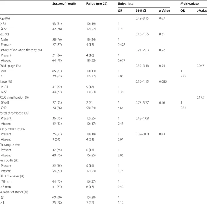
Table 2 shows the relationship between patient charac-teristics and clinical failure after successful EBD. The Child–Pugh class (odds ratio [OR] 4.66, 95% confidence interval [CI] 1.23–30.5, p = 0.021) and BCLC classifica-tion C/D (OR 3.90, 95% CI 1.47–10.4, p = 0.0046) were factors shown to be associated with clinical failure, irre-spective of successful drainage in a univariable analysis. In a multivariate analysis, only the Child–Pugh class (OR 2.85, p = 0.047) was associated with clinical failure, irre-spective of successful drainage.
The median survival duration was significantly longer in patients with clinical success than in those without success (5.0 months vs. 0.93 months; hazard ratio [HR] 3.2, 95% CI 1.87–5.37) (Fig. 2). In patients with Child– Pugh class C, the survival duration among those with clinical success was longer than among those with clini-cal failure (2.8 months vs. 0.93 months, p = 0.0235).
Table 3 shows the relationship between clinical factors and the survival duration after EBD. The Child–Pugh class (OR 1.70, 95% CI 1.04–2.71, p = 0.035), stage (HR 2.35, 95% CI 1.47–3.84, p = 0.0005), BCLC classification (HR 2.58, 95% CI 1.51–4.42, p = 0.0002), portal thrombo-sis (HR 0.44, 95% CI 0.27–0.70, p = 0.0005), HCC treat-ment after ERCP (HR 0.32, 95% CI 0.18–0.52, P < 0.0001), and clinical course (HR 3.2, 95% CI 1.87–5.37, P < 0.0001) were associated with the survival duration after EBD in a univariate analysis. The absence of portal thrombosis (HR 0.52, 95% CI 0.28–0.96, p = 0.038), HCC treatment after ERCP (HR 0.39, 95% CI 0.21–0.70, p = 0.0015), and clinical success (HR 0.39, 95% CI 0.21–0.70, p = 0.0018) were significant factors associated with a long survival in a multivariate analysis. In patients with tumor portal
Fig. 1 Clinical success was achieved in 85 (81.0%) patients. Most patients who failed to achieve clinical success at the first session were suspected of having liver failure
thrombosis and without HCC treatment after ERCP, the survival duration among those with clinical success was longer than among those with clinical failure (4.3 months vs. 1.0 months, P < 0.0001; 3.8 months vs. 0.73 months,
p = 0.0005).
Furthermore, the median survival duration was sig-nificantly longer in patients with HCC treatment after
ERCP than in those without treatment (12 months vs. 2.8 months; HR 4.3, 95% CI 4.93–21.5) (Fig. 3). Among patients without HCC treatment after ERCP, the median survival duration was significantly longer in patients with clinical success than in those with clini-cal failure (3.8 months vs. 0.73 months; HR 2.1, 95% CI 2.8–5.7) (Fig. 3).
Table 2 Univariate and multivariate analysis of factors associated with clinical success
OR: Odds ratio, CI: Confidential interval, BCLC: Barcelona Clinic Liver Cancer, IHBD: intrahepatic bile duct
Success (n = 85) Failue (n = 22) Univariate Multivariate OR 95% CI p Value OR p Value Age (%) 0.48–3.15 0.67 > 72 43 (81) 10 (19) 1 ≧72 42 (78) 12 (22) 1.23 Sex (%) 0.15–1.55 0.21 Male 58 (76) 18 (24) 1 Female 27 (87) 4 (13) 0.478
History of radiation therapy (%) 0.21–2.23 0.52
Present 21 (84) 4 (16) 1 Absent 64 (78) 18 (22) 0.677 Child–pugh (%) 0.52–3.48 0.54 0.047 A/B 65 (87) 10 (13) 1 1 C 20 (63) 12 (37) 3.90 2.85 Stage (%) 0.16–1.15 0.086 I/II/III 41 (82) 9 (18) 1 IV/V 44 (77) 13 (23) 1.35 BCLC classification (%) 0.175 0/A/B 27 (93) 2 (7) 1 0.73–5.77 0.16 1 C/D 20 (26) 58 (74) 4.66 2.84 Portal thrombosis (%) Present 36 (75) 12 (25) 1 0.13–1.08 Absent 49 (83) 10 (17) 0.43 Biliary structure (%) Present 76 (81) 18 (19) 1 0.39–3.00 0.83 Absent 9 (69) 4 (31) 2.01 Cholangitis (%) Present 37 (75) 6 (14) 1 Absent 48 (75) 16 (25) 2.06 Hemobilia (%) Present 29 (85) 5 (15) 1 Absent 56 (77) 17 (23) 1.76 IHBD diameter (%) ≦8 mm 44 (73) 16 (27) 1 > 8 mm 41 (87) 6 (13) 0.40 Number of stents (%) ≦1 60 (80) 15 (20) 1 > 1 25 (78) 7 (22) 1.12
Discussion
In this study, we showed that endoscopic biliary drainage for obstructive jaundice in patients with HCC could be performed safely with a high technical success rate. Fur-thermore, clinical success improves the survival duration, even in patients with poor prognostic factors of HCC, such as Child–Pugh score C, stage IV disease, and tumor portal thrombosis.
The technical success rate of EBD was 98.7% in our study. Reported success rates are 92.1%-100% [2, 3, 7], so our result is not inferior to previously reported findings. In our study, two patients who failed to achieve techni-cal success had hard strictures that a stent could not pass through. Both cases had a history of radiation therapy for HCC. We believe that a hard stricture due to radiation therapy is associated with a risk of treatment failure.
The clinical success rate was 83% in our study. Reported success rates are 35.7%-75.9% [2, 3, 12–15], so our result is superior to that previously reported. The presence of IHBD dilatation is significantly associated with clinical
Fig. 2 Patients with clinical success had a longer survival than those without clinical success (5.0 months vs. 0.93 months; HR 3.2, 95% CI 1.87–5.37)
Table 3 Univariate and multivariate analysis of clinical factors associated with survival time after endoscopic biliary drainage
HR Univariate Multivariate
95% CI p value HR 95% CI p value
Median age (years) ≦72 1
> 72 0.83 0.53–1.30 0.42
Sex Male 1
Female 0.87 0.52–1.42 0.59
History of radiation therapy Present 1
Absent 1.02 0.62–1.76 0.95 Child–Pugh A/B 1 1 C 1.7 1.04–2.71 0.035 1.17 0.64–2.13 0.62 Stage I/II/III 1 1 IV 2.35 1.47–3.84 0.0005 1.53 0.82–2.84 0.18 BCLC classification 0/A/B 1 1 C/D 2.58 1.51–4.42 0.0002 1.02 0.40–2.59 0.97
Portal thrombosis Present 1 1
Absent 0.44 0.27–0.70 0.0005 0.52 0.28–0.96 0.038
Biliary stricture Present 1
Absent 0.68 0.30–1.33 0.28
Cholangitis Present 1
Absent 0.82 0.53–1.30 0.41
Hemobilia Present 1
Absent 1.48 0.92–2.44 0.11
Plastic stent Single 1
Multiple 0.71 0.41–1.17 0.18
HCC treatment after ERCP Absent 1 1
Present 0.32 0.18–0.52 < 0.0001 0.39 0.21–0.70 0.0015
Clinical course Success 1 1
success [3]. However, in our study, an inclusion criterion was the presence of IHBD dilation. Jaundice and liver dysfunction without IHBD dilatation in HCC patients are thought to indicate liver failure, which is difficult to improve via biliary drainage; this may explain why our findings were superior to those of previous studies. How-ever, the clinical success rates of EBD in other malignant strictures, such as hilar cholangiocarcinoma, have been reported to range from 54% to 91.3% [16–19]. Our data are not inferior to those of previous reports on other malignant strictures, either.
Total complication rates reportedly range from 0% to 21.7% [2, 7] in HCC stricture, and 5.3% to 47.8% [19] in other malignant strictures. The complication rate was 2.8% in the present study (hyperamylasemia in 2, gall bladder hemorrhaging in 1), which was similar to the rates in other studies. The rate of post-ERCP pancreati-tis (PEP) was reported to be 18.6% [3] for cases of HCC stricture and 0%-38.4% [19, 20] for cases of other malig-nant stricture. Adler et al. reported that patients with Child–Pugh class C had a higher risk of PEP than others [21]. However, whether or not the risk of PEP in patients with liver cirrhosis is high is controversial [22, 23]. In our study, although there were no cases of post-ERCP pan-creatitis, the number of patients was small, so a larger prospective trial is required to resolve this issue.
In our study, Child–Pugh class C was significantly asso-ciated with clinical failure. The clinical success rate was 86.7% in cases of class A/B but only 62.5% in class C. In
a previous report, the 1-year cumulative survival rates were 45% (Child C), 80% (Child B) and 95% (Child A). Patients with Child–Pugh class C have a poor prognosis because of liver failure [1, 24], so biliary drainage tends to be less effective in these patients than in others. How-ever, on comparing the survival rates between the clini-cal success and cliniclini-cal failure cases among patients with Child–Pugh class C, the survival duration in the clinical success cases was longer than that in the clinical failure cases (2.8 months vs. 0.93 months, p = 0.0235). In our study, HCC treatment after ERCP was significantly asso-ciated with a long survival. In addition, among patients without HCC treatment after ERCP, the median survival duration was significantly longer in patients with clinical success than in those with clinical failure. HCC treatment after ERCP is sometimes difficult in patients who can-not achieve clinical success through endoscopic therapy. Therefore, clinical success with endoscopic therapy is thought to contribute to a long prognosis, both indirectly and directly.
Previous studies have shown that, in addition to suc-cessful drainage, subsequent treatment for HCC, the liver function, and HCC tumor stage were significant factors associated with a long survival [2, 3, 8]. Our study found that portal thrombosis and clinical suc-cess of ERCP were significant factors influencing the survival according to the multivariate analysis. Indeed, the survival duration in patients with clinical success was significantly longer than in those without clinical
Fig. 3 The median survival duration was significantly longer in patients with HCC treatment after ERCP than in those without treatment (12 months vs. 2.8 months; HR 4.3, 95% CI 4.93–21.5). Among patients without HCC treatment after ERCP, the median survival duration was significantly longer in patients with clinical success than in those with clinical failure (3.8 months vs. 0.73 months; HR 2.1, 95% CI 2.8–5.7)
success (HR 0.39, 95% CI 0.21–0.70, p = 0.0018). Suc-cessful drainage may provide HCC patients with jaun-dice an opportunity for an extended survival, not only through successful drainage itself but also through the opportunity to receive additional HCC treatment. Fur-thermore, in patients with portal thrombosis who had a poor prognosis, when clinical success was achieved, their survival was extended (HR 0.23, 95% CI 0.11– 0.51, p = 0.0005).
Several limitations associated with the present study warrant mention. First, all data was reviewed retrospec-tively. However, this was a multi-center trial, and the number of patients was over 100. To our knowledge, this report included the largest number of patients to date. Second, no data could be analyzed between the survival of the patients with successful biliary drainage in this study and that of the HCC patients without bil-iary obstruction. Such data would reveal more clearly the effectiveness of biliary drainage in the patients with biliary obstruction with HCC. Third, we used only PSs for biliary drainage. A previous report mentioned that the survival rate was significantly longer in the PS group than in the metallic stent group when patients with HCC underwent endoscopic biliary drainage [7]. Therefore, we used PSs for biliary drainage in this study. However, no study has prospectively compared the effectiveness of biliary drainage using PSs with that using metallic stents in a large number of patients. Randomized controlled tri-als in large cohorts are required to further investigate this issue. Finally, there may have been some selection bias in our study. When we decided whether or not to perform ERCP for HCC patients, we might have unknowingly excluded patients with a poor condition.
In conclusion, Child–Pugh class C is an important pre-dictor of the effectiveness of endoscopic biliary drainage in HCC patients with liver dysfunction with or without cholangitis. When clinical success is achieved, the prog-nosis is improved; therefore, we should actively promote biliary drainage for HCC jaundice.
Conclusions
EBD for obstructive jaundice and liver dysfunction in patients with HCC can be performed safely with a high technical success rate. Clinical success may extend the survival duration, even in patients expected to have a poor prognosis.
Abbreviations
HCC: Hepatocellular carcinoma; EBS: Endoscopic biliary stenting; IHBD: Intrahepatic bile duct; CRP: C‑reactive protein; ERC: Endoscopic retrograde cholangiography; PS: Plastic stent; ENBD: Endoscopic nasobiliary drainage; PTBD: Percutaneous transhepatic biliary drainage; BCLC: Barcelona clinic liver cancer; PEP: Post‑ERCP pancreatitis.
Acknowledgements
Not applicable.
Authors’ contributions
The following authors wrote the main manuscript text: AM, HK. The following authors made substantial contributions to the conception or design of the work, or performed the acquisition, analysis, or interpretation of data for the work: AM, HK, TU, EI, MT, MF, MW, TT, RH, HT, MM and HM. The following authors drafted the work or revised it critically for important intellectual con‑ tent: AM and HK. The following authors gave the final approval for the version to be published: HK and HO. All authors reviewed the manuscript.
Funding
Not declared.
Availability of data and materials
The datasets used and/or analyzed during the current study are available from the corresponding author on reasonable request.
Ethics approval and consent to participate
Informed consent was obtained via an opt‑out option on the website. The Institutional Review Board of Okayama University approved this study.
Consent for publication
Not applicable.
Competing interests
The authors declare that they have no competing interests.
Author details
1 Department of Gastroenterology and Hepatology, Okayama University
Graduate School of Medicine, Dentistry, and Pharmaceutical Sciences, 2‑5‑1 Shikata‑cho, Kita‑ku, Okayama 700‑8558, Japan. 2 Department of Internal
Medicine, Fukuyama City Hospital, Fukuyama, Japan. 3 Department of Gas‑
troenterology, Kurashiki Central Hospital, Kurashiki, Japan. 4 Department
of Internal Medicine, Japanese Red Cross Society Himeji Hospital, Himeji, Japan. 5 Department of Internal Medicine, Okayama Saiseikai General Hospital,
Okayama, Japan. 6 Department of Gastroenterology, Kagawa Prefectural Cen‑
tral Hospital, Takamatsu, Japan. 7 Department of Gastroenterology, National
Hospital Organization Fukuyama Medical Center, Fukuyama, Japan. 8 Depart‑
ment of Gastroenterology, Japanese Red Cross Okayama Hospital, Okayama, Japan. 9 Department of Internal Medicine, Tsuyama Central Hospital, Tsuyama,
Japan. 10 Department of Internal Medicine, Sumitomo Besshi Hospital,
Niihama, Japan. 11 Department of Gastroenterology, Okayama City Hospital,
Okayama, Japan.
Received: 13 October 2020 Accepted: 17 December 2020
References
1. Bruix J, Reig M, Sherman M, et al. Evidence‑based diagnosis, staging, and treatment of patients with hepatocellular carcinoma. Gastroenterology. 2016;150:835–53.
2. Sugiyama G, Okabe Y, Ishida Y, et al. Evaluation of endoscopic biliary stenting for obstructive jaundice caused by hepatocellular carcinoma. World J Gastroenterol. 2014;20:6968–73.
3. Woo HY, Han SY, Heo J, et al. Role of endoscopic biliary drainage in advanced hepatocellular carcinoma with jaundice. PLoS ONE. 2017;2:12. 4. Park S, Park JY, Chung MJ, et al. The efficacy of endoscopic palliation
of obstructive jaundice in hepatocellular carcinoma. Yonsei Med J. 2014;55:1267–72.
5. Qin LX, Tang ZY. Hepatocellular carcinoma with obstructive jaun‑ dice: diagnosis, treatment and prognosis. World J Gastroenterol. 2003;9:385–91.
6. Miyayama S, Yamashiro M, Okuda M, et al. Main bile duct stricture occur‑ ring after transcatheter arterial chemoembolization for hepatocellular carcinoma. Cardiovasc Intervent Radiol. 2010;33:1168–79.
•fast, convenient online submission
•
thorough peer review by experienced researchers in your field
• rapid publication on acceptance
• support for research data, including large and complex data types
•
gold Open Access which fosters wider collaboration and increased citations maximum visibility for your research: over 100M website views per year
•
At BMC, research is always in progress. Learn more biomedcentral.com/submissions
Ready to submit your research
Ready to submit your research ? Choose BMC and benefit from: ? Choose BMC and benefit from:
7. Chung KH, Lee SH, Park JM, et al. Self‑expandable metallic stents vs plastic stents for endoscopic biliary drainage in hepatocellular carcinoma. Endoscopy. 2015;47:508–16.
8. An J, Lee KS, Kim KM, Park DH, et al. Clinical features and outcomes of patients with hepatocellular carcinoma complicated with bile duct inva‑ sion. Clin Mol Hepatol. 2017;23:160–9.
9. Sasahira N, Tada M, Yoshida H, et al. Extrahepatic biliary obstruction after percutaneous tumour ablation for hepatocellular carcinoma: aetiology and successful treatment with endoscopic papillary balloon dilatation. Gut. 2005;54:698–702.
10. Kiriyama S, Kozaka K, Takada T, et al. Tokyo Guidelines 2018: diagnostic criteria and severity grading of acute cholangitis (with videos). J Hepato‑ biliary Pancreat Sci. 2018;25:17–30.
11. ASGE Standards of Practice Committee. Adverse events associated with ERCP. Gastrointest Endosc. 2017;85:32–47.
12. Minami Y, Kudo M. Hepatocellular carcinoma with obstructive jaundice: endoscopic and percutaneous biliary drainage. Dig Dis. 2012;30:592–7. 13. Matsueda K, Yamamoto H, Umeoka F, et al. Effectiveness of endoscopic
biliary drainage for unresectable hepatocellular carcinoma associated with obstructive jaundice. J Gastroenterol. 2001;36:173–80. 14. Martin JA, Slivka A, Rabinovitz M, et al. ERCP and stent therapy for
progressive jaundice in hepatocellular carcinoma: which patients benefit, which patients don’t? Dig Dis Sci. 1999;44:1298–302.
15. Suh YG, Kim DY, Han KH, et al. Effective biliary drainage and proper treat‑ ment improve outcomes of hepatocellular carcinoma with obstructive jaundice. Gut Liver. 2014;8:526–35.
16. Choi JM, Kim JH, Kim SS, et al. A comparative study on the efficacy of covered metal stent and plastic stent in unresectable malignant biliary obstruction. Clin Endosc. 2012;45:78–83.
17. Kim KM, Park JW, Lee JK, et al. A comparison of preoperative biliary drainage methods for perihilar cholangiocarcinoma: endoscopic versus percutaneous transhepatic biliary drainage. Gut Liver. 2015;9:791–9. 18. Kanno Y, Ito K, Fujita N, et al. Single‑session endoscopic bilateral y‑con‑
figured placement of metal stents for hilar malignant biliary obstruction. Dig Endosc. 2011;23:91–6.
19. Duan F, Cui L, Bai Y, et al. Comparison of efficacy and complications of endoscopic and percutaneous biliary drainage in malignant obstruc‑ tive jaundice: a systematic review and meta‑analysis. Cancer Imaging. 2017;17:27.
20. Freeman ML, Guda NM. Prevention of post‑ERCP pancreatitis: a compre‑ hensive review. Gastrointest Endosc. 2004;59:845–64.
21. Adler DG, Haseeb A, Francis G, et al. Efficacy and safety of therapeutic ERCP in patients with cirrhosis: a large multicenter study. Gastrointest Endosc. 2016;83:353–9.
22. Jagtap N, Nabi Z, Tandan M, et al. Is it safe to perform endoscopic retro‑ grade cholangiopancreatography in decompensated cirrhosis? J Clin Exp Hepatol. 2019;9:554–60.
23. Syrén E, Eriksson S, Enochsson L, et al. Risk factors for pancreatitis fol‑ lowing endoscopic retrograde cholangiopancreatography. BJS Open. 2019;3:485–9.
24. D’Amico G, Garcia‑Tsao G, Pagliaro L. Natural history and prognostic indicators of survival in cirrhosis: a systematic review of 118 studies. J Hepatol. 2006;44:217–31.
Publisher’s Note
Springer Nature remains neutral with regard to jurisdictional claims in pub‑ lished maps and institutional affiliations.