Abbreviations: CBF, cerebral blood flow; CSF, cerebrospinal fluid; HDT, head-down tilt; ICP, intracranial pressure; TCD, transcranial Doppler
Effects of Microgravity on Cerebral Hemodynamics
Yasuaki Kawai, Mitsuru Doi, Akira Setogawa, Reiko Shimoyama, Keigo Ueda,
Yasumasa Asai and Kyoko Tatebayashi
Division of Adaptation Physiology, Department of Functional, Morphological and Regulatory Sci-ence, School of Medicine, Tottori University Faculty of Medicine, Yonago 683-8503 Japan
After centuries of wonder, human being finally realized the decisive possibility of going to the moon a little more than 50 years ago, and in 1969 this became a reality, no longer a dream. The human body, however, is designed to live under 1 g as on Earth. Thus, it is important to understand what happens in the human body under microgravity in order to make rapid progress in space development. One of the most serious problems produced by microgravity is a fluid shift from lower to upper body. This cephalad fluid shift may change the hemodynamics in the brain. In this review, effects of actual or simulated microgravity on cerebral blood flow, intracranial pressure, formation of brain edema, and orthostatic intolerance will be discussed. These studies provide us lots of information not only to maintain the health of astronauts but also to treat senile persons who, on rare occasions, sit or stand up.
Key words: brain edema; cerebral circulation; head-down tilt; intracranial pressure; microgravity
Advances in aerospace technology in the 20th century provided human beings with an opportunity to face a microgravitational (almost 0 g) environ-ment, and this experience will further increase in 21st century. Astronauts experience weightlessness during space flight. Since the human body is de-signed to live under 1 g as on Earth, exposure to microgravity causes significant changes in body functions. Thus, it is important to understand what happens to the human body under microgravity in order to maintain health during space flight.
One of the most serious problems produced by microgravity is a fluid shift toward the upper body. This cephalad fluid shift is caused by changes in a hydrostatic pressure gradient and induces charac-teristic symptoms such as facial edema, nasal con-gestion and headache during space flight (Thornton et al., 1987) as well as orthostatic intolerance on
reentry to Earth gravity. Here we present a survey of recent findings on hemodynamic changes in the brain during and after microgravity. Effects of mi-crogravity on the cardiovascular system can be sim-ulated by head-down tilt (HDT) on Earth (Kakurin et al., 1976; Nixon et al., 1979; Hargens et al., 1983). In this review, therefore, effects of both actual and simulated microgravity in humans and also in ani-mals will be presented.
I. Cerebral Blood Flow
It has been speculated from early in the history of space development that exposure to microgravity alters cerebral blood flow (CBF). No reports, how-ever, have shown the effect of microgravity on CBF until the beginning of 1990s because of the
limita-tion in non-invasive technique for CBF measure-ment that is available for human study. The first evidence was reported by Bondar, a Canadian astro-naut, and colleagues (1991). They demonstrated a 30% increase in CBF velocity during parabolic flight using a transcranial Doppler (TCD) technique developed by Aaslid (1982). Parabolic flight is an aircraft free fall and provides 20 s of an actual microgravitational environment inside the plane. Next, Bagian and Hackett (1991) measured CBF velocity using TCD in a space shuttle and reported that CBF velocity did not change significantly after 10 h of space flight compared with the preflight baseline value. These findings led us to think that the time course of CBF changes should be more closely examined. It is difficult, however, to moni-tor CBF continuously during space flight because the astronauts usually have to do so many tasks. Thus, we planned to measure CBF velocity during HDT, simulated microgravity, in collaboration with Dr Hargens at NASA Ames Research Center.
First, we examined the effect of acute HDT on blood flow velocity in the middle cerebral artery of humans. CBF velocity was measured using a 2 MHz pulse TCD. In 6 volunteers, exposure to 6˚ HDT for 5 min increased CBF velocity by 14 ± 3% compared with the baseline value in an upright sit-ting posture (Kawai et al., 1992). An increase in CBF velocity immediately after the onset of HDT has been reported also from other laboratories (Frey et al., 1993; Savin et al., 1995). Next, we investi-gated CBF velocity before, during and after 24 h of HDT. The mean CBF velocity increased from the pre-HDT baseline value of 55.5 ± 3.7 cm/s to 61.5 ± 3.3 cm/s at 0.5 h of HDT, reached a peak value of 63.2 ± 4.1 cm/s at 3 h of HDT, remained significant-ly above the baseline for 6 h, and then began to de-crease toward the baseline after 9 h of HDT (Kawai et al., 1993).
TCD is a useful technique for measuring CBF velocity, but can not detect a change in vascular di-ameter. Since CBF is a product of CBF velocity and the cross-sectional area of the blood vessel, an increase in CBF velocity does not necessarily mean an increase in CBF. Thus, in the next study, we ex-amined brain oxygenation during postural change
using near infrared spectroscopy. Brain oxygena-tion increased by 9.4 ± 1.1% and hemoglobin concentration in the forehead increased by 4.9 ± 1.7% after going from an upright to a supine posture (Kawai et al., 1996). Together with the TCD find-ings, these results suggest that CBF increases at least in the early phase of microgravity. On the other hand, image analysis using single photon emission comput-er tomography demonstrated that a significant in-crease in CBF occurred in the basal ganglia and the cerebellum during HDT, but not in the cerebral hemisphere (Satake et al., 1994).
Direct measurements of CBF using a laser Doppler flowmeter were carried out in animals. Florence and colleagues (1998) showed that CBF increased transiently during parabolic flight in rab-bits. However, the increase in CBF was not observ-ed in rabbits exposobserv-ed to 45˚ HDT (Asai et al., 2002). Further studies will be needed to clarify the contro-versy.
II. Intracranial Pressure
Total volume of brain tissue, cerebrospinal fluid (CSF), and blood in the cranial vault is usually well regulated at a constant value. However, an increase in volume results in an elevation of intracranial pressure (ICP) because the cranium is rigid and poorly distensible. Therefore, the cephalad fluid shift due to microgravity may increase the ICP. When the ICP rises, it compresses the blood vessels and increases vascular resistance, causing a reduc-tion of cerebral blood flow. Keil and coworkers (1992) reported that ICP elevated from –6.3 mmHg in an upright sitting posture to 5.3 mmHg at 6˚ HDT in anesthetized monkeys. Murthy et al. (1992) showed an increase in ICP by 15.5 mmHg during the acute phase of 6˚ HDT in humans by a non-invasive tympanic membrane displacement technique (Marchbanks, 1984). These reports demonstrated an elevation of ICP within several minutes after the onset of HDT.
We designed animal experiments using rats and rabbits to investigate the time course of ICP during HDT. Rats were tilted head-down to 45˚ in a
hand-made jacket and remained in this position for 8 h. The ICP increased by 2 mmHg immediately after the onset of HDT (Kawai et al., 1997). This increase in ICP is attributable to both the shift of CSF from the spine toward the head (Friden and Ekstedt, 1983) and the increase of blood volume in intracranial veins (Kotani et al, 1993). After the ini-tial elevation, the ICP decreased toward the base-line over the next 3 h. This decrease is probably due to the absorption of CSF through arachnoid villi. After 3 h of HDT, ICP began to rise again. We speculated that the second rise in ICP might be due to edema formation in the brain since capillary pressure in the head increased during HDT (Parazinski et al., 1991). The increase in ICP in rats is small (Maurel et al., 1996) because of the little effect of hydrostatic pressure resulting from the animal’s small body size. Thus, in the next study, we used rabbits to examine the effect of greater hydrostatic pressure than in rats. In rabbits, ICP elevated from 4.6 ± 0.7 mmHg at horizontal prone position to 13.7 ± 1.0 mmHg immediately after the onset of 45˚ HDT and gradually reduced toward the baseline value over the next 8 h (Fig.1) (Doi and Kawai, 1998). A second rise in ICP was not observ-ed in rabbits. These measurements were carriobserv-ed out using anesthetized animals. In the next study, we measured ICP in conscious rabbits. The ICP was measured through a catheter chronically implanted
into the subarachnoid space throughout the HDT period. It increased from 4.8 ± 0.9 mmHg to 9.2 ± 1.6 mmHg immediately after the onset of 45˚ HDT, reached a peak value of 12.8 ± 2.5 mmHg at 12 h of HDT, and then decreased towards the pre-HDT baseline value during 7 days of HDT (personal com-munication).
III. Brain Edema
Facial edema is a symptom commonly observed in astronauts during space flight and also in subjects exposed to HDT. However, little is known of ede-ma forede-mation in the brain during microgravity. Parazynski and colleagues (1991) demonstrated ele-vation of capillary pressure in the head and reduc-tion of colloid osmotic pressure in human subjects during HDT. Wen and coworkers (1994) showed disruption of the blood-CSF barrier because of elevated arterial pressure during 90˚ HDT in rab-bits. These findings suggest that HDT may increase capillary filtration and produce brain edema. A morphological study showed vessel dilatation and perivascular edema in the monkey brain after 7 days of 6˚ HDT (Kaplansky et al., 1985).
We investigated brain edema in rabbits ex-posed to 8 days of HDT using physicochemical and histological techniques (Shimoyama et al., 2000).
pre 45˚ HDT post –2 0 2 4 6 8 10 (h) 15 10 5 0 –5 time ICP (mmHg)
*
*
*
*
*
Fig. 1. Change in intracranial pressure
(ICP) in anesthetized rabbits before, dur-ing and after 8 h of 45° head-down tilt (HDT) ( , n = 8). The ICP increased dur-ing the first 4 h of HDT and dropped be-low the baseline value after cessation of HDT. Rabbits in the control group ( , n = 8) remained in the horizontal prone po-sition throughout the experimental period (from Doi et al., 1998).
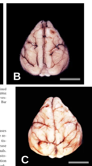
Fig. 2. Macroscopic appearances of the brain obtained
from (A) the control rabbit, (B) the 2 day HDT rabbit and (C) the 8 day HDT rabbit. Dilatation of the pial ves-sels is observed in the brain of HDT rabbits (B, C). Bar = 1 cm (from Shimoyama et al., 2000).
We hypothesized that edema formation increases water content in the brain tissue. However, the re-sults showed that the water content in the brain tis-sue of rabbits exposed to HDT did not increase significantly compared with that of control animals. Neither Evans blue staining nor immunohisto-chemical examination demonstrated extravasation of plasma constituents in the brain. Although mark-ed congestion was observmark-ed in the brain (Fig. 2), hematoxilin and eosin staining showed no edema-tous changes. In addition, transmission electron microscopy revealed that tight junctions of the cap-illary endothelium were intact in the HDT rabbits (Fig. 3). These findings suggest that either HDT up to 8 days does not produce brain edema in rabbits or it induces only a slight brain edema that is hard to dem-onstrate by only measuring water content or histo-logical examinations. Regulation of microcirculation seems to be well maintained in the brain of rabbits exposed to 45˚ HDT. A recent study of ours
show-ed that exposure to 8 days of HDT increasshow-ed the thickness of the medial layer in the rabbit basilar artery and decreased the intraluminal cross-sectional area (Fig. 4) (Ueda, 2001). Such morphological changes may play a beneficial role in preventing edema formation during HDT in rabbits. In addi-tion, although it is commonly accepted that there are no lymphatics in the brain of most animals, as much as half of the drainage of CSF is through the lymph in rabbits (Bradbury et al., 1981). Ohhashi
A
B
Fig. 3. Transmission
ele-ctron microscopy reveal-ed that the tight junction of capillary endothelium was intact and the features of brain edema, such as pinocytotic vesicles, were not seen in the HDT group (from Shimoyama et al., 2000).
Fig. 4. Typical cross-sectional
views of rabbit arteries.
A: basilar, control. B: basilar, HDT. C: popliteal, control. D: popliteal, HDT.
(from Ueda et al., 2001)
and colleagues (1993) revealed that venous valves were found in rabbit jugular veins but not in those of monkeys. These differences may explain the discrepancy between our result and that in monkeys reported by Kaplansky and coworkers (1985). Thus, we have to take account of species differences in analyzing the effect of HDT.
IV. Orthostatic Intolerance
Astronauts frequently experience orthostatic in-tolerance after coming back to Earth from space. Symptoms have ranged from increased heart rate to frank syncope. In Gemini crewmembers after 14 days of space flight, tilting them from a supine
con-A
B
C
D
50 µm 50 µm 100 µm 100 µmtrol posture to a 70˚ head-up tilt position increased heart rate by about 50 beats/min, and the test was aborted because of presyncopal symptoms (Berry and Catterson, 1967). Syncope is presumably at-tributable to decreased CBF. In our previous study, CBF velocity decreased to 87% of the pre-HDT baseline value during upright seated recovery after 24 h of HDT (Kawai et al., 1993). Although pre-syncope or pre-syncope was not observed in the study, decreased CBF velocity is likely to increase the risk of syncope during stand test or lower body negative pressure which is commonly used to quantify ortho-static deconditioning.
Several factors are involved in brain ischemia. First, diminished blood volume during space flight plays a significant role in orthostatic intolerance (Hoffler, 1977; Johnson, 1979). Bungo and col-leagues (1985) showed an efficacy of fluid loading with 1 L of normal saline to prevent symptoms of intolerance. The efficacy of this countermeasure, however, varies with flight duration. It seems to protect orthostatic tolerance only for short space flight. Second, Thornton and Hoffler (1977) sug-gested that an increase of vascular compliance in the lower extremities might play a certain role. It increases accumulation of blood in the legs and de-creases venous return, resulting in decreased cardi-ac output. Third, a disturbance in the baroceptor reflex has been proposed to be involved in the mechanism for orthostatic intolerance caused by exposure to microgravity (Convertino et al., 1990; Eckberg et al., 1992; Fritsch et al., 1992).
Microgravity is a stressful environment for humans who are adapted to the 1 g on Earth. Furthermore, we spend most of our time during day standing or sitting where the direction of gravity aligns with the long axis of the body (i.e., from head to foot). Therefore, the hydrostatic pressure gradient along the vascular tree produced by Earth gravity plays a significant role in the regulation of circulatory func-tion. Thus, we have to know what happens in our body during exposure to microgravity in order to make further progress in space development. Such
knowledge will provide us with a lot of information in other aspects of medicine such as treatment of senile persons who, on rare occasions, sit or stand up.
Acknowledgments: Some of our studies referred in this
article were carried out as a part of “Ground Research Announcement for Space Utilization” promoted by the Japanese Space Forum.
Reference
1 Aaslid R, Markwalder T-M, Nornes H. Noninvasive transcranial Doppler ultrasound recording of flow velocity in basal cerebral arteries. J Neurosurg 1982; 57:769–774.
2 Asa i Y, Inoue S, Tatebayashi K, Shiraishi Y, Kawai Y. Effects of head-down tilt on cerebral blood flow and somatosensory-evoked potentials in rabbits. Jpn J Physiol 2002;52:105–110.
3 Bagian JP, Hackett P. Cerebral blood flow: compar-ison of ground-based and spaceflight data and corre-lation with space adaptation syndrome. J Clin Phar-macol 1991;31:1036–1040.
4 Berry CA, Catterson AD. Pre-Gemini medical pre-dictions vs. Gemini flight results. In: Gemini sum-mary conference. Washington DC: Government Printing Office; 1967. p. 197–281.
5 Bondar RL, Stein F, Kassam MS, Dunphy PT, Bennett BS, Johnston KW. Cerebral blood flow ve-locities by transcranial Doppler during parabolic flight. J Clin Pharmacol 1991;31:915–919.
6 Bradbury MWB, Cserr HF, Westrop RJ. Drainage of cerebral interstitial fluid into deep cervical lymph of the rabbit. Am J Physiol 1981;240:F329–F336. 7 Bungo MW, Charles JB, Johnson PC Jr.
Cardiovas-cular deconditioning during space flight and the use of saline as a countermeasure to orthostatic intoler-ance. Aviat Space Environ Med 1985;56:985–990. 8 Convertino VA, Doerr DF, Eckberg DL, Fritsch JM,
Vernikos-Danellis J. Head-down bed rest impairs vagal baroreflex responses and provokes orthostatic hypotension. J Appl Physiol 1990;68:1458–1464. 9 Doi M, Kawai Y. Mechanisms of increased
intra-cranial pressure in rabbits exposed to head-down tilt. Jpn J Physiol 1998;48:63–69.
10 Eckberg DL, Convertino VA, Fritsch JM, Doerr DF. Reproducibility of human vagal carotid
baro-receptor-cardiac reflex responses. Am J Physiol 1992;263:R215–220.
11 Florence G, Lemenn M, Desert S, Bourron F, Serra A, Bonnier R, et al. Cerebral cortical blood flow in rabbits during parabolic flights (hypergravity and microgravity). Eur J Appl Physiol 1998;77:469– 478.
12 Frey MA, Mader TH, Bagian JP, Charles JB, Meehan RT. Cerebral blood velocity and other cardiovascular responses to 2 days of head-down tilt. J Appl Physiol 1993;74:319–325.
13 Friden HG, Ekstedt J. Volume/pressure relationship of the cerebrospinal space in humans. Neurosurgery 1983;13:351–366.
14 Fritsch JM, Charles JB, Bennet BS, Jones MM, Eckberg DL. Short-duration space flight impairs hu-man carotid baroreceptor-cardiac reflex responses. J Appl Physiol 1992;73:664–671.
15 Hargens AR, Tipton CM, Gollnick PD, Mubarak SJ, Tucker BJ, Akeson WH. Fluid shifts and muscle func-tion in humans during acute simulated weightless-ness. J Appl Physiol 1983;54:1003–1009.
16 Hoffler GW. Cardiovascular studies of U.S. space crews: an overview and perspective. In: Hwang NHC, Normann NA, eds. Cardiovascular flow dy-namics and measurements. Baltimore: University Park Press;1977.
17 Johnson PC Jr. Fluid volume changes induced by space flight. Acta Astronautica 1979;6:1335–1341. 18 Kakurin LI, Lobachik VI, Mikhailov VM, Senkevich
YuA. Antiorthostatic hypokinesia as a method of weightlessness simulation. Aviat Space Environ Med 1976;47:1083–1086.
19 Kaplansky AS, Savina YeA, Kazakova PB, Khoroshilova-Maslova IP, Kharin GM, Yakovleva VI, et al. Morphological study of antiorthostatic hypo-kinesia in monkeys. Kosm Biol Aviakosm Med 1985;19:53–60.
20 Kawai Y, Doi M, Matsuura K, Setogawa A, Hargens AR, Murthy G, et al. Cerebral hemodynamics dur-ing simulated microgravity in humans and rats. In: Sharma BK, Takeda N, Ganguly NK, Singal PK, eds. Adaptation biology and medicine. New Delhi: Narosa Publishing House; 1997. p. 155–162. 21 Kawai Y, Murthy G, Watenpaugh DE, Breit GA,
DeRoshia CW, Hargens AR. Cerebral blood flow velocity in humans exposed to 24 h of head-down tilt. J Appl Physiol 1993;74:3046–3051.
22 Kawai Y, Murthy G, Watenpaugh DE, Hargens AR. Cerebral blood flow velocity increases with acute head-down tilt of humans. Physiologist 1992;35 (Suppl):S186–S187.
23 Kawai Y, Okuda Y, Ogura K. Acute responses of brain oxygenation during postural change in hu-mans. In: Messmer K, Kubler WM, eds. Proceed-ings of the 6th World Congress for Microcirculation; 1995 December 9–12; Chandigarh. Bologna: Mondzzi Editore; 1996. p. 697–701.
24 Keil LC, McKeever KH, Skidmore MG, Hines J, Severs WB. The effect of head-down tilt and water immersion on intracranial pressure in nonhuman pri-mates. Aviat Space Environ Med 1992;63:181–185. 25 Kotani J, Adachi R, Fujita N, Sugioka S, Ueda Y. Effect of cerebral venous congestion on the pressure-volume index in the evaluation of intracranial pres-sure dynamics. J Neurosurg Anesthesiol 1993;5: 121–126.
26 Marchbanks RJ. Measurement of tympanic mem-brane displacement arising from aural cardiovascu-lar activity, swallowing, and intra-aural muscle re-flex. Acta Otolaryngol (Stockh) 1984;98:119–129. 27 Maurel D, Ixart G, Barbanel G, Mekaouche M,
Assenmacher I. Effects of acute tilt from orthostatic to head-down antiorthostatic restraint and of sustain-ed restraint on the intra-cerebroventricular pressure in rats. Brain Res 1996;736:165–173.
28 Murthy G, Marchbanks RJ, Watenpaugh DE, Meyer JU, Eliashberg N, and Hargens AR. Increased intra-cranial pressure in humans during simulated micro-gravity. Physiologist 1992;35:S184–S185.
29 Nixon JV, Murray RG, Bryant C, Johnson RL Jr, Mitchell JH, Holl OB, et al. Early cardiovascular ad-aptation to simulated zero gravity. J Appl Physiol 1979;46:541–548.
30 Ohhashi T, Morimoto-Murase K, Kitoh T. Physiolo-gy and functional anatomy of the venous system. In: Hirakawa S, Rothe CF, Shoukas AA, Tyberg JV, eds. Veins: their functional role in the circulation. Tokyo: Springer-Verlag; 1993. p. 33–47.
31 Parazynski SE, Hargens AR, Tucker B, Aratow M, Styf J, Crenshaw A. Transcapillary fluid shifts in tissues of the head and neck during and after simu-lated microgravity. J Appl Physiol 1991;7:2469– 2475.
32 Satake H, Konishi T, Kawashima T, Matsunami K, Uno T, Imai S, et al. Intracranial blood flow mea-sured with single photon emission computer tomog-raphy (SPECT) during transient –6 degrees head-down tilt. Aviat Space Environ Med 1994;65:117– 122.
33 Savin E, Bailliart O, Checoury A, Bonnin P, Grossin C, Martineaud JP. Influence of posture on middle cerebral artery mean flow velocity in humans. Eur J Appl Physiol 1995;71:161–165.
Contractile responses of the basilar artery isolated from rabbits exposed to 8-day head-down tilt. Jpn J Physiol 2001;51:209–215.
38 Wen TS, Randall DC, and Zolman JF. Protein accumulation in cerebrospinal fluid during –90˚ head-down tilt in rabbit. J Appl Physiol 1994;7: 1081–1086.
Received December 25, 2002; accepted January 10, 2003 Corresponding author: Yasuaki Kawai
34 Shimoyama R, Miyata H, Ohama E, Kawai Y. Does edema formation occur in the rabbit brain exposed to head-down tilt? Jpn J Physiol 2000;50:141–147. 35 Thornton WE, Hoffler GW. Hemodynamic studies
of the legs under weightlessness. In: Johnston RS, Dietlein LF, eds. Biomedical results from Skylab (NASA SP-377). Washington DC: Government Printing Office; 1977. p. 324–329.
36 Thornton WE, Moore TP, Pool SL. Fluid shifts in weightlessness. Aviat Space Environ Med 1987;58 (Suppl):A86–90.