Multicomponent Analysis by Surface Plasmon Resonance-based Immunosensor for Control of Food Hygiene
Tomomi Yamasaki
March 15, 2019
Contents
List of abbreviations … 1
Introduction … 3
Chapter 1 Development of an Immunosensor Based on Surface Plasmon Resonance for Simultaneous Residue Analysis of Three Pesticides
—Boscalid, Clothianidin, and Nitenpyram— in Vegetables … 6
Chapter 2 Development of a Surface Plasmon Resonance-Based Immunosensor for Detection of 10 Major O-Antigens on Shiga Toxin-Producing Escherichia coli, with a Gel Displacement Technique to Remove
Bound Bacteria …31
Chapter 3 Evaluation of a Surface Plasmon Resonance-Based Multiplex O-antigen Serogrouping for Escherichia coli using Eleven Major
Serotypes of Shiga Toxin-Producing E. coli …54
Chapter 4 Specific Detection of c-Kit Expressed on Human Cell Surface by
Immunosensor Based on Surface Plasmon Resonance …72
List of publications …81
Acknowledgements …83
1
List of abbreviations
BSA bovine serum albumin
CCD charge-coupled device
CDC Centres for Disease Control and Prevention CFU colony-forming units
DART direct analyses in real time
dcELISA direct competitive enzyme-linked immunosorbent assay
DCM dichloromethane
DMSO dimethyl sulfoxide E. coli Escherichia coli
EDC 1-ethyl-3-(3-dimethylaminopropyl)carbodiimide, hydrochloride EHEC enterohemorrhagic E. coli
ELISA enzyme-linked immunosorbent assay GdnHCl guanidine hydrochloride
HEK293T human embryonic kidney cell line HPLC high performance liquid chromatography HRP horseradish peroxidase
HUS haemolytic uremic syndrome IC20 20% inhibitory concentrations IC50 50% inhibitory concentrations IC80 80% inhibitory concentrations
IR Infrared
LC-MS iquid chromatography with mass spectrometry MEG01s human megakaryoblastic leukemia cell line
2 MoAb monoclonal antibody
MRL maximum residue limit
NHS N-hydroxysuccinimide NMR nuclear magnetic resonance PBS phosphate buffered saline PBS-T PBS containing 0.1% Tween 20 PCR polymerase chain reaction PFGE pulsed-field gel electrophoresis PoAb polyclonal antibody
RSD relative standard deviation
RU resonance unit
SDS sodium dodecyl sulfate SPR surface plasmon resonance
STEC Shiga toxin-producing Escherichia coli TFA trifluoroacetic acid
3
Introduction
Immunoassay is used for various analyses in the field of food hygiene. An immunosensor based on surface plasmon resonance (SPR immunosensor) can measure antigen-antibody reaction without labeling of antibody and allows rapid multicomponent analysis. In this study, SPR immunosensors for analyzing pesticide residues and Shiga toxin-producing Escherichia coli (STEC) have been developed. Furthermore, a SPR immunosensor for detecting surface proteins of animal cells has also been developed. These immunosensors would be applicable to other researches and analyses on food hygiene and cell surface factors in the future. Four chapters of this study are summarized below.
Chapter 1. Simultaneous analysis of three pesticides
Boscalid is a carboxamide fungicide which was introduced in 2002. Clothianidin and nitenpyram are neonicotinoid insecticides introduced in 2002 and 1995, respectively. These are representative pesticide widely used around the world. Since fungicides and insecticides are often used in the same time for agricultural crops, simultaneous analytical methods for their residues have been required. However, the enzyme-linked immunosorbent assay (ELISA), which is an immunoassay commonly used for their analysis, is usually used for a single component. We aimed to develop a SPR immunosensor capable of simultaneous measuring of these three pesticides, boscalid, clothianidin, and nitenpyram by using a monoclonal antibody to each pesticide.
A sensor chip with four independent flow channels was used. Three conjugates of derivatives of each pesticide with bovine serum albumin (BSA) and BSA as a control were immobilized on each of the flow channels, and the resultant sensor chip was attached to a SPR instrument. The monoclonal antibody against each pesticide and the standard solution of each
4
pesticide were all mixed, and the mixture was injected into the SPR instrument and measured.
Each pesticide was specifically measured in the range of 15 ng/mL to 93 ng/mL for boscalid, 6.7 ng/mL to 27 ng/ml for clothianidin, and 7.3 ng/mL to 62 ng/ml for nitenpyram. Pesticides spiked to vegetables can be measured at recovery of 72% to 105% with a high correlation to the direct competitive ELISA. It was confirmed that the SPR immunosensor developed is applicable as a practical analytical method for pesticide residues.
Chapter 2. Simultaneous serotyping of E. coli
STEC is identified by microbiology test including O antigen serotyping. Six type of O antigens, O26, O103, O111, O121, O145 and O157, are tested in Japan, and over 90% of STEC is covered. However, because there are infections by STEC with other O antigens, a method that can be serotyped simultaneously with other O antigens has been required. Then, a microarray type SPR immunosensor has been developed here, which is capable of simultaneous serotyping for ten types of O antigens including O91, O115, O128, and O159 added to the six types above.
Ten kind of antibodies were purified from commercially available rabbit antisera against each O antigens and immobilized on different positions of a sensor chip, and the resultant chip was attached to a SPR instrument. A suspension of E. coli was injected into the SPR instrument and analyzed. E. coli with respective O antigen could be serotyped within a minute.
Usually, regeneration of used chip was very difficult because of the removal of bound E.
coli without impairing the reactivity of antibody immobilized. We have succeeded in the regeneration by introducing gelatin gel. Now the sensor chip is able to be used for repeated measurement over 100 times.
Chapter 3. Clinical application of the developed SPR immunosensor
Clinical 188 isolates of STEC with eleven type of O antigens including O45 added to the
5
above 10 O antigens were examined by the SPR immunosensor, and the results were compare with it of the slide agglutination method which is the conventional method for O antigen serotyping.
The overall sensitivity of O antigen serotyping was 98.9%. The detection limits of all serotypes were distributed between 1.1×10⁶ CFU/mL and 17.6×10⁶ CFU/mL. It was concluded that the SPR immunosensor developed in chapter 2 is useful as a simultaneous and automated method for O antigen serotyping of STEC.
Chapter.4 Detection of membrane protein c-Kit of animal cells
C-Kit is a cell membrane receptor with tyrosine kinase activity, which converts extracellular signals received into intracellular signals and controls the proliferation, differentiation, survival, metabolism, and migration of cells. C-Kit is important as a tumor marker because it is known to be highly expressed in gastrointestinal stromal tumors, germinal cell tumors such as seminomas, malignant melanoma, acute myeloid leukemia, etc. SPR immunosensor is developed as a detection method of c-Kit expressed on cell surface.
A commercially available anti-c-Kit antibody was immobilized on the surface of the sensor chip, and the resultant sensor chip was attached to a SPR instrument. A suspension of human megakaryoblastic leukemia cell line (MEG01s) and human embryonic kidney cell line (HEK 293T) was injected into the SPR instrument and analyzed. Although c-Kit could be detected, nonspecific reactions were observed. However, it was greatly suppressed by adding gelatin into the antibody solution for immobilizing. In addition, gelatin was also helpful for the regeneration of a used sensor chip as a described in chapter 2.
6
Chapter 1 Development of an Immunosensor Based on Surface Plasmon Resonance for Simultaneous Residue Analysis of Three Pesticides
—Boscalid, Clothianidin, and Nitenpyram— in Vegetables
Abstract
A simultaneous immunosensor based on surface plasmon resonance (SPR) was developed for determination of 3 pesticides - boscalid, clothianidin and nitenpyram - instead of the direct competitive enzyme-linked immunosorbent assays (dcELISAs) widely used as individual determination methods. Carboxy groups that introduced compounds to their pesticides were designed, and conjugates of them and bovine serum albumin were immobilized onto separate channels of the same sensor chip. When a mixture of 3 monoclonal antibodies reacted to each pesticide, and 3 pesticides were injected into the SPR immunosensor, each channel showed specific reactivity at 15 – 93 ng/mL for boscalid, 6.7 – 27 ng/mL for clothianidin, and 7.3 – 62 ng/mL for nitenpyram. Recovery tests using vegetables spiked with a mixture of 3 pesticides showed good results: 75 – 90%, 88 – 104%, and 72 – 105%, respectively, with a high correlation to results of the dcELISAs. The SPR immunosensor would be useful for the determination of pesticide residues in vegetables.
Introduction
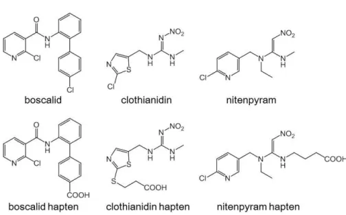
Boscalid, 2-chloro-N-(4’-chlorobiphenyl-2-yl) nicotinamide, is a carboxamide fungicide that was introduced in 2002.1 Both clothianidin [(E)-1-(2-chloro-1,3-thiazol-5-ylmethyl)-3- methyl-2-nitroguanidine] and nitenpyram [(E)-N-(6-chloro-3-pyridylmethyl)-N-ethyl-N'- methyl-2-nitrovinylidenediamine] are neonicotinoid insecticides introduced in 2002 and 1995, respectively.2,3 Clothianidin is a modified derivative of nitenpyram, although they have different ring structures; clothianidin has a chlorothiazol ring, whereas nitenpyram has a chloropyridine
7
ring, as shown in Fig. 1.4 Fungicides and insecticides are often applied simultaneously to agricultural fields when the plants are put to risks of fungal disease and insect damage, especially in hot and humid seasons. The combination of boscalid and clothianidin or
nitenpyram is also applied widely to prevent fungal diseases and insect pests. It is important to monitor their residues simultaneously in agricultural products. The maximum residue limits (MRLs) in vegetables have been set to 1 – 40 mg/kg for boscalid, 0.2 – 40 mg/kg for
clothianidin, and 0.2 – 5 mg/kg for nitenpyram in Japan, including some exceptional vegetables for which the ranges are lower.
Fig. 1 Structure of boscalid, clothianidin, nitenpyram and their haptens.
Generally, boscalid is determined by gas or liquid chromatography by mass
spectrometry.5-8 Clothianidin and/or nitenpyram are determined by high-performance liquid chromatography (HPLC) with diode-array detection or by liquid chromatography with mass spectrometry (LC-MS).9-14 Boscalid and clothianidin can be determined simultaneously by
8
multi-residue analysis using LC-MS.15-17 These instrumental analyses are sensitive and accurate;
however, these technologies are sophisticated, labor-intensive, and time-consuming.
Direct competitive enzyme-linked immunosorbent assays (dcELISAs) were developed for the monitoring of pesticide residues in vegetables as more simple and rapid methods compared to the above chromatography techniques, but of the single pesticide although simultaneous analysis is required in the field.18-20 The development of simultaneous dcELISA for three pesticides was described previously.21 Its reactivity was specific with each pesticide, but was difficult to put into practical use because optimization of the assay condition was complicated.
Immunosensors based on electrochemistry have been developed to determine pesticides in foods.22-24 Immunosensors based on surface plasmon resonance (SPR immunosensors) have also been developed to determine pesticides.20,25-27 Since SPR immunosensors especially enable real-time monitoring of the antigen-antibody interaction in contrast to the above dcELISAs and electrochemical immunosensors, they are expected to be applicable to pesticide residue analysis in fresh vegetables that will be quickly distributed. However, there have been no reports of any SPR immunosensor for the simultaneous and rapid analysis of pesticides, although it involves required techniques.
We hypothesized that a simultaneous SPR immunosensor would be developed if each of the antigen-antibody interactions showed no cross-reaction to the other target pesticides. In this study, we have described the successful development of a simultaneous SPR immunosensor to determine boscalid, clothianidin, and nitenpyram, with no cross-reaction among the pesticides, as their boscalid part is shown in Fig. 2. The design of the haptens has also been discussed in the context of developing simultaneous immunosensors.
9
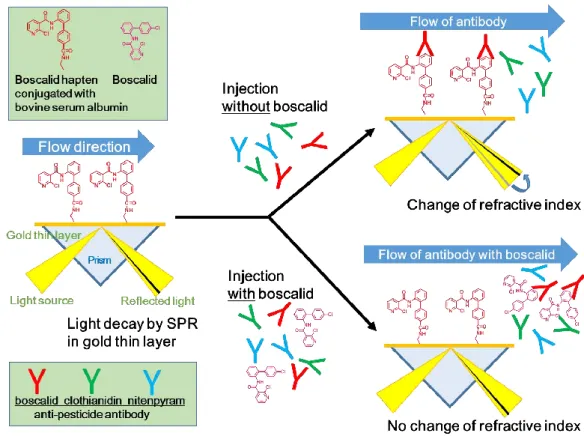
Fig. 2 Schematic illustration of boscalid determination on boscalid channel in the SPR immunosensor.
Experimental
Reagents and chemicals
Boscalid, benalaxyl, fenhexamid, tecloftalam, clothianidin, nitenpyram, imidacloprid, dinotefuran, thiacloprid, thiamethoxam were purchased from Wako Pure Chemical Industries, Ltd. (Osaka, Japan). Acetamiprid was purchased from Hayashi Pure Chemical Ind., Ltd. (Osaka, Japan). Bovine serum albumin (BSA: Prod. No. A7888) was purchased from Sigma-Aldrich Co.
(St. Louis, MO, USA). Horseradish peroxidase (HRP) was purchased from Toyobo Co., Ltd.
(Osaka, Japan). The anti-boscalid MoAb was prepared as described previously.20 The anti- clothianidin MoAb and anti-nitenpyram MoAb were provided from Horiba Ltd. (Kyoto, Japan).18 All other chemicals and reagents used were of analytical grade, and purchased from Wako Pure Chemical Industries, Ltd. or Nacalai Tesque, Inc.
10 Hapten design for boscalid and clothianidin
The haptens for boscalid and clothianidin were synthesized as described previously.18,20 Their structures were summarized in Fig. 1.
Hapten design for boscalid, clothianidin, and nitenpyram
The hapten for nitenpyram was synthesized using the following scheme, referring to previous reports.28,29 The structure was described in Fig. 1. All reactions were performed under an atmosphere of argon unless otherwise noted. Dichloromethane (CH2Cl2) was purchased from Kanto Chemical Co., Inc. (Tokyo, Japan). All reactions were monitored by thin layer
chromatography that is glass plates pre-coated with silica gel (60 F254; layer thickness, 0.2 mm) purchased from Merck KGaA (Darmstadt, Deutschland). The products were visualized using irradiation with UV light or by treatment with a solution of phosphomolybdic acid or a solution of p-anisaldehyde. Flash column chromatography was performed using silica gel (Art.
No. 7734) purchased from Merck KGaA. All other chemicals and reagents used were analytical grade, and purchased from Kanto Chemical Co., Inc..
Infrared (IR) spectra were measured on a JASCO FT/IR-4600 spectrometer. 1H nuclear magnetic resonance (NMR) (500 MHz, 400 MHz) and 13C NMR (125 MHz, 100 MHz) spectra were recorded on JEOL JNM-ECX500 and JEOL JNM-ECS400 spectrometers, respectively.
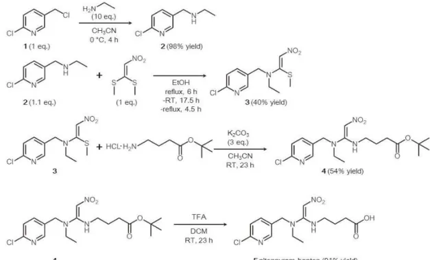
Chemical shifts were reported as δ values (ppm) relative to tetramethylsilane (0 ppm) and dimethyl sulfoxide (DMSO-d6; 2.50 ppm). Direct analyses in real time (DART) mass (positive mode) analyses were performed on a liquid chromatography-time-of-flight JMS-T100LP instrument. The overall synthesis scheme is summarized in Fig. 3.
1) Synthesis of N-((6-chloropyridin-3-yl)methyl)ethanamine (2) 28
A solution of 2-chloro-5-(chloromethyl)pyridine (1) (972.1 mg, 6.0 mmol) in CH3CN (1.5
11
mL) was slowly added to a solution of ethanamine (3.865 g, 60 mmol, 70 wt% aqua.) in CH3CN (3.0 mL) over 25 min at 0 °C (in an ice-bath). The resulting mixture was stirred for 4 h at 0 °C (in an ice-bath), followed by the addition of H2O (10 mL). The product was extracted with CH2Cl2 and dried with Na2SO4. The solvent was removed to give crude
N-((6-chloropyridin-3-yl)methyl)ethanamine (2) (1.007 g) in 98% yield. 2 was used for the next step without further purification. 2: 1H NMR (500 MHz, CDCl3) δ 8.33 (d, 1H, J = 2.29 Hz, H-2 Py), 7.67 (dd, 1H, J = 2.29, 8.03 Hz, H-4 Py), 7.29 (d, 1H, J = 8.03 Hz, H-5 Py), 3.79 (s, 2H, -PyCH2NH-), 2.67 (q, 2H, J = 7.26 Hz, -NHCH2CH3), 1.13 (t, 3H, J = 7.26 Hz, -NHCH2CH3) ppm. 13C NMR (125 MHz, CDCl3) δ 149.6, 149.0, 138.5, 134.7, 123.7, 50.0, 43.4, 14.9 ppm.
High-resolution mass spectrometry (HRMS) (DART) m/z: calcd. for C8H12ClN2 (M+1+) 171.0689, found 171.0682. IR (neat) 3418, 3087, 2968, 1458, 1104 cm-1.
2) Synthesis of
N-((6-Chloropyridin-3-yl)methyl)-N-ethyl-1-(methylthio)-2-nitroethen-1-amine (3)28 N-((6-chloropyridin-3-yl)methyl)ethanamine (2) (1.00 g, 5.9 mmol) was dissolved in anhydrous EtOH (3.0 mL), followed by the addition of
(2-nitroethene-1,1-diyl)bis(methylsulfane) (886.4 mg, 5.4 mmol). The resulting mixture was heated to reflux for 6 h, followed by continuous stirring for 17.5 h at room temperature. The mixture was refluxed again for 4.5 h and cooled to room temperature. The solvent was then removed. The residue was purified by column chromatography on silica gel (hexane/ethyl acetate = 3/1→1/1→1/3 v/v) to give
N-((6-chloropyridin-3-yl)methyl)-N-ethyl-1-(methylthio)-2-nitroethen-1-amine (3) (616.3 mg) in 40% yield. 3: 1H NMR (400 MHz, CDCl3) δ 8.27 (d, 1H, J = 2.44 Hz, H-2 Py), 7.55 (dd, 1H, J = 2.44, 8.24 Hz, H-4 Py), 7.35 (d, 1H, J = 8.24 Hz, H-5 Py), 6.80 (s, 1H, -CHNO2), 4.69 (s, 2H, -PyCH2N-), 3.49 (q, 2H, J = 7.02 Hz, -NCH2CH3), 2.47 (s, 3H, -SCH3), 1.25 (t, 3H, J = 7.02 Hz, -NCH2CH3) ppm. 13C NMR (125 MHz, CDCl3) δ 165.7, 151.2, 148.6, 137.9, 130.1,
12
124.5, 113.8, 53.1, 48.5, 17.7, 13.1 ppm. HRMS(DART) m/z: calcd. for C11H15ClN3O2S (M+1+) 288.0574, found 288.0549. IR (neat) 2976, 1519, 1388, 1258, 1106 cm-1.
3) Synthesis of tert-butyl 4-((1-(((6-chloropyridin-3-yl)methyl)(ethyl)amino)-2- nitrovinyl)amino)butanoate (4)29
A solution of tert-butyl 4-aminobutanoate hydrochloride (19.6 mg, 0.1 mmol) in CH3CN (0.1 mL) was added drop-wise to a mixture of
N-((6-chloropyridin-3-yl)methyl)-N-ethyl-1-(methylthio)-2-nitroethen-1-amine (28.8 mg, 0.1 mmol) and K2CO3 (41.5 mg, 0.3 mmol) in dry CH3CN (0.6 mL) at room temperature. The resulting mixture was stirred for 23 h at room temperature. The solvent was then removed and the residue was purified by column chromatography on silica gel (hexane/ethyl acetate = 2/1→1/1→1/0→ethyl acetate/methanol = 1/1 v/v) to give
tert-butyl 4-((1-(((6-chloropyridin-3-yl)methyl)(ethyl)amino)-2-nitrovinyl)amino)butanoate (4) (21.6 mg) in 54% yield. 4: 1H NMR (500 MHz, CDCl3) δ 9.56 (bs, 1H, -NH), 8.29 (d, 1H, J = 2.29 Hz, H-2 Py), 7.56 (dd, 1H, J = 2.68, 8.03 Hz, H-4 Py), 7.36 (d, 1H, J = 8.03 Hz, H-5 Py), 6.52 (s, 1H, -CHNO2), 4.35 (s, 2H, -PyCH2N-), 3.40 (q, 2H, J = 6.88 Hz, -NCH2CH3), 3.12 (q, 2H, J =7.26 Hz, -NCH2CH2CH2COOt-Bu), 2.36 (t, 2H, J = 6.88 Hz, -NCH2CH2CH2COOt-Bu), 1.93-1.99 (m, 2H, -NCH2CH2CH2COOt-Bu), 1.44 (s, 9H, t-Bu), 1.18 (t, 3H, J = 6.88 Hz, -NCH2CH3) ppm. 13C NMR (125 MHz, CDCl3) δ 171.8, 162.1, 151.1, 148.6, 138.0, 130.2, 124.5, 103.8, 80.7, 49.8, 44.9, 44.6, 31.9, 27.9, 25.2, 12.0 ppm. HRMS(DART) m/z: calcd. for C18H28ClN4O4 (M+1+) 399.1799, found 399.1799. IR (neat) 3452, 3131, 2977, 2934, 1724, 1517, 1366 cm-1.
4) Synthesis of nitenpyram hapten,
4-((1-(((6-chloropyridin-3-yl)methyl)(ethyl)amino)-2-nitrovinyl)amino)butanoic acid (5)29 A solution of
tert-butyl 4-((1-(((6-chloropyridin-3-yl)methyl)(ethyl)amino)-2-nitrovinyl)amino)butanoate (4)
13
(17.2 mg, 0.043 mmol) in CH2Cl2 (DCM; 0.43 mL) was treated with CF3COOH (trifluoroacetic acid (TFA); 46 μL). The mixture was stirred for 23 h at room temperature. The solvent was removed and the residue was purified by column chromatography on silica gel (ethyl acetate/methanol = 15/1→8/1 v/v) to give the desired nitenpyram hapten,
4-((1-(((6-chloropyridin-3-yl)methyl)(ethyl)amino)-2-nitrovinyl)amino)butanoic acid (5) (13.4 mg) in 91% yield. 5: 1H NMR (500 MHz, DMSO-d6) δ 12.17 (bs, 1H, -COOH), 8.67 (bs, 1H, - NH), 8.34 (d, 1H, J = 2.29 Hz, H-2 Py), 7.34 (dd, 1H, J = 2.29, 8.03 Hz, H-4 Py), 7.51 (d, 1H, J
= 8.03 Hz, H-5 Py), 6.43 (s, 1H, -CHNO2), 4.51 (s, 2H, -PyCH2N-), 3.22-3.27 (m, 4H, - NCH2CH3, -NCH2CH2CH2COOH), 2.23 (t, 2H, J = 7.26 Hz, -NCH2CH2CH2COOH), 1.70-1.77 (m, 2H, -NCH2CH2CH2COOH), 1.11 (t, 3H, J = 7.26 Hz, -NCH2CH3) ppm.
Fig. 3 Scheme of the nitenpyram hapten synthesis.
14 Preparation of hapten and protein conjugate
Boscalid, clothianidin, and nitenpyram haptens were, respectively, conjugated to BSA and HRP, as described previously.20
Constitution of SPR immunosensor
The SPR immunosensor comprised a commercially available microflow-type instrument (Biacore T200; GE Healthcare Europe, Munich, Germany), and its sensor chip had four channels coated with carboxymethyl dextran (CM5; GE Healthcare Europe), as described previously. 20,27
1) Preparation of the sensor chip for the three pesticides
The sensor chip was placed in the instrument. The chip channels were rinsed with PBS containing 0.005% tween 20 for 600 s at 5 μL/min. The carboxy groups in the channels were then activated by rinsing with a mixture of 80 μL of EDC (400 mmol/L) and 80 μL of NHS (100 mmol/L) dissolved in distilled water for 350 s at 5 μL/min. The three kinds of hapten and BSA conjugates (40 μg/mL each) dissolved in acetic acid buffer (10 mmol/L; pH 5.0) were
respectively added to their respective channels for 360 s at 5 μL/min to immobilize them onto the channels. The remaining channel was used for a negative control to permit deletion of the baseline noise. Ethanolamine (1 mol/L; pH 8.5) was allowed to flow for 350 s at 5 μL/min to inactivate the residual carboxy groups. The prepared sensor was rinsed with running buffer (50 mmol/L phosphate buffer containing 75 mmol/L NaCl, 0.1% BSA, and 5% methanol; pH 7.0), and was then ready for use.
2) Preparation of sample
The pesticide standard solutions were prepared with 10% methanol to the following concentrations: boscalid and nitenpyram (3.1 – 200 ng/mL), clothianidin (1.6 – 100 ng/mL), dinotefuran (1.6 ng/mL – 10 μg/mL), thiacloprid (160 ng/mL – 100 μg/mL), and the other
15
pesticides (10 μg/mL). The pesticides were mixed at the same final concentrations for simultaneous analysis. By contrast, the anti-boscalid, anti-clothianidin, and anti-nitenpyram MoAbs were diluted to 15 μg mL-1 with high ion strength phosphate buffered saline (modified PBS: 100 mmol/L phosphate, 150 mmol/L NaCl; pH7.0) containing 0.2% BSA. A mixture of the MoAbs was also prepared at the same final concentration. The pesticide standard solution or the diluent prepared from the vegetables (75 μL) was mixed with an equal volume of the MoAb solution (75 μL), and used as measurement samples.
3) Analysis conditions
Measurement samples were allowed to flow serially through the first boscalid channel, the second clothianidin channel, and the last nitenpyram channel immobilized with each of the corresponding haptens and BSA conjugate for 180 s at 20 μL/min. The solution was
continuously changed to the running buffer, and it was allowed to flow further for 180 s at 20 μL/min to obtain the Kd values.
4) Regeneration of the sensor-chip
After the reaction was complete, the sensor-chip was regenerated by removal of the bound MoAbs. GdnHCl (3.0 mol/L) in acetic acid (1.0 mol/L; pH 1.9) was allowed to flow initially for 60 s at 20 μL/min. After rinsing with distilled water for 60 s at 20 μL/min, the chips were rinsed with 0.2% SDS for 120 s at 20 μL/min to complete regeneration. The sensor chip was re-used after rinsing with running buffer.
5) Data processing
Raw data were output as resonance units (RUs). A 0.1 degree refractive index shift by the SPR phenomenon was defined as 1000 RUs. The actual reaction signal was represented as the delta RU value at 180 s from the start of the reaction.
16 dcELISA
dcELISA was performed as described previously.20 The specific conditions for boscalid, clothianidin, and nitenpyram are described below. The anti-pesticide MoAbs were diluted with PBS (10 mmol/L phosphate, 150 mmol/L NaCl; pH7.0) to the following concentrations: anti- boscalid MoAb (500 ng/mL), anti-clothianidin MoAb (250 ng/mL), and anti-nitenpyram MoAb (125 ng/mL). The hapten and HRP conjugates were diluted with modified PBS containing 0.2%
BSA to the following concentrations: 250 ng/mL for boscalid, 50 ng/mL for clothianidin, and 1000 ng/mL for nitenpyram. Pesticide standard solutions were prepared in 10% methanol to the following concentrations: boscalid (0.038 – 156 ng/mL), clothianidin (0.024 – 100 ng/mL), and nitenpyram (0.24 – 1000 ng/mL).
Preparation of vegetable samples
A variety of vegetables belonging to different families, (broccoli (Brassica oleracea var.
italica), cucumber (Cucumis sativus L.), lettuce (Lactuca sativa L.), spinach (Spinacia oleracea L.), tomato (Solanum lycopersicum L.), and Welsh onion (Allium fistulosum L.)) were purchased from a market in Kyoto city. dcELISAs, which are more sensitive than the SPR immunosensor, were used to confirm that they did not contain any boscalid, clothianidin, or nitenpyram. The vegetable samples (100 g) were homogenized in a blender. Pesticide mixtures dissolved in methanol (100 μL) were spiked into the homogenized samples (5.0 g) in 50 mL screw-cap tubes at the final concentrations of boscalid, clothianidin, and nitenpyram: A) 2, 0.75, and 1.5 μg/g; B) 4, 1.5, and 3 μg/g; C) 8, 3, and 6 μg/g, respectively. After standing for 30 min at room
temperature, 25 mL of methanol was added to the homogenates. The tubes were shaken vigorously on a reciprocal shaker (Shaker SA320; Yamato Scientific Co., Ltd., Tokyo, Japan) for 30 min to extract the pesticides into the liquid phase. The extracts were centrifuged at 3000 rpm for 10 min at room temperature. The supernatants were diluted to 8.5-folds with distilled
17
water to prepare 10% methanol equivalent solutions. They were further diluted with 10%
methanol to adjust the concentrations to the working range of the SPR immunosensor or the dcELISA. The diluents were used to prepare measurement samples.
Results and Discussion Hapten design
For simultaneous analysis, measurement samples containing the three MoAbs and the three pesticides were allowed to flow serially through the boscalid, clothianidin, and nitenpyram channels immobilized with each of haptens and BSA conjugate. The hapten on one channel is therefore necessary to react with the corresponding MoAb only without any cross reaction on the other channels.
As shown in Fig. 1, boscalid has a structure different from that of clothianidin, but has the same 2-chloropyridine ring structure as nitenpyram. It is present in the basic structure at the ortho-position in boscalid, but at the para-position in nitenpyram. Generally, it is easy to raise MoAbs that recognize such a difference in the angle. We presumed that the haptens for boscalid would not react with the anti-nitenpyram MoAb. By contrast, the 2-chlorobenzene ring in the basic structure of the boscalid at the para-position is similar to the structure of the 2-
chloropyridine ring in nitenpyram, which might make it difficult for a MoAb to recognize the difference. Therefore, the linker of boscalid was extended from the chlorine position of the 2- chlorobenzene ring to inhibit any possible cross-reaction.
Clothianidin and nitenpyram belong to the same neonicotinoid insecticide group.
Clothianidin has a nitroguanidine structure that is similar to the nitrovinylidenediamine structure in nitenpyram. Thus, their haptens might react with MoAb raised against another hapten,
resulting in a failure to develop a useful simultaneous SPR immunosensor. In a previous study, two kinds of haptens of the insecticide etofenprox, whose linker sites were on opposite sides (at
18
the ethoxy group and the phenoxybenzene group), induced different cross reactivity with the antibodies raised against the haptens.30 Thus, we believed that the concept would be effective to develop a simultaneous SPR immunosensor. The clothianidin hapten was synthesized based on the published structure,18 and then the linker for the nitenpyram hapten was introduced at the opposite side from clothianidin (Fig. 1).
As shown in Table 1, each of the haptens actually functioned in the constituted SPR immunosensor without any cross-reaction with the other MoAbs, when each of the MoAb solutions flowed serially through the first boscalid channel, the second clothianidin channel, and the last nitenpyram channel.
Table 1 Typical signal of anti-pesticide MoAbs onto each pesticide channel by the constituted SPR immunosensor
MoAb Channel immobilized with hapten and BSA conjugate (delta RU)
Boscalid Clothianidin Nitenpyram
Anti-boscalid 450 0 0 Anti-clothianidin 2 1500 8 Anti-nitenpyram 0 3 310
Determination of pesticides by the SPR immunosensor
The anti-boscalid MoAb, the anti-clothianidin MoAb, and the anti-nitenpyram MoAb solutions were injected separately into the SPR immunosensor. The RU value of the MoAb solutions increased in a time-dependent manner, reaching 450 RU for boscalid, 1500 RU for clothianidin, and 310 RU for nitenpyram at 180 s from the reaction start point, as shown in Fig.
4A. The signals were returned to the baseline after washing with 3 kinds of regeneration buffers.
The Kd values, calculated from the time course results, were determined as 2.9 × 10⁻¹² mol/L for boscalid, 8.1 × 10⁻¹² mol/L for clothianidin, and 7.7 × 10⁻¹² mol/L for nitenpyram. All of the
19
MoAbs showed high affinity to the corresponding hapten and BSA conjugate, as indicated by the Kd values. These high affinities are important to constitute an SPR immunosensor for the accurate determination of the pesticides.
The determination of each pesticide was initially examined using the SPR immunosensor.
The signal produced between the hapten and BSA conjugate and the MoAb was inhibited by the corresponding pesticide in a concentration-dependent manner for all three pesticides, as shown in Figs. 4B – 4D. The inhibition curves were drawn using the signal data at 180 s from the reaction start point, as shown in Fig. 5. The 20%, 50%, and 80% inhibitory concentrations (IC20, IC50, and IC80) were 15, 41, and 93 ng/mL for boscalid; 6.7, 15, and 27 ng/mL for clothianidin;
and 7.3, 24, and 62 ng/mL for nitenpyram. The quantitative working ranges, defined as between the IC20 value and IC80 value, suggested that the SPR immunosensor was sufficiently sensitive to be applied to residue analysis of the target pesticides around the MRLs in the majority of vegetables.
The constituted SPR immunosensors were combined for the simultaneous analysis of their pesticides. A mixture of the three MoAbs was added to each pesticide, but also a mixture of their pesticides, and this mixture was injected into the SPR immunosensor. As shown in Fig.
5, the inhibition curves were almost identical to the above determination results for the
individual MoAbs. The simultaneous SPR immunosensor could determine boscalid in the range of 15 – 93 ng/mL, clothianidin in the range of 6.7 – 27 ng/mL, and nitenpyram in the range of 7.3 – 62 ng/mL. This successful result could be attributed by the design of highly specific haptens that reacted only with the corresponding MoAb and by the use of high affinity MoAbs.
20
Fig. 4 Time course of anti-pesticide MoAbs reaction without the corresponding pesticides on each channel in SPR-immunosensor (A), and their signal reduction by the corresponding pesticides (ng/mL): (B) boscalid, (C) clothianidin, (D) nitenpyram. W1, W2, and W3 show regeneration steps by GdnHCl in acetic acid (pH 1.9), distilled water, and 0.2% SDS, respectively.
21
Fig. 5 Inhibition curves for each of the pesticides by SPR-immunosensor: (A) boscalid, (B) clothianidin, (C) nitenpyram. (●) shows inhibition curve for each of the MoAbs with the 1 pesticide, (○) shows inhibition curve for the mixture of 3 MoAbs with the 1 pesticide, and (▲) shows inhibition curve for the mixture of 3 MoAbs with 3 pesticides. Each data point is the mean of triplicate in independent examinations; error bars indicate ± SD.
Cross reactivity
The constituted SPR immunosensor was highly specific to boscalid, clothianidin, and nitenpyram. However, it was not clear whether other structurally related pesticides would cross- react in the SPR immunosensor. Therefore, the cross-reactivity of the anti-boscalid MoAb was examined using fenhexamid, tecloftalam, and benalaxyl, which belong to the same carboxamide
22
fungicide group. The cross-reactivity of anti-clothianidin and anti-nitenpyram MoAbs was examined using acetamiprid, imidacloprid, dinotefuran, thiacloprid, and thiamethoxam, which are all neonicotinoid insecticides. The three MoAbs were mixed, and the mixture was further mixed with each of the pesticides. After injection into the SPR immunosensor, each of the IC50
values was obtained from inhibition curves drawn from the time course signal. The cross- reactivity (%) of the MoAbs was obtained from their ratio with the target pesticide. As described in Table 2, the sensor channel for boscalid was specific to boscalid. The sensor channel for nitenpyram was also specific to nitenpyram despite acetamiprid, imidacloprid, and thiacloprid having the 2-chloropyridine ring bound on the para-position like nitenpyram. It was speculated that the anti-nitenpyram MoAb used would recognize nitrovinylidenediamine via the 2-
chloropyridine ring of nitenpyram, because the linker of the hapten was extended from the methyl amine, which exists on the opposite side from the 2-chloropyridine ring.
In contrast, the anti-clothianidin MoAb cross-reacted with dinotefuran at the same level as clothianidin. The cross-reactivity was 119%. Dinotefuran and clothianidin have a common structure: the 1,3-dimethyl-2-nitroguanidin group. The hapten linker of clothianidin was extended from the chlorine atom position of the thiazole ring, which exists on the opposite side of the nitroguanidin group, as shown in Fig. 1. We speculated that the anti-clothianidin MoAb must recognize this common structure.
Thus, the cross-reactivity examination suggested that the SPR immunosensor could determine dinotefuran in addition to boscalid, clothianidin, and nitenpyram. Dinotefuran, clothianidin, and nitenpyram, which belong to the same insecticide group, are not usually applied to an agricultural field at the same time. Thus, it was suggested that the constituted SPR immunosensor can determine boscalid and dinotefuran simultaneously, in addition to boscalid and clothianidin, or boscalid and nitenpyram.
23
Table 2 Cross-reactivity of the MoAbs with the structurally related pesticides by the SPR immunosensor Pesticides
examined
Cross-reactivity (%) of MoAbs
Anti-boscalid Anti-clothianidin Anti-nitenpyram
Boscalid 100a <0.18 <0.22
Clothianidin <0.18 100 <0.22
Nitenpyram <0.18 <0.18 100
Fenhexamid <0.18 NTb NT
Tecloftalam <0.18 NT NT
Benalaxyl <0.18 NT NT
Acetamiprid NT <0.18 <0.22
Imidacloprid NT <0.18 <0.22
Dinotefuran NT 119 <0.22
Thiacloprid NT 0.21 <0.22
Thiamethoxam NT <0.18 <0.22
a. Each data is the mean of duplicates in independent examinations.
b. NT means not tested.
Recovery of pesticides spiked in vegetables
The recovery of the pesticides by the SPR immunosensor was examined using vegetable homogenates spiked with a mixture of boscalid, clothianidin, and nitenpyram. The lower quantitative limits of the three pesticides in vegetables were estimated to be 0.77 μg/g for boscalid, 0.34 μg/g for clothianidin, and 0.37 μg/g for nitenpyram from the preparation method of vegetable samples. The sensitivity was adequate to determine their concentrations around the MRLs for Welsh onion, lettuce, cucumber, tomato, broccoli, and spinach: 5 – 40 μg/g for boscalid, 1 – 40 μg/g for clothianidin, and 5 μg/g for nitenpyram. Mixtures of three pesticides were spiked at the following concentrations of boscalid, clothianidin, and nitenpyram: A) 2,
24
0.75, and 1.5 μg/g; B) 4, 1.5, and 3 μg/g; C) 8, 3, and 6 μg/g. Table 3 shows that the recovery values were 75 – 90% for boscalid, 88 – 104% for clothianidin, and 72 – 105% for nitenpyram.
The results suggested that the SPR immunosensor could determine the pesticides simultaneously with satisfactory recovery. The relative standard deviation (RSD) values associated with the recovery tests also showed a high repeatability at 0.00 – 10.1%, except for nitenpyram, which showed RSD values of 18.3% in Welsh onion (3 μg/g), 15.0% in spinach (3 μg/g), and 18.9% in spinach (6 μg/g). The results suggested that the constituted sensor is applicable for quantitative residue analysis of boscalid and clothianidin, and for the semi- quantitative analysis of nitenpyram in vegetables.
25
Table 3 Recovery examination of pesticide mixtures spiked in vegetables by the SPR immunosensor
Spiked pesticide conc. (μg/g)
Welsh onion Lettuce Cucumber
Reca RSDa Rec RSD Rec RSD
A
Boscalid 2 79.7b 2.49 76.4 1.44 77.2 0.00
Clothinidin 0.75 92.3 1.15 100 0.77 99.8 0.38
Nitenpyram 1.5 78.0 8.63 80.8 5.07 84.1 6.91
B
Boscalid 4 87.2 2.49 78.9 1.44 89.6 2.49
Clothinidin 1.5 96.3 3.32 98.5 1.01 96.9 2.39
Nitenpyram 3 84.1 18.3 98.5 5.07 95.2 6.91
C
Boscalid 8 83.8 3.80 85.5 1.44 88.8 1.44
Clothinidin 3 98.7 1.01 87.9 0.38 99.8 1.38
Nitenpyram 6 90.7 1.91 94.1 1.92 101 5.07
Spiked pesticide conc. (μg/g)
Tomato Broccoli Spinach
Rec RSD Rec RSD Rec RSD
A
Boscalid 2 75.5 3.80 78.9 1.44 74.7 2.49
Clothinidin 0.75 96.7 3.66 97.4 1.92 95.6 2.39
Nitenpyram 1.5 85.2 5.07 74.1 1.92 71.9 5.07
B
Boscalid 4 82.2 4.98 82.2 2.49 81.3 7.61
Clothinidin 1.5 94.5 2.99 99.6 1.33 98.7 4.32
Nitenpyram 3 89.6 3.32 94.1 8.36 94.1 15.0
C
Boscalid 8 78.0 10.1 83.0 3.80 77.2 2.49
Clothinidin 3 100 2.39 104 1.15 95.8 1.53
Nitenpyram 6 98.5 8.36 101 8.36 105 18.9
a. Rec shows recovery (%) and RSD shows relative standard deviation (%).
b. Each Rec. is the mean of triplicate in independent examinations.
26
Correlation results between the dcELISA and the SPR immunosensor
The applicability of the SPR immunosensor was confirmed by comparing the results obtained from the immunosensor with those of the individual dcELISAs, which, except for nitenpyram, had been evaluated by previous studies.18,20 As shown in Fig. 6, the SPR
immunosensor results correlated highly with those of the dcELISAs: R2 = 0.98 for boscalid, R2
= 1.00 for clothianidin, and R2 = 0.98 for nitenpyram, with a slight bias of their slope: 0.77 for boscalid, 1.27 for clothianidin, and 1.12 for nitenpyram. It was confirmed that the developed SPR immunosensor could determine the three kinds of pesticides residues simultaneously in vegetables.
Fig. 6 Correlation of pesticide concentrations determined in cucumber (○) and tomato (●) samples spiked with mixture of 3 pesticides, between dcELISA and SPR-sensor: (A) boscalid, (B) clothianidin, and (C) nitenpyram. Each data point is the mean of triplicate in independent examinations; error bars indicate ± SD.
Conclusions
Our study indicated that the developed SPR immunosensor could be applied to the simultaneous analysis of the three pesticides: boscalid, clothianidin, and nitenpyram. The individual sensitivities were adequate to determine the concentrations around the MRLs of the
27
tested vegetables. The SPR immunosensor is applicable to a wide range of vegetables, such as Welsh onions, lettuce, cucumber, tomato, broccoli, and spinach, which belong to different families. The results also indicate that further development of simultaneous immunosensors is possible using the highly specific haptens and the high-affinity MoAbs. Such simultaneous SPR immunosensors would be useful for rapid, accurate, and simultaneous pesticide analyses.
References
1. C. MacBean, “The Pesticide Manual, In: Boscalid, 16th ed.”, 2012, British Crop Protection Council, Hampshire, 122.
2. C. MacBean, “The Pesticide Manual, In: clothianidin, 16th ed.”, 2012, British Crop Protection Council, Hampshire, 225.
3. C. MacBean, “The Pesticide Manual, In: nitenpyram, 16th ed.”, 2012, British Crop Protection Council, Hampshire, 809.
4. H. Uneme. Chemistry of clothianidin and related compounds. J. Agric. Food Chem., 2011, 59, 2932.
5. X. Liu, F. Dong, D. Qin, and Y. Zheng. Residue analysis of kresoxim-methyl and boscalid in fruits, vegetables and soil using liquid-liquid extraction and gas chromatography-mass spectrometry. Biomed. Chromatogr., 2010, 24, 367.
6. L. Lagunas-Allué, J. Sanz-Asensio, and M. T. Martínez-Soria. Comparison of four extraction methods for the determination of fungicide residues in grapes through gas chromatography-mass spectrometry. J. Chromatogr. A, 2012, 1270, 62.
7. A. Gulkowska, I. J. Buerge, and T. Poiger.Online solid phase extraction LC-MS/MS method for the analysis of succinate dehydrogenase inhibitor fungicides and its applicability to surface water samples. Anal. Bioanal. Chem., 2014, 406, 6419.
8. A. Abad-Fuentes, E. Ceballos-Alcantarilla, J. V. Mercader, C. Agulló, A. Abad-Somovilla,
28
and F. A. Esteve-Turrillas. Determination of succinate-dehydrogenase-inhibitor fungicide residues in fruits and vegetables by liquid chromatography-tandem mass spectrometry.
Anal. Bioanal. Chem., 2015, 407, 4207.
9. H. Obana, M. Okihashi, K. Akutsu, Y. Kitagawa, and S. Hori. Determination of acetamiprid, imidacloprid, and nitenpyram residues in vegetables and fruits by high- performance liquid chromatography with diode-array detection. J. Agric. Food Chem., 2002, 50, 4464.
10. H. Obana, M. Okihashi, K. Akutsu, Y. Kitagawa, and S. Hori. Determination of
neonicotinoid pesticide residues in vegetables and fruits with solid phase extraction and liquid chromatography mass spectrometry. J. Agric. Food Chem., 2003, 51, 2501.
11. E. Watanabe, K. Baba, and H. Eun. Simultaneous determination of neonicotinoid insecticides in agricultural samples by solid-phase extraction cleanup and liquid chromatography equipped with diode-array detection. J. Agric. Food Chem., 2007, 55, 3798.
12. R. Hou, W. Jiao, X. Qian, X. Wang, Y. Xiao, and X. Xiao-Chun Wan.Effective extraction method for determination of neonicotinoid residues in tea. J. Agric. Food Chem., 2013, 61, 12565.
13. R. Raina-Fulton.Determination of neonicotinoid insecticides and strobilurin fungicides in particle phase atmospheric samples by liquid chromatography-tandem mass spectrometry.
J. Agric. Food Chem., 2015, 63, 5152.
14. M. Gbylik-Sikorska, T. Sniegocki, and A. Posyniak. Determination of neonicotinoid insecticides and their metabolites in honey bee and honey by liquid chromatography tandem mass spectrometry. J. Chromatogr. B, 2015, 990, 132.
15. Y. Akiyama, T. Matsuoka, N. Yoshioka, S. Akamatsu, and T. Mitsuhashi. Pesticide residues in domestic agricultural products monitored in Hyogo Prefecture, Japan, FY 1995–2009. J.
29 Pest. Sci., 2011, 36, 66.
16. B. D. Morris and R. B. Schriner. Development of an automated column solid-phase extraction cleanup of QuEChERS extracts, using a zirconia-based sorbent, for pesticide residue analyses by LC-MS/MS. J. Agric. Food Chem., 2015, 63, 5107.
17. A. David, C. Botías, A. Abdul-Sada, D. Goulson, and E. M. Hill. Sensitive determination of mixtures of neonicotinoid and fungicide residues in pollen and single bumblebees using a scaled down QuEChERS method for exposure assessment. Anal. Bioanal. Chem., 2015, 407, 8151.
18. M. Uchigashima, E. Watanabe, S. Ito, S. Iwasa, and S. Miyake. Development of
immunoassay based on monoclonal antibody reacted with the neonicotinoid insecticides clothianidin and dinotefuran. Sensors, 2012, 12, 15858.
19. E. Watanabe, S. Miyake, and Y. Yogo. Review of enzyme-linked immunosorbent assays (ELISAs) for analyses of neonicotinoid insecticides in agro-environments. J. Agric. Food Chem., 2013, 61, 12459.
20. Y. Hirakawa, T. Yamasaki, A. Harada, T. Ohtake, K. Adachi, S. Iwasa, N. Narita, and S.
Miyake.Analysis of the fungicide boscalid in horticultural crops using an enzyme-linked immunosorbent assay and an immunosensor based on surface plasmon resonance. J. Agric.
Food Chem., 2015, 63, 8075.
21. S. Miyake, Y. Ishii, Y. Yamaguchi, K. Ohde, M. Motoki, M. Kawata, S. Ito, Y. Yuasa, and H. Ohkawa, “Environmental Fate and Effects of Pesticides”, ed. J. R. Coats and H.
Yamamoto, 2003, 853, American Chemical Society, Washington, 124.
22. M. Tomassetti, E. Martini, and L. Campanella. New immunosensors operating in organic phase (OPIEs) for analysis of triazinic pesticides in olive oil. Electroanalysis, 2012, 24, 842.
23. R. Garcia-Febrero, E. Valera, A. Muriano, M. Pividori, F. Sanchez-Baeza, and M. Marco.
30
An electrochemical magneto immunosensor (EMIS) for the determination of paraquat residues in potato samples. Anal. Bioanal. Chem., 2013, 405, 7841.
24. C. March, J. Manclús, Y. Jiménez, A. Arnau, and A. Montoya.A piezoelectric
immunosensor for the determination of pesticide residues and metabolites in fruit juices.
Talanta, 2009, 78, 827.
25. E. Mauriz, C. García-Fernández, J. Mercader, A. Abad-Fuentes, A. Escuela, and L.
Lechuga. Direct surface plasmon resonance immunosensing of pyraclostrobin residues in untreated fruit juices. Anal. Bioanal. Chem., 2012, 404, 2877.
26. M. Tomassetti, E. Martini, L. Campanella, G. Favero, G. Sanzó, and F. Mazzei. A new surface plasmon resonance immunosensor for triazine pesticide determination in bovine milk: a comparison with conventional amperometric and screen-printed immunodevices.
Sensors, 2015, 15, 10255.
27. Y. Hirakawa, T. Yamasaki, E. Watanabe, F. Okazaki, Y. Murakami-Yamaguchi, M. Oda, S.
Iwasa, H. Narita, and S. Miyake. Development of an immunosensor for determination of the fungicide chlorothalonil in vegetables, using surface plasmon resonance. J. Agric. Food Chem., 2015, 63, 6325.
28. S. Lu, X. Shao, Z. Li, Z. Xu, S. Zhao, Y. Wu, and X. Xu. Design, synthesis, and particular biological behaviors of chain-opening nitromethylene neonicotinoids with cis
configuration. J. Agric. Food Chem., 2012, 60, 322.
29. H. Yamashita, M. Takai, M. Uchigashima, and S. Ito, Japan. Kokai Tokkyo Koho 2006, 2006282547 (in Japanese).
30. S. Miyake, A. Hayashi, T. Kumeta, K. Kitajima, H. Kita, and H. Ohkawa. Effectiveness of polyclonal and monoclonal antibodies prepared for an immunoassay of the etofenprox insecticide. Biosci. Biotechnol. Biochem., 1998, 62, 1001.
31
Chapter 2 Development of a Surface Plasmon Resonance-Based
Immunosensor for Detection of 10 Major O-antigens on Shiga Toxin Producing Escherichia coli, with Gel Displacement Technique to Remove Bound Bacteria
Abstract
A surface plasmon resonance-based immunosensor (SPR-immunosensor) was developed for the detection of Shiga toxin-producing Escherichia coli (STEC) belonging to the O-antigen groups O26, O91, O103, O111, O115, O121, O128, O145, O157, and O159. The polyclonal antibodies (PoAbs) generated against each of the STEC O-antigen types in rabbits were purified and were immobilized on the sensor chip at 0.5 mg/mL. The limit of detection for STEC O157 by the SPR-immunosensor was found to be 6.3 × 10⁴ cells for 75 s. Each of the examined 10 O- antigens on the STECs was detected by the corresponding PoAb with almost no reaction to the other PoAbs. The detected STECs were sufficiently removed from the PoAbs using gelatin or agarose gel without deactivation of the PoAbs, enabling repeatable use of the sensor chip. The developed SPR-immunosensor can be applied for the detection of multiple STEC O-antigens.
Furthermore, the new antigen removal technique using the gel displacement approach can be utilized with various immunosensors to improve the detection of pathogens in clinical and public health settings.
Introduction
Shiga toxin-producing Escherichia coli (STEC) is an intestinal bacterium that infects humans who consume contaminated foods and drinks. STEC pervades widely through the food chain across a broad geographical area at low temperatures, resulting in a high number of infections annually. Patients infected with STEC often progress to hemorrhagic colitis and
32
hemolytic uremic syndrome because of delayed treatment, sometimes resulting in kidney failure or death.1 Rapid detection of the pathogen is important for treating such patients. Serotyping of the bacterial O-antigens is generally used to detect STEC infection.2 Serotyping has been carried out using the agglutination reaction between the pathogens and antisera, but it is difficult to digitalize the observation. PCR is also used for serotyping, but it is time-consuming and labor- intensive. Thus, an alternative detection method with a greater reliability and shorter detection time is required for use in clinical and public health settings.
While O157 is the major STEC O-antigen among about 180 different O-antigen types, many other O-antigens have been isolated from contaminated patients, foods, and drinks.3 The following O-antigen types accounted for 53% of the O-antigens detected between 2000 and 2012 in Japan: O26 (35%), O111 (8%), O103 (4%), O121 (3%), O145 (2%), and others (1%).4 A method for detecting O157 and the five other most common O-antigens in foods was announced by the Ministry of Health, Labour, and Welfare in Japan in 2014. Additionally, in the USA, non- O157 O-antigens accounted for 51% of STEC infections in 2012 according to the Foodborne Diseases Active Surveillance Network.5 Approaches for regulating O26, O45, O103, O111, O121, O145, and O157 in raw beef were announced by the USDA in 2012.6 In addition to these O- antigens, more than 90 other O-antigen types have been isolated from patients.7-14 Therefore, a method for the simultaneous detection of multiple O-antigens on STECs would be useful in clinical and public health settings.
Immunosensors have attracted attention as automatic and reliable detection methods for E.
coli O157. Various electrochemical immunosensors were successfully developed for the detection of E. coli O157 using indium tin oxide, interdigitated array, screen-printed carbon, and gold electrodes.15-18 A piezoelectric immunosensor was developed using a quartz crystal microbalance.19 Surface plasmon resonance-based immunosensors (SPR-immunosensors) have also been developed by utilizing an anti-E. coli O157:H7 antibody immobilized to a thin gold
33
film surface on a glass prism.20,21 An SPR-immunosensor was reported to be highly sensitive for detecting E. coli O157 (the reported sensitivity was 2.8 colony-forming units [cfu]/mL) combined with the on-chip culture technique.22 For the detection of multiple bacterial antigens, an SPR- immunosensor was developed to detect a maximum of 4 of the following bacteria using micro- channels: E. coli O157, Salmonella choleraesuis, Listeria monocytogenes, and Campylobacter jejuni.23 Another SPR-immunosensor designed as a batch system, in which the sensor chip is sterilized with a diluted bleach solution after each use, was developed to detect 7 O-antigens on STECs.24 However, multi-detection methods for O-antigens on STECs that allow for the continuous loading of a series of contaminated samples have not been reported, although there is a need for this technology. This may be because of difficulties in removing bacterial cells bound to the immobilized antibody on the sensor surface without deactivating the antibody. Alternative methods that do not involve antibodies have been proposed, including using bacteriophages, lectins, and carbohydrates.25-27 However, it is difficult to achieve both multiple and specific detection of STEC O-antigens using these methods.
In this study, we developed a reliable multi-detection method for major STEC O-antigens by continuously loading a series of contaminated samples using an SPR-immunosensor with a microarray-type sensor chip, on which 10 types of polyclonal antibodies (PoAbs) corresponding to individual STEC O-antigens were immobilized. Furthermore, we developed a versatile method for removing a wide range of pathogenic bacterial cells bound to the antibodies without deactivating the antibodies with a new “displacement” technique using a gel. This is the first report of a reliable multi-detection method for bacterial antigens by continuously loading samples, including major STEC O-antigens, using an immunosensor and a sensor chip that can be reused.
Experimental Materials
34
Rabbit antisera specific to each of the E. coli O-antigens O26, O91, O103, O111, O115, O121, O128, O145, O157, and O159 were purchased from Denka Seiken Co., Ltd. (Tokyo, Japan).
For the preparation of the sera by Denka Seiken Co., Ltd., each rabbit was immunized with the corresponding bacterial cells inactivated by formaldehyde, cross reactive PoAbs were removed from the sera using an absorption technique using inactivated E. coli of various O-antigen types, and the specified antisera were adjusted to the same titer after an agglutination test.28 Protein G beads (Sepharose 4 Fast Flow) were purchased from GE Healthcare (Little Chalfont, UK). The polyvinylidene difluoride membrane (0.2 µm) was purchased from Atto Co. (Tokyo, Japan).
Horseradish-peroxidase (HRP)-labeled goat anti-rabbit immunoglobulin (IgG) (H+L) antibody was purchased from Abcam plc. (Cambridge, UK). The chemiluminescent substrate of HRP for western blot analysis was purchased from Thermo Fisher Scientific (Waltham, MA, USA). Rabbit anti-mouse IgG PoAb was purchased from Dako (Glostrup, Denmark). Bovine serum albumin (BSA) was purchased from Sigma-Aldrich Co. (St. Louis, MO, USA). Gelatin, agarose, and all other chemicals and reagents of analytical grade were purchased from Nacalai Tesque (Kyoto, Japan).
STEC isolates
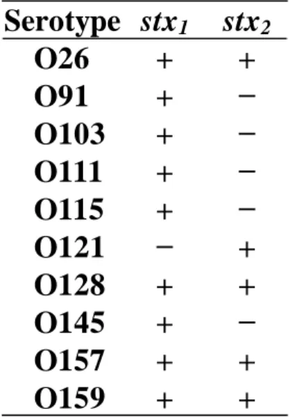
STEC strains isolated from patients used in this study are listed in Table 1. They were collected by the Kobe Institute of Health (Kobe, Japan), and the species were identified by the conventional indole test, methyl red test, Voges–Proskauer test, and citrate test. The O-antigen serotype was confirmed by the conventional agglutination test with rabbit antisera against each of the STEC O-antigens.2 The presence of the Shiga toxin gene was confirmed by conventional PCR.29 The STEC strains were cultured overnight at 35°C on LB agar plates or in 2 mL of LB medium. Bacterial colonies produced on the plate were suspended in the running buffer of the SPR-immunosensor described below, and the suspension was immediately examined using the
35
sensor. The cultured bacterial cells in LB medium were directly used for examination. The bacterial cell numbers were counted as cfu after incubation on LB agar plates overnight at 35°C or by measuring optical density at 530 nm using a Vi-spec 2 turbidimeter (Kyokuto Pharmaceutical Industrial Co., Ltd, Tokyo, Japan). The colonies were alternatively resuspended in 0.2% formaldehyde overnight at 4°C and subsequently heated for 20 min at 80°C to fix the bacterial cells. The fixed cells were then used for western blot analysis.
Table 1 STEC strains isolated from patients
Preparation of O-antigen specific PoAbs
Anti-O-antigen PoAbs were purified from rabbit antisera to the STEC O-antigens (O26, O91, O103, O111, O115, O121, O128, O145, O157, and O159) using Protein G beads. Each 100 µL of beads was suspended in 100 µL of phosphate-buffered saline (PBS; 10 mM phosphate, 150 mM NaCl; pH 7.0), and then added to 1 mL of antisera. The mixture was gently stirred overnight at 4°C. After the beads were washed 3 times with 1 mL of PBS, the supernatant was removed by centrifugation at 9393 × g for 1 min. The PoAbs bound to the beads were eluted with 100 µL of
Serotype stx
1stx
2O26 + +
O91 + −
O103 + −
O111 + −
O115 + −
O121 − +
O128 + +
O145 + −
O157 + +
O159 + +
36
100 mM citrate solution (pH 2.5). The eluted PoAb solution was immediately neutralized with 15 µL of 1.0 M trisodium phosphate and used for SPR-immunosensor constitution. The prepared PoAbs were abbreviated as anti-O26, anti-O91, anti-O103, anti-O111, anti-O115, anti-O121, anti- O128, anti-O145, anti-O157, and anti-O159. The protein concentration of each PoAb was determined by a protein assay using Coomassie brilliant blue solution (Nacalai Tesque) according to the manufacturer’s instruction manual.
Western blot analysis
Fixed cells of STEC O26, O91, O103, O111, O115, O121, O128, O145, O157, and O159 (1.0 × 10⁷ cells) were suspended in 20 μL of sample buffer (62.5 mM Tris-HCl, pH 7.0 modified with 1% sodium dodecyl sulfate (SDS), 0.01% bromophenol blue, and 10% glycerol), and then incubated for 15 min at 25°C to lyse the cells. After centrifugation at 9393 × g for 10 min, the supernatant was boiled for 5 min and separated by SDS-polyacrylamide gel electrophoresis with a 15% polyacrylamide running gel. The O-antigens were blotted onto the polyvinylidene difluoride membrane using a semi-dry blotter (NA-1512; Nihon Eido Co. Ltd., Japan) at 0.5 mA/cm2 for 2.5 h in transfer buffer (62.5 mM Tris base, 200 mM glycine, and 20% ethanol). The membrane was blocked with PBS containing 0.1% Tween® 20 (PBS-T) containing 5% skim milk at 4°C overnight, and then washed once with PBS-T. Membranes were transferred to anti-O26, anti-O111, or anti-O157 PoAb (each 1.0 μg/mL) solution in PBS-T containing 0.3% skim milk, and then incubated at 25°C for 1 h. After washing 4 times with PBS-T, the membrane was further incubated with HRP-labeled anti-rabbit IgG antibody (0.5 μg/mL) in PBS-T containing 0.3% skim milk at 25°C for 1 h. After washing 4 times with PBS-T, the antigen bands formed from reaction with the anti-O-antigen PoAbs were developed using the chemiluminescent substrate of HRP according to the manufacturer’s instruction manual. The band on the membrane was detected using a gel imager (ImageQuant™ LAS 4000; GE Healthcare).
37 SPR-immunosensor
The surface plasmon resonance imaging system was purchased from HORIBA Scientific (OpenPlex; Palaiseau, France). The biochip consisted of a prism and a gold thin-layer, the surface of which was modified with molecules terminated in a carboxyl group (SPRi-Biochip CS-HD, HORIBA Scientific). Since the carboxyl groups were pre-activated by esterification with N- hydroxysuccinimide, the target antibodies could bind directly to the biochip.
1) Preparation of sensor chip
Purified anti-O-antigen PoAbs and anti-mouse IgG PoAb (control) were diluted in PBS (0.10–0.75 mg/mL). To immobilize the PoAb on the chip by covalent bonding, these PoAb solutions were respectively spotted onto the chip surface at 10 nL/spot using a spotter (Spot Master; Musashi Engineering, Tokyo, Japan) in 80% relative humidity. The spotted sensor chip was incubated at 4°C for 16 h in 80% relative humidity. The chip surface was then washed 3 times with PBS. The surface was blocked with PBS containing 1% BSA at 25°C for 30 min and washed 3 times with PBS. Unreacted carboxyl groups were deactivated with 1 M ethanolamine solution (pH 8.5) at 25°C for 30 min, and then washed 3 times with PBS. The prepared sensor chip was set into the instrument according to the manufacturer’s instruction manual. The reaction chamber for detecting STEC O-antigens was composed of a flow cell (volume ~ 80 μL) attached to the sensor chip surface through a hexagonal gasket.
2) Flow conditions for the reaction
All reactions were carried out at 25°C. Running buffer (PBS containing with 0.2% BSA and 0.02% Tween® 20) was degassed using a degasser (DEGASi® Classic; Biotech, Onsala, Sweden), and was set up to continuously flow into the reaction chamber, driven by a peristaltic pump (MINIPULS® 3; Gilson, Inc., Middleton, WI, USA) at 50 μL/min. The prepared bacterial cell suspension was preserved in a sample loop (volume = 200 µL) and was set up to flow into
38
the chamber when the valve was switched to the injection mode.
3) Detection of STEC O-antigens
The sensor chip was irradiated by a light-emitting diode (λ = 810 nm) and the light was reflected at the interface between the prism and the gold thin layer with attenuation by SPR phenomena. The intensity of the reflected light (reflectivity) was acquired by an 8-bit charge- coupled device (CCD) camera every 3 s. After 200 μL of the bacterial cell suspension was injected, the reflectivity of each spot changed depending on the quantity of the bacterial cell bound to the PoAbs. The percent change of the reflectivity (%ΔR) every 3 s was calculated based on the CCD signal, and was averaged within a pre-defined detection area on each spot of the PoAbs. The data for each test antibody were normalized by subtracting the reflectivity for anti-mouse IgG PoAb.
The %ΔR was visualized as a bright spot that could easily be distinguished from the background by the CCD camera.
4) Regeneration of the sensor chip
Gelatin (3.0 g) was dissolved with 100 mL of distilled water at 80°C, and was gelated by cooling at 4°C for 7 days. Agarose (0.2 g) was dissolved with 100 mL of boiled distilled water, and was gelated by cooling at 25°C for 1 h. After the detection of each O-antigen, 50 μL of 10 mM NaOH, 100 mM glycine buffer (pH 2.0), or the above gel suspension (3% gelatin gel kept on ice immediately before use or 0.2% agarose gel) was set to flow into the reaction chamber for 1 min at 50 μL/min. The residual gel was flushed out from the chamber with running buffer at 1 mL/min. The efficiency of removal of the bound cells was confirmed by monitoring the SPR signals. The sensor chip was reused for the next detection of the STEC O-antigens without any other treatments after the signals had sufficiently returned to baseline.
Results and Discussion
Reactivity of anti-O-antigen PoAbs
39
The anti-O26, anti-O111, and anti-O157 PoAbs were examined to confirm that they specifically reacted with each corresponding O-antigen on STEC by western blot analysis, as shown in Fig. 1. When the blotted membrane was reacted with anti-O26 PoAb, a characteristic ladder band of the O-antigen was located at 14–28 kDa and 48–64 kDa in the O26 antigen lane as shown in Fig. 1A. In the reaction with anti-O111 PoAb, a band was located at 32–90 kDa in the O111 antigen lane, as shown in Fig. 1B. In the reaction with anti-O157 PoAb, a band was located at 42–87 kDa of the O157 antigen lane, as shown in Fig. 1C. Characteristic ladder bands are produced by repeat oligosaccharide structures containing 2–7 sugar residues. The bands reacted with anti-O26, anti-O111, and anti-O157 PoAbs and showed the same patterns as observed in previous studies.30,31 We confirmed that the 3 PoAbs examined specifically reacted with their corresponding O-antigens. The other PoAbs were also specific for their corresponding O-antigens (data not shown).
Figure 1.
B
1 2 3 4 5 6 7 8 9 10 C
1 2 3 4 5 6 7 8 9 10
10 28 16 70
A
1 2 3 4 5 6 7 8 9 10
MW (kDa)
54
Fig. 1 Reactivity of prepared PoAbs with 10 O-antigens on STECs by western blot analysis. Results of reactivity with A) PoAb O26, B) PoAb O111, and C) PoAb O157 are shown. Lanes 1–10 show the O26, O91, O103, O111, O115, O121, O128, O145, O157, and O159 antigens, respectively.
Anti-O111 PoAb did not react with common antigens in the STECs examined. However,
40
anti-O26 and anti-O157 PoAbs reacted with common antigens, including 11, 18, and 37 kDa proteins. These results are as would be expected, given that the examined PoAbs were prepared using inactivated whole bacteria cells.28 However, these proteins are not present on the surface of bacterial cells and differ from the O-antigens. Thus, the reactivity of the PoAbs with common antigens may have only a negligible influence on the direct detection of O-antigen in viable cells.
All of the prepared PoAbs were used for SPR-immunosensor constitution.
Immobilization of PoAbs to the sensor chip surface
Immobilization of anti-O157 PoAb was examined. The PoAb solution was serially diluted with PBS to 0.10, 0.25, 0.50, and 0.75 mg/mL and then spotted onto the sensor chip surface containing carboxylic acid pre-activated with N-hydroxysuccinimide. The amino groups on lysine residues of the PoAb were covalently bound without treatment. The PoAb-immobilized chip was placed in the SPR-immunosensor and STEC O157 cells suspended in running buffer were injected at 107 cfu/mL. The reaction signal (%ΔR) increased in a time-dependent manner and reached a plateau (0.7) by 300 s at more than 0.5 mg/mL of PoAb concentration, as shown in Fig. 2. We assumed that lysine residues immobilized with carboxylic acid would be present in the same numbers in the constant region of all PoAbs. A concentration 0.5 mg/mL was used to immobilize the PoAbs.