Fukushima Medical University
福島県立医科大学 学術機関リポジトリ
This document is downloaded at: 2021-11-07T23:08:51Z
Title A case of transgastric gallbladder puncture as a complication during endoscopic ultrasound-guided drainage of a pancreatic pseudocyst
Author(s)
Hikichi, Takuto; Irisawa, Atsushi; Takagi, Tadayuki;
Shibukawa, Goro; Wakatsuki, Takeru; Imamura, Hidemichi;
Takahashi, Yuta; Sato, Ai; Sato, Masaki; Oyama, Hitoshi; Sato, Naoto; Yamamoto, Go; Mokonuma, Tatsuyuki; Obara,
Katsutoshi; Ohira, Hiromasa
Citation Fukushima Journal of Medical Science. 53(1): 11-18
Issue Date 2007-06
URL http://ir.fmu.ac.jp/dspace/handle/123456789/203
Rights © 2007 The Fukushima Society of Medical Science
DOI
Text Version publisher
Fukushima J. Med. Sci., Vol. 53, No.1, 2007
[Case Report]
A CASE OF TRANS GASTRIC GALLBLADDER PUNCTURE AS A COMPLICATION DURING ENDOSCOPIC
ULTRASOUND-GUIDED DRAINAGE OF A PANCREATIC PSEUDOCYST
T AKUTO HIKICHP), A TSUSHI IRISA W
N),T ADA YUKI T AKAGPl, GORO SHIBUKA W
N),T AKERU W AKATSUKPl, HIDEMICHI IMAMURN),
YUTA TAKAHASHP), AI SAT02), MASAKI SAT02), HITOSHI OYAMNl, NAOTO SAT02), GO YAMAMOT02), TATSUYUKI MOKONUMN),
KATSUTOSHI OBARN) and HIROMASA OHIRN)
l)Department
0/
Endoscopy, Fukushima Medical University Hospital, 960-1295, Japan 2)Department0/
Internal Medicine IL Fukushima Medical University School0/
Medicine,960-1295, Japan
(Received July 18, 2006, accepted November 13, 2006)
Abstract: A 43-year-old man, a regular drinker, developed a pseudocyst in the pancreatic tail as a result of acutely worsening chronic pancreatitis. Because the pseudocyst,
10em in diameter, did not disappear despite conservative treatment, an internal drainage stent was placed transgastrically under endoscopic ultrasound (EUS) guidance. However, cyst infection occurred, and EUS-guided drainage was performed, when the gallbladder was punctured inadvertently. Immediately a nasocystic drain was placed in the gallbladder. Owing to this timely measure, only mild and localized peritonitis developed. Conservative treatment with fasting and an antibiotic was administered, and peritonitis subsided quickly. On the same day, another nasocystic drain was placed for the pancreatic pseudocyst, and it disappear- ed. As far as we know, this is the first case in which gallbladder puncture was inadvertently performed during EUS-guided drainage of a pancreatic pseudocyst.
Key words: gallbladder puncture, pancreatic pseudocyst, EUS-FN A, EUS-guided drainage, complication
i3I:1t!!:ffiA, A~~ii!;:, il1Ii*,~z, t.IlfJlI'I'HAA,~:tm ~, ~t-.t*J1!:,
il1Ii:!it**,
{tI:§ ~,{tI:§OCll[l,:kill t, {tI:§~A, 111* ~, ai!lm~z, 'Nlii:Jmlil1, :k:>f5b1E
Correspondence to: Takuto Hikichi, Department of Endoscopy, Fukushima Medical Univer- sity Hospital, Fukushima City 960-1295, Japan.
E-mail: [email protected]
11
12 T. HIKICHI et al.
INTRODUCTION
A pancreatic pseudocyst is a nonepithelial cyst formed after acute deterioration of acute and chronic pancreatitis, and injury to and resection of the pancreas. It often resolves spontaneously with conservative treatment.
Ifit does not, complica- tions, such as cyst infection and bleeding and rupture of the digestive tract, may ensue, threatening life. Therefore, in the past it has been removed surgically or drained percutaneously. Recently, with the advances in endoscopic therapy, endos- copic transmural drainage has become possiblel-
3).However, the problem with this method using a conventional endoscope was that puncture under direct vision had to be blind puncture, causing complications, such as bleeding and digestive tract perforation. In recent years, endoscopic ultrasound (EUS)-guided transmural drain- age has been developed, and its safety and usefulness are estimated to be superior to those of the conventional method
4 - 12).Complications of EUS-guided pancreatic pseudocyst drainage are divided into two groups: early complications associated with surgical techniques, and late complications occurring in the course of recovery. Common early complications include bleeding and perforation, and late complications are mostly cyst infections due to obstruction of a drainage tube
4 ).Kruger et
at.5)reported that no early complications occurred in the 35 patients they had seen, and Giovannini et
at. 6)reported that only 1 (2.9%) of their 35 patients developed mild pneumoperitoneum, which resolved quickly. Vosoghi et
at. 7),however, reported that in 1 (7%) of their 14 patients, bleeding occurred 2 days after the procedure, requiring surgery. Thus, in performing EUS-guided drainage, although it is regarded as safer than the conventional method, the knowledge of how to prevent and how to cope with complications is important. We inadvertently injured the gallbladder of a patient with pancreatic pseudocyst during EUS-guided drainage. We report here how we coped with it and how it could have been prevented.
CASE REPORT
A 43-year-old man, a regular drinker, presented to our hospital complaining of
an abdominal pain in April 2001. Because a CT scan showed a 10-cm pseudocyst
and bleeding into the cyst in the pancreatic tail, he was hospitalized and underwent
conservative treatment for 56 days. However, the pseudocyst did not reduce, and so
he was rehospitalized to undergo EUS-guided drainage in December 2001. On
admission, although there was no abdominal pain, and a blood test showed no
inflammation reaction or elevated tumor marker, CT scan showed a 10-cm
pseudocyst in the pancreatic tail (Fig. 1); therefore, EUS-guided cystogastrostomy
was performed on the fifth hospital day. A curved linear-array echo endoscope
(FG-36UX; Pentax Corp., Tokyo, Japan) was inserted into the stomach, and an
EUS-GUIDED PAl CREATIC PSEUDOCYST DRAINAGE
Fig.1. Contrast-enhanced abdominal CT image. A dilated pancreatic duct and a 10 cm diameter cyst were seen in the pancreatic tail (arrow). The cyst was consid- ered a pseudocyst because there was no evidence of malignancies, such as mural nodules at the cyst wall or a tumor in the pancreas.
13
ultrasound imaging device att ached t o it (EUB- 6000 ; Hitachi Ltd., T ok yo, Ja pa n) depi c ted a pancre atic pseud ocyst as a uni form gr anul a r echo patte rn (Fig . 2). After the pseudocyst was punctured wit h a 19G needle (ECHOTIP ; Wilso n-Cook Me dica l Inc., Osaka, Japa n) without electric ity, the stomach wall (uppe r gastri c body) a nd cyst wall of th e punctur e site were stretched with a dilat or, and a Zimm on Biliary Stent 7Fr, 4 cm (Wilson- Cook Medi cal Inc., Osaka, Japan) was pl aced (Fig. 3).
H owever, the pseudocyst did not resolve; t he temper ature a nd inflamm a tion reac-
ti on increa sed, suggesting cyst infection due to s tent clogging. The pa tien t's condi-
ti on remained unimproved with conservative treatment, and on th e 34th hospi ta l day,
EUS-guided nasocysti c dra in age (EN CD) of the pseudocyst was pe rformed tra ns-
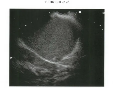
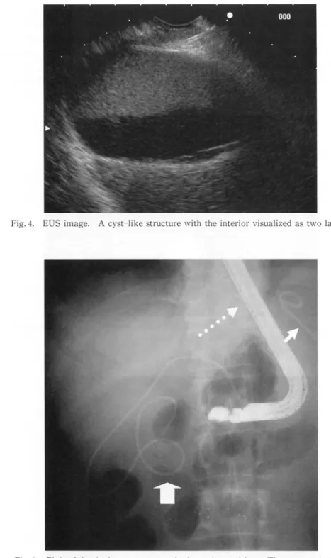
gastricall y. Before punc ture, tra nsgastri c EUS ha d confirmed a cyst- lik e struc ture
whose interior wa s visua li zed as two layers, i.e., a n echoic and a nechoic areas (Fig .
4). This cyst
-like structure was punctured under EUS g uidance, a nd a 5 Fr end os-
copi c nasobiliary drain age (EN BD ) tub e (Flexim a ; Bost on Scie ntific Co rp., T okyo,
Japan) wa s placed. Fluor oscopic observati on of the patient in th e supine positi on
revea led the tube in the right side of the abdomen, from which it was evide nt tha t
the ga llbladde r had been punctured, not the pancreatic pseud ocyst (Fig. 5). Ca reful
review showed that the puncture site was in the lower gastr ic body when it sho uld
have been in the upper gastri c body. The tube
wasleft in the gallbl a dder so tha t
bile wo uld not leak into the a bdomina l cavity causing bile peri tonitis. The
pseud ocyst was drained by r einserting the EUS and plac ing a 5Fr EN BD tube
(E NBD- 5; Wilson
-Cook Medical Inc.,Osak a, Japan) in the cyst. The flui d was
milk
ywhite.
14 T. HIK[CHI et al.
Fig. 2. EUS image. The pancreatic pseudocyst showed no wall thickening and had a granular echo pattern with relatively uniform inner echo density.
Fig. 3. Endoscopic image. A stent was inserted into the pancreatic pseudocyst from the posterior wall of the greater curvature of the stomach.
EUS-GUIDED PANCREATIC PSEUDOCYST DRAINAGE
Fig. 4. EUS image. A cyst-like structure with the interior visualized as two layers.
Fig. 5. Plain abdominal roentgenogram in the supine position. The nasocystic drain (broad arrow) is located away from the stent (arrow) placed in the pancreatic pseudocyst. The dotted arrow indicates the ultrasonic endoscope used.
15
16 1'. I-IIKICI-ll el al.
Abdominal roentgenogram taken on the following day showed that the tip of the tube in the gallbladder had moved slightly. A CT scan performed 2 days later confirmed that it had slipped out of the gallbladder into the abdominal cavity (Fig.
6). Three days later, that part of the tube that had slipped out of the gallbladder was removed under fluoroscopic observation, taking care that the rest of the pseudocyst did not fall out. To prevent leakage of gastric juice from the puncture
Fig. 6. Plain abdominal CT image. The tip of the tube inserted into the gallbladder was slipped out of it into the abdominal cavity.
Fig. 7. Plain abdominal CT image. The pancreatic pseudocyst subsided considerably, and inside
it was the tube that had been placed {arrow}.
EUS-GUIDED PANCREATIC PSEUDOCYST DRAINAGE 17
site into the abdominal cavity, a tube was inserted into the stomach through the nose.
Continuous administration of antibiotics during fasting controlled the inflammatory reaction. A CT scan performed 7 days later showed that the pseudocyst subsided considerably (Fig. 7). A restricted diet was started on the 48th hospital day, 14 days after the first drainage, and the ENCD tube in the pseudocyst was removed on the 54th hospital day, 19 days after the first drainage. Even after the tube was removed, the inflammatory reaction remained controlled, and a CT scan showed no infection or enlargement of the cyst, and so the patient was released from the hospital on the 65th hospital day.
DISCUSSION
Grimm et al.
8 )first reported EUS-guided transmural drainage of pancreatic pseudocysts in 1992. Before then, pseudocysts had been punctured and drained under direct vision using a regular endoscope. The traditional method, however, involved blind puncture and was inapplicable to patients without a luminal bulge in the digestive tract
9).Furthermore, there were risks of bleeding and perforation as complications. The new technique, using the EUS image as a guide, is safe as many investigators report
4 - 12)and permits puncture in real time. Vosoghi et al.
7 ),who reviewed the complication rates of various treatments, reported the highest safety of EUS-guided transmural drainage as follows: the complication rates of surgical treatment, percutaneous drainage, transpapillary drainage, endoscopic puncture drainage under direct vision and EUS-guided puncture drainage were 28%, 18%, 12%, 15% and 1.5%, respectively.
Thus, EUS-guided transmural drainage is the first-choice treatment for patients with pancreatic pseudocyst, if they can tolerate the endoscopic procedure. How- ever, although its complication rate is lower than that of any other treatment, those who perform this procedure should have a thorough knowledge of the complications that can arise from it. The early complications associated with this technique include bleeding, perforation and intraperitoneal emphysema
5,7,lO-12).Binmoeller et
aP)
reported a case (4.1%) of inadvertent puncture of the gallbladder during the attempted puncture of a pancreatic pseudocyst under direct vision. In their case, bile leaked, and a surgical operation was required. Fortunately, in our case, no surgery was needed. After inadvertent puncture of the gallbladder and subsequent stretch of the puncture site, a small amount of bile leaked into the abdominal cavity.
However, an ENCD tube reduced the pressure in the gallbladder, eventually stopping
the bile leak and eliminating the need for surgery. The factors considered to have
led to inadvertent puncture of the gallbladder in this case are: 1) The gallbladder
that enlarged because of long fasting was unusually visible from the stomach. 2)
Because there was a large amount of debris in the gallbladder, it was mistaken for
an infected pancreatic pseudocyst (pancreatic abscess). 3) Because the entire
procedure was performed with the patient in the left lateral decubitus position, the
18 T. HIKICHI et at.
pancreatic pseudocyst and gallbladder overlapped on the x-ray fluoroscopic image.
To prevent similar complications, it is essential to accurately identify a pancreatic pseudocyst on EUS images and carefully choose a puncture site in the stomach on endoscopic images. Moreover, the pancreatic pseudocyst should also be confirmed under X -ray fluoroscopy with the patient's position changed from left decubitus, taken when the endoscope is inserted, to supine.
In conclusion, EUS-guided transmural drainage of pancreatic pseudocysts is safer than the blind puncture technique using a regular endoscope. However, endoscopists should realize again that because many different objects surrounding the digestive tract are visible, the target can be mistaken, leading to a complication.
Those who perform this procedure need to fully understand the possibility of a complication such as ours, and keeping in mind how to cope with it, they should strive for accuracy in diagnosis and every aspect of this procedure.
REFERENCES
1. Cremer M, Deviere J, Engelholm L. Endoscopic management of cysts and pseudocysts in chronic pancreatitis: long-term follow up after 7 years of experience. Gastrointest Endosc, 35: 1-9, 1989.
2. Sahel J, Bastid C, Pellat B, Schurgeres F, Sarles H. Endoscopic cystogastrostomy of cysts of chronic calcifying pancreatitis: a report of 20 cases. Pancreas, 2: 447-453, 1987.
3. Binmoeller KF, Seifert H, Walter A, Soehendra N. Transpapillary and transmural drainage of pancreatic pseudo cysts. Gastrointest Endosc, 42: 219-224, 1995.
4. Fockens P. EUS in drainage of pancreatic pseudocysts. Gastrointestinal Endosc, 56(Suppl): S93-97, 2002.
5. Kruger M, Schneider AS, Manns MP, Meier PN. Endoscopic management of pancreatic pseudocysts or abscesses after an EUS-guided I-step procedure for initial access.
Gastrointestinal Endosc, 63: 409-416, 2002.
6. Giovannini M, Pesenti C, Rolland AL, Moutardier V, Delpero JR. Endoscopic ultra·
sound-guided drainage of pancreatic pseudo cysts or pancreatic abscess using a therapeu·
tic echo endoscope. Endoscopy, 33: 473-477, 2001. .
7. Vosoghi M, Sial S, Garrett B, Feng J, Lee T, Stabile BE, Eysselein VE. EUS-guided pancreatic pseudocyst drainage: review and experience at Harbor-UCLA Medical Center. Med Gen Med, 4: 2, 2002.
8. Grimm H, Binmoeller KF, Soehendra M. Endosonography-guided drainage of a pancre·
atic pseudocysts. Gastrointest Endosc, 38: 170-171, 1992.
9. ASGE guideline: the role of endoscopy in the diagnosis and the management of cystic lesions and inflammatory fluid collections of the pancreas. Gastrointest Endosc, 61:
363-370, 2005.
10. Binmoeller KF, Soehendra N. Endoscopic ultrasonography in the diagnosis and treat·
ment of pancreatic pseudocysts. Gastrointest Endosc Clin N Am, 5: 805-816, 1995.
11. AntiIIon MR, Shah RJ, Stiegmann G, Chen YK. Single-step EUS-guided transmural drainage of simple and complicated pancreatic pseudocysts. Gastrointest Endosc, 63:
797 -803, 2006.
12. Giovannini M, Binmoeller K, Seifert H. Endoscopic ultrasound-guided cystogastros·
to my. Endoscopy, 35: 239-245, 2003.