Fukushima Medical University
福島県立医科大学 学術機関リポジトリ
This document is downloaded at: 2021-11-07T23:17:43Z
Title Bilirubin and its oxidative metabolite biopyrrins in patients with acute myocardial infarction
Author(s) Kunii, Hiroyuki; Ishikawa, Kazunobu; Yamaguchi, Tokio;
Komatsu, Nobuo; Ichihara, Toshikatsu; Maruyama, Yukio
Citation Fukushima Journal of Medical Science. 55(2): 39-51
Issue Date 2009-12
URL http://ir.fmu.ac.jp/dspace/handle/123456789/238
Rights © 2009 The Fukushima Society of Medical Science
DOI
Text Version publisher
Vol. 55, No.2, 2009 [Original Article]
BILIRUBIN AND ITS OXIDATIVE METABOLITE BIOPYRRINS IN PATIENTS WITH ACUTE MYOCARDIAL INFARCTION
HIROYUKI KUNIP), KAZUNOBU ISHIKA W
N,4),TOKIO YAMAGUCHP), NOBUO KOMATSU3), TOSHIKATSU ICHIHARN)
and YUKIO MARUYAMN)
1)
First Department of Internal Medicine, Fukushima Medical University, Fukushima, japan, 2)Department of Biochemical Genetics, Medical Research Institute, Tokyo Medical and Dental University, Tokyo, japan,
3)Division of Cardiology, the Iwaki Kyoritsu General Hospital, Iwaki, Fukushima, japan, ')Center for Medical Education and Career Development, Fukushima Medical University
(Received December 2, 2008, accepted May 12, 2009)
Abstract: This study aimed to assess the involvement of bilirubin and its oxidative metabolite biopyrrin in patients with acute myocardial infarction (AMI) and to determine the responsible organs that overproduce these molecules. One hundred thirteen consecutive patients hospitalized for AMI were analyzed. Levels of serum bilirubin, plasma and urinary biopyrrins were measured on the day of admission, day 2, 3, 7 and 14. Expressions of biopyrrins and heme oxygenase-1 (HO-1), a stress- responsive bilirubin-producing enzyme, in heart, aorta, kidney, liver and lung were immunostained with autopsied specimens. Serum bilirubin, plasma and urinary biopyrrins were increased within 24 hr, formed a peak on day 3 and then decreased by day 14. These three parameters were well correlated to each other. The maximum biopyrrin elevation was higher in death cases and associated with impair- ed left ventricular function. Immunohistochemical analyses revealed biopyrrin accumulation and HO-1 expression in the infracted myocardium. Immunoreactive HO-1 and biopyrrins were also observed in renal tubular cells, aortic wall and lung.
Serum biiirubin and its metabolite biopyrrins were elevated in patients with AMI.
Plasma and urinary biopyrrin elevation were associated with mortality and morbid- ity. Induction of anti-oxidative enzyme HO-1 seemed to be involved in the activa- tion of bilirubin/biopyrrin pathway.
Key words: biopyrrin, bilirubin, heme oxygenase, acute myocardial infarction, oxidative stress
E!!I#mz, EJllfDffl,
WD~W:t;:,'N'l11L:t;:,
$JJj{flj)J;j,AW$:t;:
Corresponding author: Kazunobu Ishikawa, MD, PhD, FAHA. E-mail: [email protected] Tokio Yamaguchi, PhD. E-mail: [email protected]
http://fmu.ac.jp/home/lib/F -igaku/ http://www.sasappa.co.jp/online/
39
40 H. KUNII
et at.
INTRODUCTION
Acute myocardial infarction (AMI) provoked by coronary atheromatous plaque erosion/rupture and thrombus formation continues to be a major life-threatening health problem in western countries even after the recent prevalence of percutaneous coronary interventional reperfusion therapyl-3). Although numerous effects have been made to find serological markers in patients with AMI for risk stratification during acute phase management and for the prediction of prognosis of these patients
4 -8), please conform the citation standard of this journal establishment of more sensitive and specific serological markers is a demanding problem.
Bilirubin has been considered as a toxic waste product from heme catabolism.
However, recent studies have shown that bilirubin possesses antioxidant activity, which scavenges hydroperoxy radicals as effectively as vitamin E9,lO). Under vari- ous pathophysiological conditions such as ischemic reperfusion injuries, bilirubin has been reported to exhibit significant protective effects as an antioxidant in animal studies
l l -13). Bilirubin reacts with reactive oxygen species (ROS) and its reaction product is metabolized into oxidized metabolites referred to as "biopyrrins"13-15).
The increased production of biopyrrin has been reported under a variety of condi- tions such as post-laparotomy state16), sepsis
17),Alzheimer's disease18) and hepatic ischemic reperfusion injuries13), all of which seem to be caused by oxidative stress.
In addition, the elevation of urinary biopyrrins has been recently reported in patients with several cardiovascular disorders such as congestive heart failure, AMI and vasospastic angina19- 21) although these studies are based on the discrete and limited observation of biopyrrins in urine. However, it seems difficult to use urinary biopyrrins for rapid diagnosis of AMI because it requires adjustment for urinary creatinine, and because it is often difficult to obtain urine samples from patients with AMI, who go into cardiogenic shock. Therefore, it is desirable to establish a serological marker. Besides, no previous studies have explored the relationship between bilirubin and biopyrrin levels. Tissues responsible for the elevation of biopyrrins are also unknown.
In this study, we applied biopyrrin measurement to plasma samples for the first time, and examined its correlation among serum bilirubin and urinary biopyrrins.
We also examined whether the plasma biopyrrin associates with acute phase
mortality and cardiac performance. Heme oxygenase-l (HO-l) expression, an
enzyme predominantly responsible for the intrinsic production of bilirubin as well as
biopyrrin accumulation, was examined by immunohistochemistry with autopsy
specimens from patients with AMI.
METHODS
Study Objects
We studied 113 consecutive patients hospitalized for AMI in our hospital from
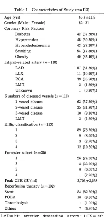
Table 1. Characteristics of Study (n=1l3) Age (yrs)
Gender (Male: Female) Coronary Risk Factors
Diabetes Hypertension Hypercholesteremia Smoking
Obesity
Inf~rct-related artery (n=110) LAD
LCX RCA LMT Unknown
Numbers of diseased vessels (n=llO) I-vessel disease
2-vessel disease 3-vessel disease LMT
Killip classification (n=1l3) 1
2 3 4
Forrester subset (n=35) 1
2 3
4
Peak CPK (IU/ml)
Reperfusion therapy (n=102) Stent
POBA Thrombolysis Others
65.9±1l.8 82: 31
42 (37.20%) 45 (39.80%) 42 (37.20%) 54 (47.80%) 40 (35.40%)
. 57 (51.80%) 11 (10.00%) 39 (35.50%) 2 (1.80%) 1 (0.90%)
63 (57.30%) 35 (31.80%) 10 (9.10%) 2 (1.80%)
89 (78.70%) 9 (8.00%) 3 (2.70%) 12 (10.60%)
26 (74.20%) 8 (22.90%)
o
(0.00%) 1 (2.90%) 2.702±2.53884 (82.30%) 10 (9.80%) 1 (1.00%) 7 (6.90%) LAD= left anterior descending artery; LCX = left circumflex artery; LMT=left main trunk; POBA=plain old balloon angioplasty; RCA=nght coronary artery.
42 H. KUNII
et ai.
November 2001 to March 2005 (Table 1). Diagnostic criteria for AMI were as follows: persistent chest pain over 30 minutes, electrocardiographic changes (ST- segment elevation at least 0.2 m V or new Q-wave) and elevation of plasma cardiac enzymes, such as troponin-T, creatinine kinase, aspartate aminotransferase and lactate dehydrogenase. Exclusion criteria included any history of cerebrovascular disease and evidence of ongoing systemic or cardiac inflammatory processes. Since there is a possibility that underlying liver and biliary tract diseases affect bilirubin metabolism, we also excluded these patients. After admission, patients were intravenously administered heparin (5,000 U bolus followed by 10,000"'12,000 U/day) and nitroglycerin for 3 to 5 days. Patients who received a coronary stent for reperfusion therapy were administered with 200 mg/day ticlopidine for at least 4 weeks. Usage of other pharmaceutical agents such as angiotensin converting enzyme inhibitors, angiotensin receptor blockers, P-blockers and diuretics were not excluded.
Cardiac Catheterization and Reperfusion Therapy
Within 30 min after the diagnosis of AMI, emergent coronary angiography was performed. There were three cases unable to achieve cardiac catherization because of cardiogenic shock. The culprit arteries responsible for AMI are shown in Table 1. There was a single case in which the culprit artery could not be determined.
Emergent percutaneous coronary intervention (PCI) for reperfusion was performed in 102 cases (90.3%) and successful in all cases. The mean peak elevation of creatine phosphokinase (CK) was 2,702±2,538 IU/L. Forrester subset was assessed by thermodilution method using a Swan-Ganz catheter if applicable when emergent PCI was conducted.
Study Protocol
Levels of bilirubin oxidative metabolites, biopyrrins, in urine and plasma were measured by enzyme-linked immunosorbent assay (ELISA) with 24G7 monoclonal antibody (Shino test, Inc., Tokyo, Japan)22). Urine samples were obtained on admis- sion and in the early morning after day 2. Urinary biopyrrins were adjusted for urinary creatinine (Unit/gram-creatinine). Serum total bilirubin levels were deter- mined using bilirubin oxidase methods. Samples were collected on the day of admission, day 2, day 3, day 7 and day 14 after admission. The study protocol was approved by the regional ethics committee in our institution, and informed consent for this study was obtained from all patients.
Immunohistochemical Analysis
Serial sections from autopsy specimens in patients with AMI were stained with
Masson-Trichrome to determine infarct and non-infarct myocardium in heart
tissues. Sections of heart, aorta, kidney, liver and lung were also stained with anti-
HO-1 (StressGen, Vancouver, Canada) and anti-biopyrrin (24G7) antibodies22).
Endogenous peroxidase activity was blocked by 0.3% H
20
2in methanol. Control staining was performed with nonimmune sera. Immunohistochemical staining was performed using immunoperoxidase technique (VECT AST AIN® elite ABC kit), visualized with a 3-amino-9-ethyl carbazole and counterstained with hematoxylin.
Statistical Analysis
All data are presented as mean±SD. The comparisons of continuous data were performed with a one-way analysis of variance. When these results were statistically significant, they were analyzed by Scheffe's F test. Categorical vari- ables were tested by use of
X2,Mann-Whitney U or Kruskal Wallis test and Spearman rank test.
RESULTS
Elevation 0/ Bilirubin and Its Oxidative Metabolite Biopyrrins in Patients with AMI To examine whether the level of bilirubin, one of the HO-I reaction products, is altered in patients with AMI, we first measured the levels of serum bilirubin on the day of admission, day 2, day 3, day 7 and day 14 after admission (Fig.IA). We observed a significant elevation of serum bilirubin on the second and third hospital days. Moreover, as shown in Fig.IB and IC, significant elevations of biopyrrin in plasma and urine were observed on day 2 and day 3. Compared to serum bilirubin and plasma biopyrrin, we observed earlier elevation of urinary biopyrrin level, which were already elevated on the day of admission (Fig. IC). However, peak elevation of bilirubin in serum and biopyrrins in plasma and urine were similar on day 3. In
A B
•
0.9 0.8 :;:::. 0.7
~ !. 0.6
:;
~ 0.5J
0.'0.3
0.2 10
0.1
7 14 180 2 3 7 14 ISO
c
'ii" 5.0 .S
.!
~4.0
~
.S.!!! 3.0 .~
~
1
2.0=-
.S :!; 1.0
• •
•
7 14 180
Fig. 1. Elevation of bilirubin and its oxidative metabolite biopyrrins in patients with acute myocardial infarction (AMI). Serum bilirubin and biopyrrins in plasma and urine were measured on hospital days I, 2, 3, 7 and 14 after admission. The level of urinary biopyrrins was normalized by urinary creatinine concentration.
Serial changes of serum bilirubin (A), plasma biopyrrins (B) and urinary biopyr·
rins (C). Kruskal-Wallis test. • P <0.01, •• P <0.0001.
44
A
2.5
12.0
s 1.5
~
i
1.0 M 0.5B
350 300
• j250
!200
s jl50
~:1 )1:
H. KUNII
et al.
•
pa<O.OOCU r-=O.862
o w ~ ~ ~ ~ ~ W ~
Udllll'J'biapJrrlD./~(U!lhJa:CII' _ _ ia.)
o -I-=!:-'~~-.-~~
o 10 ~ ~ ~ ~ M W ~ Urtur)' blopyrriD I crcaliDlao (\Jab '" crcatfalDo)
c
2.5
0.5
••
pDCO.OOOI raD.n5
o 50 100 ISO 200 250 300 350 P ... biopJn'ID.(UD1&IaaI)
Fig. 2. Correlations among the maximum levels of serum bilirubin, biopyrrins in plasma and urine in patients with acute myocardial infarction.
addition, the peak levels of these markers showed a significant correlation to each other (Fig. 2).
Elevation of Biopyrrins Reflects Acute-phase Mortality and Morbidity
We had 7 death cases by hospital day 14 (6 cases: acute heart failure; 1 case:
ventricular fibrillation). Although we did not observe significant difference in the maximal levels of serum bilirubin on the hospital day 3 (Fig. 3A), biopyrrins both in plasma (Fig. 3B) and urine (Fig. 3C) were significantly higher in death cases than those of survivors (P<O.05). The levels of these markers were not affected by coronary risk factors shown in the Table (data not shown).
To test whether the elevations of bilirubin and biopyrrins reflect impaired cardiac performance after the onset of AMI, we analyzed these levels on the day 3 and divided the patients into two groups: low or high cardiac index (CI) (Fig.4A- 4C), and low or high pulmonary capillary wedge pressure (Pcwp) (Fig.4D-4F).
Since most of the study objects were classified in Forrester subset I and II in this cohort (Table 1), we divided these groups by median value: CI, 2.7 L/min/m
2 ;Pcwp, 18 mmHg, As shown in Fig. 3A-3C, patients with lower CI exhibited higher peak serum bilirubin and biopyrrins both in plasma and urine. In addition, patients with higher Pcwp exhibited higher peak biopyrrins in plasma and urine (Figs. 4E, 4F).
A B C
2.5 350 BO
~ 2 ••
i"
lOH"
II.,
I
~:.•
s "'!'40I,·· I . j15. la
J •. , j 1: !
. 1 20•
~• •
-. "'""C_ "" ... "'""C_
krYi¥Cll"...c_
(a-I06)
..
.."Fig. 3. Peak levels of biopyrrin in plasma and urine reflect acute-phase mortality.
Serum bilirubin (A), plasma biopyrrins (B) and urinary biopyrrins (C) on hospital
day 3. Urinary biopyrrin level was adjusted by urinary creatinine concentration
(C). Mann-Whitney test. ·P<o.o5.
A 2.S
~2.0
.! :J 1.5
:i
91.0S O.S
D 2.0
1.6
•
I! : : •
.... -
* •
•
Lowel (1:1-17)
Low_
(1:1-17)
B
pOO
3S0~ 250
t
200! ISO
*
•
• •
J
100 SO • II
o "'---"---=---
E 3S0 j300 -1i 2S0
hoo I
ISORishel
I
•
•
i 100
II •
Lowel
*
f SO
I
o '---''--_----'L--_
Low_
c
F 80 70
! 60
11
SO~! 40 i~ 30
':.!20
~
10 I•
•
*
*
Fig. 4. Peak biopyrrin elevation reflects impaired cardiac performance. Serum bilirubin, plasma biopyrrins and urinary biopyrrins on hospital day 3 were compared in the two groups divided by cardiac index (CI) (A-C) and by pulmo- nary capillary wedge pressure (Pcwp) (D-F). Mann-Whitney test. ·P<O.05.
Activation 0/ heme oxygenase-J (HO-J) and Bilirubin/ biopyrrin Pathway in Patients with AMI
We next tried to elucidate why transient elevations of bilirubin/biopyrrins occur during the acute phase of AMI. Elevation of serum bilirubin results from either its overproduction or reduced excretion. For this reason, we excluded the patients with liver and biliary tract diseases in this study. On the contrary, overproduction of bilirubin is mediated via the transcriptional activation of HO-I in response to various pathophysiological stresses
23,24).To examine whether stress-inducible HO- I expression is associated with the activation of the bilirubin/biopyrrin pathway, we performed immunohistochemical analyses using tissues from three autopsied patients who died of AMI. Fig. 5 shows representative microphotographs of the myocardium in an autopsied patient who died of left ventricular free wall rupture 3 days after the onset of AMI. We also examined the accumulation of biopyrrins to determine their tissue localization by immunohistochemistry. HO-I and biopyrrins were predominantly expressed in the infarct myocardium (Figs. 5C, 5D), whereas their expressions were scarce in the myocardium of non-infarct areas (Figs. 5G, 5H).
The localization of HO-I was consistent with that of biopyrrins.
We next examined expression of HO-I and biopyrrins in autopsied specimens from other major organs (Fig. 6). Such expression was observed in the aortic wall (Figs. 6E, 61). Renal tubular cells also highly expressed HO-I and biopyrrins (Figs.
6F, 61). In contrast, apparent expressions were not observed in liver (Figs. 6G, 6K).
In lungs, HO-I and biopyrrins were expressed in alveolus epithelium and
inflammatory cells (Figs. 6H, 6L). These findings shown in Fig. 5 and 6 were also
46 H. KUNII el al.
Fig. 5. Representative microphotographs of the myocardium in an autopsied patient, who died of left ventricular free wall rupture 3 days after the onset of AMI. (A- D) Infarct area. (E-I-I) Non-infarct area. (A, E) Masson-Trichrome stains.
Serial sections were stained with anti-HO-l antibody (C, G), anti-biopyrrin (24G7) antibody (D, I-I). Control staining with nonimmune sera (B, F). Immunohisto- chemical stains (brown color) were performed using immunoperoxidase technique and then counterstained with hematoxylin. Scales bars=25 ,um.
Fig. 6. Representative expressions of HO-l and biopyrrins in the aorta (A, E, I), kidney (B, F, J), liver (C, G, K) and lung (D, I-l, L). Organs harvested from the same patient examined in Fig. 4 were serially stained with anti-I-10-1 antibody (E-1-I) and anti-biopyrrin (24G7) monoclonal antibody (1-L). Control staining with nonimll1une sera (A-D). Scales bars=20 ,um.
observed in other two autopsied c ases, who di ed of AMI by the 14 hospital day.
DISCUSSION
In this study, we observed increased level of bilirubin, one of the products of
antioxidant enzyme HO- 1, a nd its oxidative metabolite biopyrrins in patients with
AMI. All of the serum bilirubin, plasma and urinary biopyrrins exhibited excellent
correlati ons to each other a nd showed ma x imal e levation on da y 3 after the onset of
AMI (Fig. 1). Among them, however, the highe r levels of plasma and urinary
biopyrrins correlated with the incidence of mortality within 14 days and the impair- ment of cardiac function (Figs. 3, 4). Immunohistochemical analyses revealed expressions of HO-l and biopyrrins in the infarct myocardium, artery wall, kidney and lung (Figs. 5, 6).
Bilirubin is known as an end-product of heme degradation
23•24).Heme from hemeproteins is catalyzed into biliverdin via the action of HO-l. Subsequently, biliverdin is converted to bilirubin via the action of biliverdin reductase. Although these bile compounds are physiologically produced in spleen and bone marrow, where senescent red blood cells are catabolized, most types of cells have a heme degradation pathway since heme-containing enzymes such as cytochrome P450, catalase and guanylate cyclases are essential for their survival.
Although bilirubin has been considered as a toxic agent that can potentially cause neonatal jaundice, accumulating studies have shown the beneficial properties of bilirubin under various pathophysiological conditions
9,lo,12,25,26).Protective roles of HO-l during myocardial ischemia and reperfusion injury have been attributed to the action of bilirubin
ll ,13,27).Moreover, serum bilirubin concentration has been reported to be a negative risk factor against myocardial infarction
28 - 30)and carotid plaque
31).In this study, we observed a significant increase in the serum bilirubin concentration with a peak on day 3 after the onset of AMI (Fig.lA). We cannot explain the exact mechanism by which this elevation peaks on day 3. However, the peak level of biopyrrin on day 3 may reflect the responses against the accumulating oxidative stresses resulting from myocardial ischemia, necrosis and reperfusion injuries. Congestion and hypo-perfusion of major organs such as kidney, liver and lungs resulting in tissue hypoxia may also increase oxidative insults during the acute phase of AMI. Since a remarkable induction of HO-l has been reported in the impaired myocardium
ll )as well as lungs
32 ),liver
13),kidney
33)and blood vessels
26 ),increased production of bilirubin may reflect systemic adaptive responses under acute heart failure.
In this study, we also observed the increased level of biopyrrins in both plasma and urine. The peak formation of plasma and urinary biopyrrins was well accorded with that of serum bilirubin on day 3 after the onset of AMI (Figs. lB, lC). This association suggests the possibility that prompt conversion of bilirubin to biopyrrins occurs during the acute phase of AMI. This may reflect the consumption of bilirubin as an antioxidant under this pathophysiological condition. Although a recent study has reported that urinary biopyrrins elevate with a peak around 4 hrs after the onset of AMFO), this previous study did not examine biopyrrins levels between 24 hrs and 7 days after the onset of AMI. Taking into account that continuation of myocardial and systemic HO-l activation for 1 week has been reported in experimental studies
34),our serial measurements of bilirubin and biopyr- rins levels produced more reliable results.
Although previous studies
15,16,19-21)have reported urinary biopyrrins as a marker
against oxidative stress, we have evaluated plasma biopyrrin measurement for the
48
H. KUNIIet al.
first time. Through applying measurement systems with blood samples, rapid and more reliable biopyrrin measurement has become possible because obtaining urine samples is often difficult in oliguric patients with cardiogenic shock. In addition, correction for urinary creatinine is not necessary for blood samples.
Importantly, the extent of the maximal elevations of serum and urinary biopyr- rins on the day 3 was associated with the incidence of acute phase mortality and morbidity (Fig. 3). The elevation of biopyrrins was significantly higher in the group with lower cardiac index and higher precapillary wedge pressure (Fig. 4). Although most of our subjects were classified in Forrester subset I, patients with inferior cardiac performance exhibited higher bilirubin oxidation. Since the generation of ROS in ischemic myocardium has been reported to involve the deteriorating process of AMp5.36), the elevation of bilirubin oxidation products may reflect the magnitude of the myocardial injury. In contrast, however, elevation of serum bilirubin during acute phase of AMI (Fig. lA) was not statistically associated with acute phase mortality (Fig. 3A) and morbidity (Fig. 4D). This may be attributed to the smaller change of serum bilirubin compared to those of biopyrrins in plasma and urine.
Further studies seem to be necessary to determine the different roles of biopyrrins and bilirubin as clinical markers for AMI.
We assume that the elevations of bilirubin and biopyrrins in patients with AMI result from the transcriptional activation of HO-l, which strongly responds to pathophysiochemical and inflammatory stresses39). The extent of HO-l induction via these enhancer domains correlates with the severity of pro-inflammatory stres- ses, and the elevation of bilirubin and biopyrrins reflects the extent of oxidative insults. To identify the tissue localization of bilirubin production and its oxidation, we performed immunohistochemical analyses of HO-l and biopyrrins expression using specimens from the autopsied patients who died of AMI. We have confirmed the expressions of HO-l and biopyrrin in artery wall, kidney and lung (Fig.
6)as well as in the infarcted myocardium (Fig. 5). Adaptive responses via HO-l and bilir- ubin/biopyrrin pathway may be activated in these organs. Ischemia-reper- fusion 13.27), hemodynamic changes40), hypoxia41) and pro-inflammatory cytokines42) may be involved in the induction of HO-l during the acute phase of AMI. However, excessive handling of heme from myoglobin presumably derived from infarcted myocardium during this period may also be responsible for these changes. Further studies are necessary to elucidate the pathophysiological roles of HO-l and bilir- ubin/biopyrrin pathway in this setting. There are two study limitations in the present study. First, we excluded patients with liver and biliary tract diseases since these patients may have different bilirubin kinetics. Second, we could not sufficiently analyze the bilirubin and biopyrrin levels in patients with seriously impaired cardiac performance since most of our subjects belonged to Forrester subset I.
We observed elevations of serum bilirubin and its oxidative metabolites, plasma
and urinary biopyrrins in patients with AMI with a peak 3 days after onset. Plasma
and urinary biopyrrin elevation were associated with acute phase mortality and morbidity within 14 days. Activation of stress-responsive enzyme HO-1 in the infarcted myocardium, artery wall, kidney and lung are involved in the increase of bilirubin and its oxidative metabolite biopyrrins.
ACKNOWLEDGMENTS
This, to K.1. work was in part supported by Grants-in-Aid for Scientific Research (16590703) from the Ministry of Education, Culture, Sports, Science and Technology of Japan.
REFERENCES