Acta Med. Nagasaki 43:48-56
Platelet Serotonin (5-HT)2A Receptor Binding Sites in Affective Disorders:
A Quantitative Receptor Autoradiographic Study with [ 125I ] Lysergic
Acid Diethylamide
Toru TSUJIMURA 1), Tadafumi Asou 1), Masaki HAYASHIDA 1), Akihiko HIMENO2), Yoshibumi NAKANE1)
1) Department of Neuropsychiatry, Nagasaki University School of Medicine 2) Department of Pharmacology 1, Nagasaki University School of Medicine
We used the quantitative receptor autoradiographic method with a radioligand of [ 125I ]lysergic acid diethylamide
([125I]LSD) to quantitate platelet serotonin (5-HT)2A recep- tors in affective disorders. Specific binding of [125I]LSD to human platelet pellet sections was saturable, and of high affinity and single. Both ketanserin and spiperone, 5-HT2A selective ligands, inhibited [125I]LSD binding to human plate- let pellets with high potency (IC50 values of 0.15 and 0.19 nM, respectively), whereas 5-HT and paroxetine, selective 5- HT re-uptake inhibitors, inhibited binding with a very low potency. These data confirmed that binding sites of human plate- let pellets specifically labelled by [125I] LSD were 5-HT2A receptors.
The number of 5-HT2A receptors (Bmax of [ 125I] LSD bind- ing) of human platelets obtained from drug-free depressed patients was significantly higher than those of healthy vol- unteers. There were no statistical differences in the number of 5-HT2A receptors between depressed patients with and without suicidal behaviors. The increased number in platelet 5-HT2A receptotrs may indicate a hyperfunction of the central 5-HT2A receptors. The method with human platelets pellet sections we used is simple and sensitive for investigating platelet 5-HT2A receptors, a diagnostic and therapeutic marker in depressive disorders, in the clinical research.
Key words : 5-HT2A receptors, platelet, affective disorders, quantitative receptor autoradiography, [ 125I] LSD
Introduction
Affective disorders are thought to be associated Address Correspondence :
Toru Tsujimura, M.D. Department of Neuropsychiatry, Nagasaki University School of Medicine, 1-7-1 Sakamoto, Nagasaki 852-8501, Japan
with dysfuctions in the central serotonergic neurons.
Concentrations of serotonin (5-HT) and 5-hydroxyindole- acetic acid (5-HIAA) was reduced in the cerebrospinal fluid of drug-free depressive patients'), and in the post- mortem brains of depressive and suicidal patients"). A decreased number of 5-HT transporter binding sites was also noted in the postmortem brains of depressive and suicidal patients") . Interestingly, Arora and Meltzer found an increase in the density of 5-HT2A re- ceptor binding sites in the postmortem brains of de- pressive and suicidal patients').
As a similarity in binding charcteristics with 5-HT2A ligands between human blood platelets and frontal cor- tex tissues was reported"),") and a nucleotide sequence of human platelet 5-HT2A cDNA was found to be iden- tical to that of human frontal cortex'), human platelets are attracting attention as a model for the central 5- HT neurons. Changes in the density of 5-HT2A recep- tors and 5-HT transpoter binding sites were detected in membrane preparations of blood platelets obtained from patients with affective disorders' 1),12),19). The 5-HT2A ligand-binding method with membrane preparations seems inadequate to investigate 5-HT2A receptor bind- ing sites in human platelets of patients with affetive disorders, since it needs a large volume of blood (i.e, 30 ml). In fact, the binding characteristic in human platelets may vary from human blood-sampling processes""'). Himeno and Saavedra found that the quantitaive receptor autoradiographic method with patelet-pellet sections can detect 5-HT receptor binding sites in platelets obtained from a small volume of human blood (1.0 ml)11),17). Therefore, taking advantage of the high sensitivity, to characterize platelet 5-HT2A receptors of patients with affective disorders, we used the quantitaitive receptor autoradiographic method with a radioligand of [125I] lysergic acid diethylamide
([1211]LSD) in the present study.
Materials and Methods
Preliminary experiments for characterizing human platelet 5-HT2A receptors
Basic experiments to quantitate human platelet 5-HT2A receptors were performed with five healthy volunteers with no histories of psychiatric and neurologic disor- ders. All subjects were in good physical health, and were free from psychotropic medications. Informed
consent of healthy volunteers was obtained after the study procedures had been fully explained.
Depressive patients and normal controls
Outpatients with depressive disorder were recruited in our University-affiliated clinics. All patients enrolled in the protocol met ICD-10 classification of mental and behavioral disorders: diagnostic criteria for research"' ICD-10/DCR for mood (affective) disorders as deter- mined by interviews with plural psychiatrists. All de- pressed patients were determined to be physically healthy on the basis of their medical histories, physi- cal examination, and routine laboratory tests. Clinical ratings were performed in close juxtaposition to the venipuncture by staff who were blind to the binding results. In addition, binding experiments were also per- formed by other staff who were blind to the clinical ratings. Patients diagnosed with depressive disorder according to ICD-10/DCR criteria were studied. Eight depressive patients(male: 6, female: 2) with no history of neurologic disorders ranged in age from 26 to 69 years. The mean age of the subjects was 45.1±5.2 (mean± SEM) years. Depressive patients were in good physical health, and were free for at least 6 months from psychotropic medication. Subjects for normal controls (male: 4, female: 1) with no history of psychi- atric or neurologic disorders ranged in age from 25 to 45 years. The mean age of the subjects was 36.8-L3.4 years. Control subjects were in good physical health and were free from psychotropic medication. There was no statistical differences in age and sex between the two groups. Informed consent of depressive pa- tients and normal controls was obtained after the study procedures had been fully explained.
The categories of ICD-10 system for these patients were F31.5 bipolar affective disorder, current episode of severe depression with psychotic symptoms, F32.1 moderate depressive episode, F33.1 recurrent depres- sive disorder, current episode moderate, F33.2 recur- rent depressive disorder, current episode severe with- out psychotic symptoms, and F33.3 recurrent depressive
disorder, current episode severe with psychotic symp- toms.
Clinical assessment of the severity of patient's current de- pressive symptoms
The severity of the patient's current depressive symptoms was assessed using the Hamilton Depression Scale's' (HAMD).
Quantitaitive receptor autoradiographic method with [125I]LSD
We drew whole blood from normal subjects via a 21-gauge needle into 5 ml vacuum glass tubes con- taining EDTA-2Na. Platelet-rich plasma (PRP) was ob- tained by centrifugation of blood at 180 X g for 15 min at room temperature. Following centrifugation, we re- moved PRP to conical polypropylene tubes containing 20 u 1 of M-1 embedding matrix. We centrifuged the tubes at 1200 X g for 8 min at room temperature. After discarding the supernatant, we slowly added 250 u 1 of M-1 embedding matrix on top of the pellet, together with a thin wooden stick. The tubes were frozen in isopentane on dry ice. We separated the pellets by lightly warming the tubes and pulling the stick. The frozen platelet pellet was mounted on a cryostat chuck (-20°C) with mounting medium and was sectioned at 20 u m thickness.
Triplicate sections were preincubated for 15 min at room temperature in buffer solution(50 mM Tris-HCI, 120 mM NaCl, 5 mM, 1 MM MgC12, 0.05% ascorbate, pH 7.4). After preincubation, sections were incubated with [12-51]LSD (0.08 nM-3.64 nM) in the presence or absence of competing ligands for 15 min, 30 min, 60 min or 180 min, at 4 °C, 23°Cor 37Cin buffer solution (50 mM Tris-HCI, 120 mM NaCl, 5 mM, 1 mM MgC12, 0.05% ascorbate, pH 7.4). Slices were then washed at room temperature, dipped three times for 1 min each in fresh ice-cold buffer(50 mM Tris-HCI, 120 mM NaCl, 5 mM, 1 MM MgC12, 0.05% ascorbate, pH 7.4) or three times for 3 min each in fresh ice-cold buffer and for 1 sec in ice-cold distilled water. Slide sections were then dried under a stream of cold air, and the radioactivities of these sections were analyzed using the imaging plate system') . Autoradiographic [1251]micro-scales(Amersham) were used for standards.
A Scatchard analysis was performed by the method of least squares.
In the inhibition study we used a single 0.35 nM [ 1251] LSD concentration, and six concentrations( 10-'° to 10-5 M) of unlabeled ketanserin, spioerone, 5-HT, and
paroxetine. Ketanserin and spiperone are the 5-HT,, se- lective ligands, and paroxetine is a selective 5-HT reuptake inhibitor.
Data analysis
The two-tailed Student's t-test was used to contrast values for the 5-HT2A receptor binding parameters be- tween patients and normal controls or patient sub- groups.
Results
Experimental conditions for in vitro quantitation of [1251]
LSD binding in human platelets
The incubation temperatures for the [125I]LSD bind- ing were set at either VC, 23C, or 371C (Fig.1).
Specific binding of [ 1251] LSD at an incubation tempera- ture of 37°C was two to three times higher than that at 4°C or 23°C. The ratio of specific/total binding of
1211]LSD at 37°C was 50-60 %.
The incubation times for the [ 1211 ]LSD binding were at 15 min, 30 min, 60 min, or 180 min (Fig.2).
Specific binding of [ 1251] LSD at an incubation time of 60 min was much higher than that at 15 min, 30 min, or 180 min. The ratio of specific/total binding of[ 1251]
LSD at an incubation time of 60 min was also highest.
Fig.2. Incubation time of in vitro quantitation of [125I]LSD binding in human platelet pellet sections.
Sections were incubated with [125I]LSD (0.22 nM) in the pres- ence or absence of competing ligands for 15 min, 30 min, 60 min or 180 min, at 37C in buffer solution. Each point repre- sents the specific binding. Specific binding of [125I]LSD at an incubation time of 60 min was much higher than that at 15 min, 30 min, or 180 min . The ratio of specific/total binding of [ 1251] LSD at an incubation time of 60 min was also high- est.
Two different washing times were used. A washing time of 3 min in fresh ice-cold buffer showed a much higher specific binding of [ 125I] LSD than a washing time of 9 min (Fig.3).
Fig.l. Incubation temperature of in vitro quantitation of [125I]LSD binding in human platelet pellet sections. Sections were incubated with [1251]LSD (0.22 nM) in the presence (nonspecific binding)or absence(total binding) of competing ligands for 60 min , at 4 C, 23Cor 371C in buffer solution(50 mM Tris-HCI, 120 mM NaCl, 5 mM, 1 MM MgCl2, 0.05%
ascorbate, pH 7.4). Specific binding of [125I]LSD at an incuba- tion temperature of 37°C was two to three times higher than that at 4°C or 23°C. The ratio of specific/total binding of
[1211]LSD at 37Cwas 50-60 %.
Fig.3. Washing time of in vitro quantitation of [125I]LSD binding in human platelet pellet sections. Sections were in- cubated with [ 1251] LSD (0.22 nM) in the presence or absence
of competing ligands for 60 min, at 37 °C in buffer solution.
Slices were then washed at room temperature, dipped three times for 1 min each in fresh ice-cold buffer(50 mM Tris-HCI, 120 mM NaCI, 5 mM, 1 MM MgC12, 0.05% ascorbate, pH 7.4) or three times for 3 min each in fresh ice-cold buffer and for 1 sec in ice-cold distilled water. A washing time of 3 min in fresh ice-cold buffer showed a much higher specific binding of [ 125I] LSD than a washing time of 9 min.
Fig.4. Autoradiographic images of 11LSD binding inhuman platelet pellet sections
Autoradiographic images of sections incubated with 0.35 nM [1251]LSD in the absence(A) or presence(B) of 1 u M unlabeled ketanserin.
Based on these results, we chose a [19]LSD binding condition of incubation temperature of 37°C and incu- bation time of 60 min, with three washes of 1 min
each in fresh ice-cold buffer and then 1 sec in ice-cold distilled water. Under the binding condition, we ob- served considerable amounts of specific [125I]LSD bind-
ing to pellet sections of human platelets (Fig. 4).
Saturation study and Scatchard analysis of [125I]-LSD binding to human platelet pellet sections
After preincubation, sections were incubated with ['211]LSD (0.08 nM-3.64 nM) in the presence or ab- sence of 1 p M ketanserin for 60 min at 37°C in buffer solution. Specific binding of [1251]LSD to human plate- let pellet sections was saturable and of high affinity.
rn E 2.0
5.0
W
o ed z
1.0 z O
. m m °
0
i 0 BOUND, f mol/mg 4.0
Ln N
0 0.5 1.0 1.5
12 I -LSD
, nM
Fig.5. Saturation study and Scatchard analysis of [1211]LSD binding to human platelet pellet sections.
Specific binding of [125I]LSD to human platelet pellet sections was saturable and of high affinity. Each point represents the specific binding. The Scatchard analysis of these data demon- strated a correlation coefficient close to unity, indicating a single binding site.
The Scatchard analysis of these data demonstrated a correlation coefficient close to unity, indicating a sin- gle binding site (Fig.5).
Inhibition of specific binding of [125I]LSD to human platelet pellet sections (Table 1)
Both ketanserin and spiperone, the 5-HT,, selective ligands, inhibited [125I]LSD binding to human platelet pellets with a high potency, with IC50 values of 0.15 and 0.19 nM, respectively, whereas 5-HT and paroxetine (selective 5-HT reuptake inhibitor) inhib- ited binding with a very low potency (IC, values
>1000 nM). This data confirms that the binding sites of human platelet pellets labelled by [1151 ] LSD are 5- HT,,, receptors.
Table 1. Inhibition of specific binding of [t25I]LSD to human platelet pellet sections.
Inhibitor IC5o (nM)
ketanserin 0.15
spiperone 0.19
serotonin >1000
paroxetine >1000
In the inhibition study we used a single 0.35 nM [125I]LSD concentration, and six concentrations( 10` to 10-5 M)of unlabeled ketanserin, spiperone, 5-HT, and paroxetine.
Ketanserin and spiperone are the 5-HT,, selective ligands, and paroxetine is a selective 5-HT reuptake inhibitor.
Clinical assesment of the severity of patient's current de- pressive symptoms (Table 2)
The severity of the depressive state of these patients at the time of blood sampling before treatment was just over moderate severity, with patients receiving a HAMD score (17 items) of 17-37 points (25.4± 2.9, me an±SEM).
Binding parameters(Kd and Bmax)of platelet [125I]LSD binding in the depressive patient group and the control group were calculated by Scatchard analysis
The binding parameters of platelet [1211 ] LSD binding in these two groups at the time of blood sampling be- fore treatment were calculated by Scatchard analysis.
The mean (±SEM) Kd of ['21I]LSD binding for the control group was 0.87±0.06 nM (n=5), which was not significantly different from that of depressive
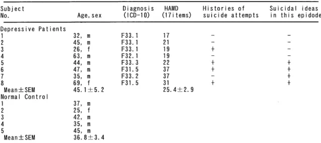
Table 2. Clincal Characteristics of Depressive Patients and Normal Controls
Subject Diagnosis HAMD Histories of Suicidal ideas
No. Age, sex (ICD-10) (17i terns) suicide attempts in this epidode Depressive Patients
1 32, m F33.1 17 - -
2 45, m F33.1 21 - -
3 26, f F33.1 19 + -
4 63, m F32.1 19 - -
5 44, m F33.3 22 + +
6 47, m F31.5 37 + +
7 35, m F33.2 37 - +
8 69, f F31.5 31 + +
Mean±SEM 45.1 ±5. 2 25.4±2. 9
Normal Control
1 37, m
2 25, f
3 42, m
4 35, m
5 45, m
Mean±SEM 36.8±3.4
F31.5: bipolar affective disorder, current episode of severe depression with psychotic symptoms,F32.1: mod- erate depressive episode, F33.1: recurrent depressive disorder, current episode moderate, F33.2: recurrent depressive disorder, current episode severe without psychotic symptoms, F33.3: recurrent depressive disor- der, current edisode severe with psychotic symptoms, HAMD: Hamilton Depression Scale
patient group, 1.04 ± 0.08 nM (n = 8). The Bmax of ['25I] LSD binding was significantly increased in the patient group (5.52±0.46 fmol/mg) compared to the control group (3.13±0.20 fmol/mg, p < 0.01) (Table 3, Fig.6).
Bmax of [125I]LSD binding and the HAMD score for eight depressive patients
A comparison of the Bmax of [ 125I] LSD binding for the eight depressive patients free from psychotropic medication for at least 6 months with the HAMD scores at the time of blood sampling before treatment showed no significant correlation (r= 0.16, p=0.56, Fig.7).
Fig.6. Binding parameters(Kd and Bmax)of platelet [125I]LSD binding in depressive patients free from psychotropic medica- tion for at least 6 months and the normal controls. The bind- ing parameters of platelet [125I]LSD binding in these two groups at the time of blood sampling before treatment were calculated by Scatchard analysis. The mean (±SEM) Kd of ['25I] LSD binding for the control group was 0.87±0.06 nM (n=5), which was not significantly different from the that of depressive patient group, 1.04±0.08 nM (n=8). The Bmax of [1251]LSD binding was significantly increased in the patient group (5.52±0.46 fmol/mg) compared to the control group (
3.13±0.20 fmol/mg, p<0.01).
Fig.7. The Bmax of [125I]LSD binding and the HAMD scores for eight depressive patients free from psychotropic medica- tion for at least 6 months. A comparison of the Bmax of [1211]LSD binding for the eight depressive patients free from psychotropic medication for at least 6 months with the HAMD scores at the time of blood sampling before treatment showed no significant correlation(r=0.16, p=0.56).
Table 3. Binding parameters(Kd and Bmax)of platelet [125I]LSD binding in the depressive patients, free from psychotropic medication at leastsix months and the normal controls
Subject Depressive Patients Normal Controls
No. Kd Bmax Kd Bmax
(nM) (f mo l /mg) OM) (f mo l /mg)
1 0.89 5.08 0.77 2.70
2 1.16 3.85 0.93 3.10
3 1.09 7.50 0.77 3.86
4. 0.68 4.94 1.04 2.90
5 1.22 6.40 0.84 3.10
6 0.94 4.70
7 0.96 7.10
8 1.39 4.60
Mean±SEM 1.04± 0.08 5.52± 0.46" 0.87± 0.05 3.13-±- 0.20 L ** p (0.01, two-tailed Student's t-test, as compared to normal controls
Bmax of [125I]LSD binding for patients with suicidal be- havior and without suicidal behavior(Fig.8)
In the eight depressive patients, four had past histo- ries of suicidal attempts (Table 2). In addition, four of the patients had suicidal ideas, show over position 2 in the item "suicide" of HAMD, at the time of blood sam- pling (Table 2).
The Bmax for the patient subgroup with histories of suicidal attempts was 5.80±0.70 fmol/mg (mean ± SEM, n=4), which was not significantly different from the patient subgroup without histories of suicidal attempts 5.24±0.68 fmol/mg(mean±SEM, n=4). The Bmax for the patient subgroup with suicidal ideas at the time of blood sampling was 5.70 ± 0.62 fmol/mg(mean ± SEM, n=4), which was also not significantly different from the patient subgroup without suicidal ideas at the time of blood sampling 5.34±0.77 fmol / mg (mean ± SEM, n=4). The Bmax for every patient subgroup was sig-
nificantly increased compared to the con- trol group (p <0.05).
The Kd for the patient subgroup with histories of suicidal attempts was 1.16 ± 0.10 nM (mean ± SEM, n=4), which was not significantly different from the patient sub- group without histories of suicidal attempts 0.92±0.10 nM(mean ±SEM, n=4). The Kd for the patient subgroup with suicidal ideas at the time of blood sampling was 1.13±
0.11 nM(mean ±SEM, n=4), which was also not significantly different from the pa- tient subgroup without suicidal idea at the time of blood sampling 0.96±0.11 nM (mean
±SEM, n=4).
Platelet 5-HT2A receptor binding and scores on Hamilton's rating scale for depression (HAMD) after treatment in two cases
Case No.5 44-year-old man , head of the office Diagnosis: F33.3 recurrent depressive disorder, current episode severe with psychotic symptoms
The patient had his first depressive episode at age 25, and at age 27, he had his second depressive epi- sode. Since then, he has had some minor depressive episodes without any difficulties in business. He had no history of psychiatric treatment or psychotropic medication. In the present episode, at age 44, after he was very busy on business, he had depressive symp- toms including a depressive mood, insomnia, and appe- tite loss. One month later, his follower caused a traffic
Fig.8. The Bmax of [125I] LSD binding for patients with suicidal behavior and without suicidal behavior.
The Bmax for the patient subgroup with histories of suicidal attempts was 5.80±0.70 fmol/ mg (mean ±SEM, n=4), which was not significantly different from the patient subgroup without histories of suicidal attempts 5.
24±0.68 fmol/mg(mean ±SEM, n=4). The Bmax for the patient subgroup with suicidal ideas at the time of blood sampling was 5.70 ±0.62 fmol/mg(mean ±SEM, n=4), which was also not significantly different from the patient subgroup without suicidal ideas at the time of blood sampling 5.34 ±0.77 fmol/mg(mean ±SEM, n=4).
Depression(SA): the patient subgroup with histories of suicidal attempts, Depression(-SA): the patient subgroup without histories of suicidal attempts. Depression(SI): the patient subgroup with suicidal ideas at the time of blood sampling, Depression(-SI): the patient subgroup without suicidal ideas at the time of blood sampling.
HAMD Bmax(fmol/mg)
25 7
f HAMD
~-Bmax 6
20
5
15 4
10 3
---•
. 2 5
1
0 r 0
0 1 3 4 5 week
HAMD and Bmax (patient No. 5)
HAMD Bmax (fmo I /mg)
20 6
5
15 .~
' 4
3 10 `
- • HAMD s
+ Bmax 2
5
1
0 0
2 3 4 6 8 week
HAMD and Bmax (patient No. 9)
Bmax(fmol/mg)
7 Q7 6
5 8
4 ,4
3 r=0.99 2
1
0 0 5 10 15 20 25
HAMD
HAMD and Bmax (patient No. 5)
Fig.9. Repeated analysis of platelet 5-HT2A receptor binding and the HAMD rating after treatment in the case of depres- sive patient No.5. His depressive symptoms subsided in re- sponse to the antidepressant treatment. The Bmax of platelet [125I]LSD binding showed a gradually decrease, corresponding to the decreased HAMD number, which was also in response to the antidepressant treatment(maprotiline maximum dose 60 mg/day). A comparison of the Bmax of [1211]LSD binding and the HAMD score demonstrated a significant linear corre- lation (r= 0.99, p=0.00002).
accident. He blamed himself for that accident and felt severe guilt. He also felt that he was subject to a po- liceman's superintendence. One month later he made a suicidal attempt (hanging), and he visited our psychi- atric clinic and was admitted to our psychiatric ward the same day. After admission, his HAMD score was evaluated and a platelet [ 125I] LSD binding assay was performed every week. At the first week of admission, flunitrazepam was prescribed. In addition, at the sec- ond week, an antidepressant treatment(maprotiline 30 mg/day) was started. His depressive symptoms sub- sided in response to the antidepressant treatment. The Bmax of platelet [125I]LSD binding showed a gradually decrease, corresponding to the decreased HAMD score, which was also in response to the antidepressant treatment(maprotiline maximum dose 60 mg/day). A comparison of the Bmax of [ 125I] LSD binding and the HAMD score demonstrated a significant linear correla- tion (r=0.99, p=0.00002, Fig.9.).
Bmax (fmo I /mg) 6
5 '
4 ,
3 r=0.71
2 1 0
0 5 10 15 20
HAMD
HAMD and Bmax (patient No. 9)
Fig.10. Repeated analysis of platelet 5-HT2A receptor binding and the HAMD rating after treatment in the case of depres- sive patient No.9. After the start of treatment with an experi- mental new drug(double blind study), his depressive symp- toms improved initially, but from the sixth week of admission they became worse. Correlatively, the Bmax of platelet [125I] LSD binding decreased at first and then in- creased. A comparison of the Bmax of [ 125I] LSD binding and the HAMD scores also demonstrated a significant linear cor- relation (r=0.71, p=0.02).
Case No.9 35-year-old man, cook
Diagnosis: F31.3 bipolar affective disorder, current epi- sode mild or moderate depression
The patient had his first depressive episode at age 23, and at age 24, he had first hypomanic episode. At age 25, he had his second depressive episode, and at age 27, he had his third depressive episode. In the pre- sent episode, at age 34, he had depressive symptoms that included a depressive mood and low self confi- dence, because he was worried about his prearranged marriage. His depressive state gradually deteriorated.
Four month later he broke off his engagement. At that time he also had suicidal ideas and tried to commit suicide. One month later he visited our psychiatric clinic and was admitted to our psychiatric ward the same day. After admission, his HAMD score was evalu- ated and a platelet [ 125I] LSD binding assay was per- formed every week. After the start of treatment with an experimental new drug (double blind study, both active drugs), his depressive symptoms improved
initially, but from the sixth week of admission they became worse. Correlatively, the Bmax of platelet ["'I]
LSD binding decreased at first and then increased. A comparison of the Bmax of [125I]LSD binding and the HAMD scores also demonstrated a significant linear correlation (r=0.71, p=0.02, Fig.10).
The significant linear correlation between the Bmax of ['25I] LSD binding and the HAMD scores in these cases suggests that there is a possible relationship be- tween increased platelet 5-HT2A receptor concentrations and the severity of depressive symptoms in the clini- cal course.
Discussion
In recent years, serotonin receptors were divided into 14 different subtypesincluding 5-HT2 family of receptors, 5-HT2A, 5-HT2B and 5-HT2C. There appears to be a striking similarity in the pharmacological character- istics of platelet and brain 5-HT2A receptors2>,12>. In addi- tion, 5-HT2A receptors are linked to the phosphoinocitide second messenger system in both platelets") and brain".
And the nucleotide sequence of human platelet 5-HT2A cDNA is identical to that reported for the human fron- tal cortex 5-HT2A receptor". These results suggest that the regulation of 5-HT2A receptors at the gene level may be the same both platelets and brain. In the pre- sent study, we applied in vitro receptor autoradiography to characterize [ 125 I] LSD binding to human platelet pellet sections. The present method re- vealed a single class of high affinity binding sites for [ 125I] LSD in human platelets. Both ketanserin and spiperone, the 5-HT2A selective ligands, inhibited ["'I]- LSD binding to human platelet pellets with high po- tency (IC50 values of 0.15 and 0.19 nM, respectively), whereas 5-HT and paroxetine (selective 5-HT reuptake inhibitor) inhibited binding with a very low potency.
These data confirmed that the binding sites of human platelet pellets labelled by [ 1251] LSD using this method revealed 5-HT2A receptors. This method required a much smaller volume of blood (5ml) than a classical membrane binding assay (30m1).
In a comparison of [125I]LSD binding to human platelet pellets in patients with depressive disorder be- fore treatment in this episode, in patients who had been drug free for at least six months, and in normal controls, Bmax was significantly higher in depressive patients before treatment than in the controls. Patients were considered to show over moderate severity by which showed 17-37 points (25.4±2.9, mean±SEM) in the HAMD (17 items). In the depressive group, the comparison of the Bmax of [125I]LSD binding for eight
depressive patients who had been free from psychotropic medication at least 6 months and the HAMD at the time of blood sampling before treatment demonstrated no significant correlation. The small
number of depressive patients may have influenced this result.
We recorded the platelet 5-HT2A measurements and the scores on the HAMD repeatedly after treatment in
two cases. The comparison of the Bmax of [ 1211 ]LSD binding and the HAMD demonstrated significant lin- ear correlation in these cases (Fig.9, Fig.10). There is a possible relationship between the increased plate- let 5-HT2A receptor concentrations and the symptoms of depression. In the first case (No.5) , the Bmax of
[ 125 I] LSD binding showed gradual decreases, corre- sponding to the decreased HAMD scores in response to antidepressants treatment. It was considered that the decreased numbers of platelet 5-HT2A receptors in re- sponse to the antidepressants treatment might result in a down-regulation. In the second case (No.9), soon after the start of treatment with an experimental new drug(double blind study), the depressive symptoms began to improve, but after the sixth week, symptoms began to worse again. Correlatively, the Bmax de- creased at first and then increased again. This could not be fully explained by the antidepressant treatment alone.
It is very interesting that the density of platelet 5-HT2A receptors in depressive patients who had been free from psychotropic medication for at least 6 months was higher than that in normal controls. This finding showing increased values for the density of platelet 5-HT2A receptors in depressive patients com- pared to normal controls is in agreement with the study of Biegon et al'),", but in disagreement with those of Cowen et al.'), and McBride et al."', all of whom found similar mean values for the density of platelet 5-HT2A receptors in depressive patients and normal controls. On the other hand, Pandey et al." ),22) have showed increased platelet 5-HT2A receptors in sui-
cidal patients independent of psychiatric diagnosis. In our experiments there was no statistical difference be-
tween the depressive patients with suicidal behavior and the depressive patients without suicidal behavior.
Discrepancies in the results of the platelet 5-HT2A re- ceptor binding studies in depressive disorders may be a reflection of number of factors, including the effects of the length of the psychotropic medication free inter- val on platelet 5-HT2A receptor binding201. In our ex- periments, the drug-free interval of at least 6 months was longer than that in other studies. The effects of exposure to psychotropic medication on platelet 5-HT2A receptor binding in our experiments, therefore
![Table 1. Inhibition of specific binding of [t25I]LSD to human platelet pellet sections](https://thumb-ap.123doks.com/thumbv2/123deta/10138984.1972863/4.889.90.430.175.342/table-inhibition-specific-binding-human-platelet-pellet-sections.webp)
![Table 3. Binding parameters(Kd and Bmax)of platelet [125I]LSD binding in the depressive patients, free from psychotropic medication at leastsix months and the normal controls](https://thumb-ap.123doks.com/thumbv2/123deta/10138984.1972863/6.889.79.512.203.361/binding-parameters-platelet-depressive-patients-psychotropic-medication-leastsix.webp)
