New Fecal Biomarker, α1-acid Glycoprotein, for Evaluation of Inflammatory Bowel Disease: Comparison with Calprotectin
and Lactoferrin
Takashi WATANABE1), Kunihiko AOYAGI1), Satoshi NIMURA2), Koichi EGUCHI1), Yoshitaka TOMIOKA1), and Shotaro SAKISAKA1)
1) Department of Gastroenterology and Medicine, Faculty of Medicine, Fukuoka University
2) Department of Pathology, Faculty of Medicine, Fukuoka University
Abstract
Objective: Fecal leukocytes biomarkers such as calprotectin (Cal) and lactoferrin (LF) have been shown to reflect the disease activity of inflammatory bowel disease (IBD). Here, we evaluated fecal α1-acid glycoprotein (AG), acute-phase reactant protein, as a new fecal biomarker.
Methods: Thirty six patients with ulcerative colitis (UC) and Crohn’s disease (CD) were analyzed. Active or inactive conditions determined to clinical activity index (CAI), Mayo endoscopic subscore and Matts’
histopathological grade in UC, while by Crohn’s disease activity index (CDAI) and simple endoscopic score for Crohn’s disease in CD. The fecal levels of biomarkers were measured by enzyme-linked immunosorbent assay.
Results: All three fecal biomarkers were significantly higher in the CAI active than in the inactive group, but not in the CDAI active compared with the inactive group. These biomarkers were significantly elevated in endoscopically active compared to the inactive UC and CD, respectively. These biomarkers were also significantly higher in the histologically active than in the inactive UC.
Conclusions: Fecal biomarkers, AG as well as Cal and LF, could differentiate active from inactive UC and CD. Our results strongly suggest that the fecal AG may be valuable noninvasive diagnostic tools for evaluation of the activity of IBD.
Key words: Fecal α1-acid glycoprotein, Calprotectin, Lactoferrin, Inflammatory bowel disease
Introduction
Inflammatory bowel disease (IBD) such as ulcerative colitis (UC) and Crohn’s disease (CD) are chronic inf lammator y disorders of gastrointestinal tract characterized by relapse and remission. It is important to evaluate the activity of the disease for the treatment of IBD. The evaluations of the activity of IBD have been mainly achieved by clinical, laborator y and endoscopic disease activity indices. C-reactive protein
(CRP), erythrocyte sedimentation rate and white blood
cells are widely used as noninvasive parameters for IBD. These laboratory markers do not appropriately reflect the activity of the intestinal tract because of a summation of systemic host responses rather than being specific for intestinal inflammation in IBD patients [1, 2]. Subsequently, they have insufficient sensitivity and specificity for intestinal inflammation [3]. Therefore, more reliable biological markers are required to confirm the disease activity.
Current gold standard for assessing intestinal inflammation has been considered to be endoscopic evaluation. Endoscopy allows visual determination of
Correspondence to : Kunihiko Aoyagi, MD, PhD., Department of Gastroenterology and Medicine, Faculty of Medicine, Fukuoka University, 7-45-1 Nanakkuma, Jonan-ku, Fukuoka, 814-0180 Japan
Tel:+81-92-801-1011 Fax:+81-92-874-2663 E-mail:[email protected]
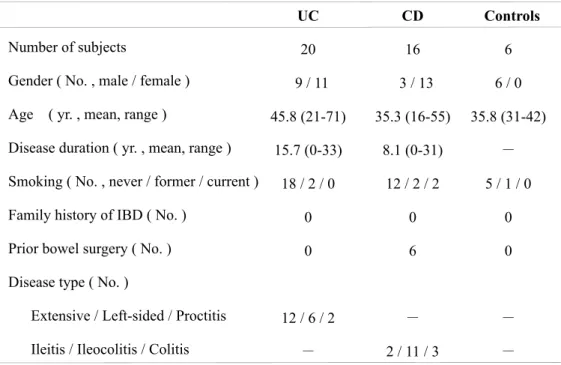
Table 1 Characteristics of subjects
disease severity and extent, and then mucosal biopsy could provide further information. However, endoscopic procedure is invasive and requires an uncomfortable preparation. The active gut inflammation is closely associated with the migration of neutrophils into the gut. A number of neutrophil-derived proteins in stools have been studied, including fecal calprotectin
(Cal), lactoferrin (L F), lysoz yme, elastase, and myeloperoxidase [2]. There has been increasing evidence that fecal biomarkers are valuable tools because of a simple, rapid, sensitive, specific, inexpensive and noninvasive to detect and monitor intestinal inflammation in IBD [4]. Recently, fecal Cal and LF have been shown to be excellent markers of intestinal inflammation, as it reflects the migration of neutrophils through the inflamed bowel wall to the mucosa [3, 5, 6].
α1-acid glycoprotein (AG)(orosomucoid) is an acute phase protein synthesized predominantly by hepatocytes in response to tissue injury, inflammation or infection [7]. During an acute phase condition, the concentration rises several times, making it one of the predominant proteins in serum [8]. Serologically, serum AG has been proved to have a stronger correlation with CDEIS, endoscopic disease score, compared with CRP [9]. The serum level of AG has been assessed as a standard disease activity index, although a long half life limits its usefulness [7]. Recently, urinary AG has been proposed as a potential biomarker in CD [10]. However, there has been no report
evaluating a disease of IBD using fecal AG. The aim of this study was to investigate the clinical utility of fecal AG as the evaluation of IBD activity. Here we identified validity of fecal biomarkers in the evaluation of IBD activity compared with various indices.
Subjects and Methods
Subjects
Thirt y-six IBD patients (UC, 20; CD, 16) were included at the Fukuoka University Hospital, Fukuoka, Japan. Control group consisted of six healthy subjects who showed no ev idence of abnor ma l it y under ileocolonoscopy. Their demographic characteristics are shown in Table 1. Six patients with CD had undergone prior ileocecal resection. Informed consent was obtained from all patients and control subjects. Study protocol was approved by the Ethics Committee of the Fukuoka University Hospital (09-5-09).
Evaluation of clinical, endoscopic and pathological disease activity in UC
As the clinical parameter, clinical activity index
(CAI) was used [11]. Endoscopic activity indices were based on Mayo endoscopic subscore [12]. Pathological evaluation of biopsy specimens was performed after stained with hematoxylin and eosin. A single experienced gastrointestinal pathologist (S.N.), who was completely
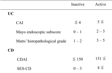
Table 2 Assessment of activity in UC and CD blinded to t he endoscopic f indings, scored t he
pathological findings according to the scoring system of Matts’ histopathological grade [13]. The endoscopic and pathological evaluations were performed regarding the most severely diseased area.
Evaluation of clinical and endoscopic disease activity in CD Crohn’s disease activity index (CDAI) was used as a clinical index [14]. Endoscopic activity indices were based on simple endoscopic score for Crohn’s disease (SES- CD)[15].
Assessment of activity in UC and CD
As shown in Table 2, CAI, Mayo endoscopic subscore, Matts’ histopathological grade, CDAI and SES-CD were divided into active and inactive conditions [11, 14, 16, 17]. Endoscopically active conditions in UC were defined as subscore of 2-3, while inactive conditions were defined as subscore of 0-1, which was previously considered as mucosal healing (MH)[17].
Collection of fecal samples and fecal analysis
Fecal samples were taken within one week before or after endoscopic examination in the absence of the change of treatments, and then stocked at -30˚C within 5 hours after sampling until analysis. Cal was measured by enzyme linked immunosorbent assay(ELISA)
method (Calprotectin ELISA Kit, Immundiagnostik AG, Bensheim, Germany). Moreover, LF and AG were similarly assayed using ELISA method at the Kyoto
Institute of Medical Science(Kyoto Medical Science Laboratory, Kyoto, Japan).
Statistical analysis
The data were analyzed using the Mann-Whitney U-test and the Spearman rank correlation coefficient.
For all analyses, P–values less than 0.05 were considered statistically significant.
Results
Comparison of fecal biomarkers (AG, Cal and LF) among UC, CD and control groups
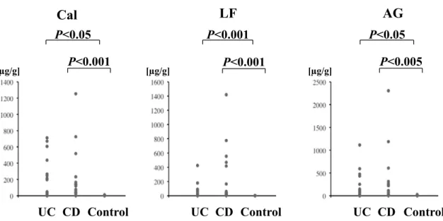
Fecal Cal, LF and AG levels were significantly higher in both UC and CD groups than in the control group (Cal, UC P<0.05, CD P<0.001; LF, UC P<0.001, CD P<0.001; AG, UC P<0.05, CD P<0.005)(Figure 1).
Correlation of fecal biomarkers (Cal, LF and AG) with CAI in UC
All fecal biomarkers were significantly correlated with CAI (Cal, r=0.789, P<0.001; LF, r=0.647, P<0.01;
AG, r=0.777, P<0.001). Fecal Cal, LF and AG levels were significantly higher in the active phase than in the inactive phase (Cal, P<0.001; LF, P<0.001; AG, P<0.001)
(Figure 2).
Comparison of fecal biomarkers (Cal, LF and AG) with endoscopic activity indices in UC
Endoscopically active UC patients showed significantly higher fecal Cal, LF and AG levels than inactive UC
UC CD Control
P<0.001 P<0.001 P<0.005
P<0.001
UC CD Control UC CD Control
P<0.05 P<0.05
Cal LF AG
[µg/g] [µg/g] [µg/g]
Inactive Active P<0.001 Cal
Inactive Active P<0.001
LF
Inactive Active P<0.001
AG
800 700 600 500 400 300 200 100 0
450 400 350 300 250 200 150 100 50 0
1200 1000 800 600 400 200 0
[µg/g] [µg/g] [µg/g]
Figure 1 Comparison of fecal biomarkers (Cal, LF and AG) among UC, CD and control groups.
In both UC and CD groups, Cal, LF and AG levels are significantly higher compared with the control group, respectively (Cal, P<0.05, P<0.001; LF, P<0.001, P<0.001; AG, P<0.001, P<0.001).
Figure 2 Comparison of fecal biomarkers (Cal, LF and AG) with CAI in UC.
The fecal Cal, LF and AG levels were significantly higher in the active phase than the inactive phase groups (Cal, P<0.001; LF, P<0.001; AG, P<0.001).
patients by Mayo endoscopic subscore (Cal, P<0.001; LF, P<0.05; AG, P<0.001)(Figure 3).
Comparison of fecal biomarkers (Cal, LF and AG) with histopathological grade in UC
Fecal Cal, LF and AG levels were significantly higher in the active conditions than in the inactive conditions by Matts histopathological grade (Cal, P<0.005; LF, P<0.05;
AG, P<0.001)(Figure 4).
Correlation of fecal biomarkers (Cal, LF and AG) with CDAI in CD
Fecal Cal and AG were poorly correlated with CDAI
(Cal, r=0.603, P<0.05; AG, r=0.503, P<0.05), but LF was not (LF, r=0.298, P=0.262,). There were no significant differences in fecal Cal, LF and AG levels between inactive and active CDAI (Figure 5).
Comparison of fecal biomarkers (Cal, LF and AG) with SES-CD in CD
Endoscopically active CD patients showed significantly
P<0.001 Cal P<0.05 LF AG P<0.001
Inactive Active Inactive Active Inactive Active
800 700 600 500 400 300 200 100 0
1600 1400 1200 1000 800 600 400 200 0
2500
2000
1500
1000
500
0
[µg/g] [µg/g] [µg/g]
[µg/g] [µg/g]
[µg/g]
Cal LF AG
P<0.005 P<0.05 P<0.001
Inactive Active Inactive Active Inactive Active
Figure 3 Comparison of fecal biomarkers (Cal, LF and AG) with endoscopic activity indices in UC.
A significant difference was observed in fecal Cal, LF and AG when dividing into inactive and active conditions (Cal, P<0.001; LF, P<0.05; AG, P<0.001).
Figure 4 Comparison of fecal biomarkers (Cal, LF and AG) with histopathological grade in UC.
A significant difference was observed in fecal Cal and LF when dividing into inactive and active conditions (Cal, P<0.005; LF, P<0.05; AG, P<0.001).
higher fecal Cal, LF and AG levels than inactive CD patients by SES-CD (Cal, P<0.001; LF, P<0.001; AG, P<0.001)(Figure 6).
Discussion
The present study first demonstrated that fecal AG in similar to fecal Cal and LF was determined to be significantly higher in both UC and CD groups than in the control group, and to associate with endoscopic disease activity scores in UC and CD. In UC, all three fecal
biomarkers were significantly higher in the CAI active group than in the inactive group, but not in the CDAI active group compared with the inactive group in CD.
These fecal biomarkers were also significantly higher in the histologically active group than in the inactive group in UC.
The previous studies have indicated that fecal Cal and LF assay are useful markers for identifying IBD from irritable bowel syndrome or healthy controls, and that these markers provide better correlations with endoscopic disease activities [3, 16, 18, 19]. Our results are in accordance
NS AG NS
LF NS
Cal
Inactive Active Inactive Active Inactive Active
1600 1400 1200 1000 800 600 400 200 0 1400
1200 1000 800 600 400 200 0
2500
2000
1500
1000
500
0
[µg/g] [µg/g] [µg/g]
Inactive Active Inactive Active Inactive Active
Cal LF AG
P<0.001 P<0.001
P<0.001
[µg/g] [µg/g] [µg/g]
Figure 5 Comparison of fecal biomarkers (Cal, LF and AG) with CDAI in CD.
There were no significant differences in fecal Cal, LF and AG levels between inactive and active CDAI.
Figure 6 Comparison of fecal biomarkers (Cal, LF and AG) with SES-CD in CD.
Fecal Cal, LF and AG assay were significantly higher in the active group compared with the inactive group by SES-CD (Cal, P<0.001; LF, P<0.001; AG, P<0.001).
with their findings regarding the good performance of fecal Cal and LF to discriminate between different inflammatory activities. In this study, fecal AG levels also have good diagnostic accuracy in distinguishing IBD from healthy controls. Furthermore, here we identified that fecal AG levels were well correlated with the endoscopic activity in UC and CD. It is considered that these results showed fecal AG, in addition to Cal and LF, reflect the activity of intestinal mucosa in IBD.
Serum AG is an acute phase protein which has been determined to be correlated well with disease activity[7].
The leakage of elevated serum AG reflects both of the increase in serum AG and the mucosal defect. In addition to fecal excretion of AG, several reports have shown that urinary excretion of AG significantly increases in lupus nephritis patients with active renal disease compared with inactive disease [20], and that urinary AG is associated with diabetic nephropathy, in which urinary AG is an independent risk factor [21]. Thus AG is appeared to be a promising fecal and urinary biomarker.
From our study, fecal biomarkers (Cal, AF and AG)
were associated with the discrimination between active
and inactive clinical indices in UC, but not in CD. The similar discrepancy has also been documented for the correlation between symptoms and endoscopic appearance [22, 23]. In CD, evaluation of remissions and relapses are usually based on the CDAI value, which represents a subjective and indirect assessment of gut inflammatory activity because it includes certain variables and symptoms not directly correlated with active inflammation and expression of severity rather than activity of disease [24]. Thus the fecal biomarkers might be correlated with endoscopic activities rather than clinical indices, suggesting that fecal biomarkers are more reliable for assessing the disease activity in IBD.
Recently, the treatment in IBD has aimed at MH on endoscopy since MH after 1 year of treatment is predictive of reduced subsequent disease activity and decreased need for active treatment such as colectomy [25]. Furthermore, endoscopic monitoring after treatment has now become of significance [26]. However, endoscopic examinations have the disadvantage of being invasive, time-consuming, expensive, and sometimes uncomfortable for patients [16]. The definition of MH is ill-defined in the small intestine, compared with the large intestine. Therefore, there is a growing importance of fecal biomarkers, which is closely correlated with endoscopic disease activity and MH.
Our study has some limitations. Although serum AG has been shown to be well correlated with disease activity, its longer half life, compared with CRP, have been considered to make its limit in clinical practice [2, 7]. However, for the evaluation of fecal excretion, the longer half life may be advantage. Second, the sensitivity and specificity of fecal AG were not obtained because of the small number of patients. Further studies are required to evaluate of fecal AG assay in a large number of IBD patients.
In conclusion, the present study first demonstrated the clinical usefulness of fecal AG, in addition to Cal and LF, in IBD. The close associations are demonstrated between fecal biomarkers and CAI, endoscopic activity and histological activity in UC, and between those and endoscopic activity in CD. These results suggest that fecal AG reflect the degree of intestinal inflammation, and could be a promising noninvasive diagnostic tool for evaluation of the activity in patients with IBD.
Acknowledgement
The authors thank Mr. Ryoichi Matsuse (Kyoto Medical Science Laboratory, Kyoto, Japan) for his excellent support in assays of fecal samples.
References
[1] Foell D, Wittkowski H, Roth J: Monitoring disease activity by stool analyses: from occult blood to molecular markers of intestinal inflammation and damage. Gut 58: 859-868, 2009.
[2] Vermeire S, Van Assche G, Rutgeerts P: Laboratory markers in IBD: Useful, magic, or unnecessary toys?
Gut 55: 426-431, 2006.
[3] Mao R, Xiao YL, Gao X, Chen BL, He Y, Yang L, Hu PJ, Chen MH: Fecal calprotectin in predicting relapse of inflammatory bowel diseases: a meta- analysis of prospective studies. Inflamm Bowel Dis 18: 1894-1899, 2012.
[4] Konikoff MR, Denson LA: Role of fecal calprotectin as a biomarker of intestinal inf lammation in inflammatory bowel disease. Inflamm Bowel Dis 12:
524-534, 2006.
[5] Roseth AG, Schmidt PN, Fagerhol MK: Correlation between faecal excretion of indium-111-labelled granulocytes and calprotectin, a granulocyte marker protein, in patient with inflammatory bowel disease.
Scand J Gastroenterol 34: 50-54, 1999.
[6] Sipponen T, Savilahti E, Kolho K, Nuutinen H, Turunen U, Färkkilä M: Crohn’s disease activity assessed by fecal calprotectin and lactoferrin:
correlation with Crohn’s disease activity index and endoscopic findings. Inflamm Bowel Dis 14: 40-46, 2008.
[7] Desai D, Faubion WA, Sandborn WJ: Review article:
biological activity markers in inflammatory bowel disease. Aliment Pharmacol Ther 25: 247-255, 2007.
[8] Hochepied T, Wullaert A, Berger FG, Baumann H, Brouckaert P, Steidler L, Libert C: Overexpression of alpha(1)-acid glycoprotein in transgenic mice leads to sensitisation to acute colitis. Gut 51: 398-404, 2002.
[9] Winterkamp S, Weidenhiller M, Otte P, Stolper J, Schwab D, Hahn EG, Raithel M: Urinary excretion of N-methylhistamine as a marker of disease activity in inflammatory bowel disease. Am J Gastroenterol 97:
3071-3077, 2002.
[10] Markó L, Szigeti N, Szabó Z, Böddi K, Takátsy A, Ludány A, Koszegi T, Molnár GA, Wittmann I:
Potential urinary biomarkers of disease activity in Crohn’s disease. Scand J Gastroenterol 45: 1440- 1448, 2010.
[11] Rachmilewitz D: Coated mesalazine (5-aminosalicylic acid) versus sulphasalazine in the treatment of active ulcerative colitis: a randomised trial. BMJ 298: 82- 86, 1989.
[12] Rutgeerts P, Sandborn WJ, Feagan BG, Reinisch W, Olson A, Johanns J, Travers S, Rachmilewitz D, Hanauer SB, Lichtenstein GR, de Villiers WJ, Present D, Sands BE, Colombel JF: Infliximab for induction and maintenance therapy for ulcerative colitis. N Engl J Med 353: 2462-2476, 2005.
[13] Matts SG: The value of rectal biopsy in the diagnosis of ulcerative colitis. Q J Med 30: 393-407, 1961.
[14] Best WR, Becktel JM, Singleton JW, Kern F Jr:
Development of a Crohn’s disease activity index.
National Cooperative Crohn’s Disease Study.
Gastroenterology 70: 439-44, 1976.
[15] Daperno M, D’Haens G, Van Assche G, Baert F, Bulois P, Maunoury V, Sostegni R, Rocca R, Pera A, Gevers A, Mary JY, Colombel JF, Rutgeerts P:
Development and validation of a new, simplified endoscopic activity score for Crohn’s disease: the SES-CD. Gastrointest Endosc 60: 505-512, 2004.
[16] S choepfer A M , B egl i nger C, S t r au ma n n A , Trummler M, Vavricka SR, Bruegger LE, et al. Fecal calprotectin correlates more closely with the Simple Endoscopic Score for Crohn’s disease (SES-CD)
than CRP, blood leukocytes, and the CDAI. Am J Gastroenterol 105:162-169, 2010.
[17] Rutgeerts P, Sandborn WJ, Feagan BG, Reinisch W, Olson A, Johanns J, Travers S, Rachmilewitz D, Hanauer SB, Lichtenstein GR, de Villiers WJ, Present D, Sands BE, Colombel JF: Infliximab for induction and maintenance therapy for ulcerative colitis. N Engl J Med 353: 2462-2476, 2005.
[18] Roseth AG, Aadland E, Jahnsen J, Raknerud N:
Asessment of disease activity in ulcerative colitis by faecal calprotectin, a novel granulocyte marker protein. Digestion 58: 176-180, 1997.
[19] Langhorst J, Elsenbruch S, Mueller T, Rueffer A, Spahn G, Michalsen A, Dobos GJ: Comparison of 4 neutrophil derived proteins in feces as indicators of disease activity in ulcerative colitis. Inflamm Bowel Dis 11: 1085-1091, 2005.
[20] Watson L , Midgley A , Pilkington C, Tullus K, Marks S, Holt R, Jones C, Beresford M: Urinary monocyte chemoattractant protein 1 and alpha 1 acid glycoprotein as biomarkers of renal disease activity in juvenile-onset systemic lupus erythematosus.
Lupus 21: 496-501, 2012.
[21] Jiang H, Guan G, Zhang R, Liu G, Liu H, Hou X, Cheng J. Increased urinary excretion of orosomucoid is a r isk predictor of diabet ic neph ropat hy.
Nephrology (Carlton) 14: 332-337, 2009.
[22] D’Haens G, Geboes K, Ponette E, Penninckx F, Rutgeerts P. Healing of severe recurrent ileitis with azathioprine therapy in patients with Crohn’s disease. Gastroenterology 112: 1475-1481, 1997.
[23] D’Haens G, Ferrante M, Vermeire S, Baert F, Noman M, Moortgat L, Geens P, Iwens D, Aerden I, Van Assche G, Van Olmen G, Rutgeerts P: Fecal calprotectin is a surrogate marker for endoscopic lesions in inflammatory bowel disease. Inflamm Bowel Dis 18: 2218-2224, 2012.
[24] Costa F, Mumolo MG, Cessarelli L , Bellini M, Romano MR, Sterpi C, Ricchiuti A, Marchi S, Bottai M: Calprotectin is a stronger predictive marker of relapse in ulcerative colits than in Crohn’s disease.
Gut 54: 364-368, 2005.
[25] Froslie KF, Jahnsen J, Moum BA, Vatn MH; IBSEN Group: Mucosal healing in inflammatory bowel disease: results from a Norwegian population-based cohort. Gastroenterology 133: 412-422, 2007.
[26] Rutgeer ts P, Diamond R H, Bala M, Olson A , Lichtenstein GR, Bao W, Patel K, Wolf DC, Safdi M, Colombel JF, Lashner B, Hanauer SB: Scheduled maintenance treatment with infliximab is superior to episodic treatment for the healing of mucosal ulcerat ion associated w it h Crohn’s disease.
Gastrointest Endosc 63: 433-442, 2006.
(平成25.7.10受付,平成25.10.10受理)