ORIGINAL ARTICLE
AN INITIAL INSTITUTIONAL APPRAISAL OF LAPAROSCOPE-ASSISTED COLECTOMY FOR EARLY COLORECTAL CANCER
Hajime Morohashi,Akihiko Murata,Motoi Koyama,Yoshiyuki Sakamoto,
Shinji Tsutsumi,Shinnosuke Yonaiyama,Hiroshi Ogasawara,Toru Yoshikawa,
Takahiro Muroya,Makoto Nakai,Keinosuke Ishido,
Naoki Wajima and Kenichi Hakamada
Abstract Introduction: The employment of laparoscopic resection for the treatment of colorectal cancer has been expanding rapidly. However, its indication is still on debate. We have been actively employing laparoscope-assisted colectomy (LAC) for patients with early colorectal cancer. Here we retrospectively review the 71 cases of early colorectal cancer treated with LAC and analyzed the initial institutional surgical outcome.
Results: LAC was indicated as an additional surgical resection after endoscopic treatment in 27 cases, while it was employed as a definitive non-endoscopic treatment in 44 cases. Mean operative time was 163 min, and mean blood loss was 57 ml. Complications were encountered in 8 cases (11.3%). Advanced lesions invading to the muscularis propria or the serosa were more included in the group initially treated with LAC alone (p=0.04). The reasons for additional LAC were the depth of submucosal invasion >1000 µm in 15 cases and lymphatic or microvascular invasion in 5 cases. Lymph node metastases were detected pathologically in 9 patients, in which one suffered relapse after surgery.
Conclusion: Implementation of LAC for early colorectal cancer seems an acceptable strategy in terms of both radicality and less invasiveness. Cooperation between endoscopists and laparoscopic surgeons is important to apply LAC appropriately for colorectal cancer.
Hirosaki Med.J. 62:173―179,2011
Key words: laparoscope-assisted colectomy; early colorectal cancer.
原 著
当科における早期大腸癌に対する腹腔鏡下大腸切除症例の検討
諸 橋 一 村 田 暁 彦 小 山 基 坂 本 義 之 堤 伸 二 米内山 真之介 小笠原 紘 吉 川 徹 室 谷 隆 裕 中 井 款 石 戸 圭之輔 和 嶋 直 紀
袴 田 健 一
抄録 【目的】大腸癌に対する腹腔鏡手術は急速に拡大しているが,その適応については議論が多い.当科では早期大腸 癌症例に対して積極的に腹腔鏡下大腸切除術を導入している.そこで,術前早期大腸癌と診断され腹腔鏡補助下大腸切 除術を施行した71症例を対象として当施設の初期治療成績を後方視的に検討した.【結果】内視鏡的切除後腹腔鏡下大腸 追加切除施行群は27例,内視鏡切除非施行腹腔鏡下大腸切術群は44例であった.平均手術時間は163分,平均出血量は 57 ml,合併症は 8 例(11.3%)であった.深達度は内視鏡切除非施行群で術後筋層以深と判明した浸潤癌が多く認められ た(p=0.04).内視鏡切除後腹腔鏡下大腸追加切除理由は,1000 µm 以上の sm 浸潤癌が14例,脈管浸襲陽性が 5 例であっ た.術後のリンパ節転移陽性例は全体で 9 例あり 1 例に再発が認められた.【結語】早期大腸癌に対する腹腔鏡下大腸切 除は,根治性と低侵襲性の両面から妥当と考えられた.この場合,特に内視鏡医と腹腔鏡手術医との連携が重要である.
弘前医学 62:173―179,2011
キーワード:腹腔鏡補助下大腸切除;早期大腸癌.
Department of Gastrointestinal Surgery, Hirosaki University Graduate School of Medicine
Correspondence: K. Hakamada
Received for publication, January 24, 2011 Accepted for publication, September 30, 2011
弘前大学大学院医学研究科消化器外科学講座 別刷請求先:袴田健一
平成23年 1 月24日受付 平成23年 9 月30日受理
Introduction
Along with advancement of endoscopic diagnostics for colorectal cancer and its wider use in both medical check-ups and clinical settings, the incidence of colorectal cancer found in earlier stage has been increasing in both Japan and USA
1). The latest 2010 version of the Guidelines for the Treatment of Colorectal Cancer by the Japanese Society for Cancer of the Colon and Rectum
2)now recommends that endoscopic excisional biopsy, either endoscopic mucosal resection (EMR) or endoscopic submucosal dissection (ESD), should be done as a first-step treatment modality if the lesions are estimated to be limited to mucosa or within submucosa to some extent
3). However, in cases that endoscopic treatment seems technically difficult from anatomical reasons or in cases that endoscopic excision seems not feasible because pretreatment endoscopy demonstrates the sign of massive invasion to the submucosa or deeper layer
4), surgery still has its role as the definitive treatment modality. Moreover, in cases that the depth of lesion is underestimated by the initial endoscopic evaluation and later found to exceed the submucosa pathologically, additional surgical resection is adopted as a rescue therapy because lymph node metastasis occurs in as high as 10%
if the lesions invades to the deeper submucosal layer
5).
As for surgical procedure, laparotomy is still the standard approach of radical resection for colorectal cancer. Recently, various kinds of minimally invasive surgeries like laparoscope- assisted colectomy
6, 7)and hand-assisted laparoscopic colectomy
8)have been developed to reduce patientsʼ burden associated with damage to the abdominal wall
8). These procedures are preferred especially in cases with earlier stage in which cancer is not exposed to the serosal surface and complete lymph node clearance can be achievable. However, the
efficacy of laparoscopic approach in comparison to open surgery is still on debate. We have been actively employing laparoscope-assisted colectomy (LAC) for early colorectal cancer together with open colectomy. We herein review the medical records of patients undergoing LAC for early colorectal cancer retrospectively and analyzed the initial institutional surgical outcome during the learning curve.
Patients and Methods
Between October 2001 and November 2010, 71 patients diagnosed as having early colorectal cancer preoperatively underwent LAC at Hirosaki University Hospital. Early colorectal cancer was defined as the lesion limited to the mucosa or within the submucosa and that without any distant metastasis. The preoperative diagnostic methods included colorectal endoscopy and muti-ditector row computed tomography in all cases. Other image studies like PET-scan or MRI were adopted in some patients when indicated.
The clinical and pathological records of all 71 patients were reviewed in terms of gender, age, tumor location, tumor size, implementation of preoperative endoscopic treatment (EMR or ESD), operative time, blood loss, degree of lymph node clearance, histological differentiation, depth of invasion, presence of vascular invasion, presence of lymph node metastasis, operative morbidity and mortality, and length of hospital stay. Late recurrence was also evaluated.
Principally, the lesions were evaluated by
the endoscopists first. They allocated each
case to either endoscopic treatment or surgical
resection as an initial intervention according
to the technical feasibility and the tumor
extension. Additional surgical resection after
endoscopic treatment was indicated for cases
with positive specimen margin, presence of
microscopic vascular or lymphatic invasion, or
depth of submucosal invasion more than >1000 µm according the Guideline
2). In all cases, the detailed reasons why LAC was performed as an initial step without endoscopic intervention or as additional surgical resection were analyzed.
Results
Of the 71 patients who underwent LAC, 3 4 w e r e m a l e a n d 3 7 w e r e f e m a l e . T h e mean patient age was 63.6 years. Fifty-one patients (71.8%) had colon cancer and 20
(28.2%) had rectal cancer. Sigmoid colon was the most prevalent site (28.2%). Forty-four
(62.0%) patients underwent LAC as an initial treatment, while 27 (38%) were converted to LAC according the results from EMR or ESD.
Degree of lymph node clearance was D1 in 15 cases (21.1%) and D2 in 54 cases (76.1%). Mean operative time was 163 (53-455) min, with a mean blood loss of 57 (0-514) ml. Postoperative complications were encountered in 8 cases
(11.3%); wound infection in 4, intestinal obstruction in 2, intra-pelvic abscess in 1, and
anastomotic failure in 1 case (Table 1).
The depth of invasion in patients diagnosed as having early cancer preoperatively and undergoing LAC as a first step treatment was underestimated in 13 (29.5%), in which the lesions invaded to muscularis propria in 9 and to subserosa in 4 cases. Consequently, stages of the two groups were different according to the implementation of preoperative endoscopic treatment or not. However, histological features including cancer differentiation, presence of vascular or lymphatic invasion, and presence of lymph node metastasis were not different between the two groups (Table 2).The reasons why endoscopic treatments were skipped before LAC were signs of massive submucosal invasion in 27 patients (61.4%), a tumor size of >20 mm in 13 (29.5%), technical difficulty in 2 (4.5%), and unfavorable histology of undifferentiated adenocarcinoma in 1 (2.3%)
(Table 3). The reasons for surgical resection after endoscopic treatment included submucosal invasion exceeding 1000 µm in 14 cases (51.9%), and microvascular and/or lymphatic invasion
Table 1 Patient Data (N = 71)
Gender Male 34 (47.9%), Female 37 (52.1 %)
Age 63.6 (39-85)
Tumor location
Cecum 6(8.5%)
Ascending colon 15(21.1%)
Transverase colon 8(11.3%)
Descending colon 2(2.8%)
Sigmoid colon 20(28.2%)
Rectosigmoid colon 14(19.7%)
Rectum above the peritoneal reflection 3(4.2%)
Rectum below the peritoneal reflection 3(4.2%)
Preoperative EMR or ESD (+) 27 (38.0%), (-) 44 (62.0%)
Operation
Degree of lymph node dissection* D1 15, D2 54, D3 1
Operative time 163 min (53-455min)
Blood loss 57 ml (0 ‒ 514ml)
Outcome
Morbidity (+) 8 (11.3%), (-) 63 (88.7%)
Mortality 0
Length of hospital stay 10.0 days (3-21 days)
*According to the Japanese Classification of Colorectal Carcinoma
EMR : endoscopic mucosal resection ESD : endoscopic submucosal dissection
in 5 cases (18.5%). Four patients (14.8%) were specimen margin-positive, 3 patients (11.1%)
suffered from local recurrence after EMR, and 1 patient (3.7%) had undifferentiated adenocarcinoma (Table 4).
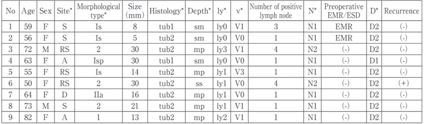
Lymph node metastases were detected pathologically in 9 patients with their extent being N1 in 7 and N2 in 2. Histological features of their primary tumors were moderately differentiated adenocarcinomas (tub2) in 7
(77.8%), presence of lymphatic invasion in
6 (66.7%) and microvascular invasion in 5
(55.6%). During the observation period, about 10 months after surgery, recurrence occurred in one patient whose cancer had invaded to the subserosal layer of the rectosigmoid colon and metastasized to N2 lymph node (Table 5).
Discussion
Laparoscopic surgery has been applied to the treatment of almost all colorectal diseases,
Table 2 Tumor profile in patients undergoing LAC with or without preoperative endoscopic treatment.
LAC after endoscopic treatment LAC alone P value
N=27 (Colon : 16 Rectum : 11) N=44 (Colon : 20 Rectum : 24)
Macroscopic type* Ip : 4 Is :5 Isp :10 IIa :4 Is : 9 Isp:9 IIa : 3 IIa+IIc:5 0.48
Maximum size 13.6 (4‒28) mm 22.1 (7‒58) mm 0.09
Cancer differentiation tub1: 16 tub2: 10 tub1: 13 tub2: 10 0.36
Depth of invasion sm:27 m : 8 sm:23 mp : 9 ss : 4 0.04
<1000 µm: 4 (14.8%) >1000 µm:23 (85.2%)
Positive margin with endoscopic treatment
(+): 4 (14.8%) (-): 23 (85.2%)
Lymphatic invasion* ly0: 18 ly1 : 3 ly2 : 1 ly0 : 27 ly1 :10 ly2 : 4 ly3 :3 0.17
Vascular invasion* v0: 16 v1 : 5 v2 : 2 v0: 30 v1: 9 v2: 3 v3: 2 0.45
Lymph node involvement (+): 2 (7.4%) (-): 25 (92.6%) (+): 7 (15.9%) (-): 37 (84.1%) 0.21
*According to the Japanese Classification of Colorectal Carcinoma
tub1: well-differentiate tubular adenocarcinoma, tub2: moderately differentiated tubular adenocarcinoma sm: submucosa, m: mucosa, mp: muscularis propria, ss: subserosa
Table 3 The reason for LAC without preoperative endoscopic treatment(N=44)
Number Sign suggestive of deep submucosal invasion 27(61.4%)
Larger size (>20mm) 13(29.5%)
Undifferentiated carcinoma 1(2.3%)
Technical difficulty 2(4.5%)
Others 1(2.3%)
Table 4 The reasons for LAC after endoscopic treatment(N=27)
Number
sm invasion >1000 µm 14(51.9%)
Microvascular and/or lymphatic invasion 5(18.5%)
Positive specimen margin 4(14.8%)
Undifferentiated carcinoma 1(3.7%)
Local recurrence after EMR 3(11.1%)
either benign or malignant, since Jacobs et al.
9)first introduced it as an alternative procedure to the conventional open surgery in 1991. Indeed, minimum invasiveness to the abdominal wall has an absolute cosmetic benefit and probably shows accelerated postoperative recovery due to less pain
6, 7). However, this approach has also been criticized for technical difficulties which require much more operative time and may predispose an increased risk of procedure- specific complications.
I n o u r i n s t i t u t i o n a l l e a r n i n g c u r v e , operative time elongated to a mean of 163 min, while blood loss was as minimum as 57 ml. Operative morbidity of 11% with zero mortality and a mean hospital stay of 10 days were comparable to those of open surgery at the same period (data not shown). Lourenco et al.
10)also reported shorter length of hospital stay and earlier recovery associated with less blood loss, although the operative time became longer than that of open surgery.
They described no significant difference in the incidence of anastomotic failure and wound infection between the two arms. Hasegawa et al.
11)reported the operative morbidity of 14.3%, with the incidences of wound infection, intestinal obstruction, intraabdominal abscess and anastomotic failure being 8.2%, 2.2%, 1.1%,
and 2.9%, respectively, which were very similar to those in our hospital. So far, we have not experienced late occurrence of incisional hernia at the port sites, which are unique to the laparoscopic approach
12).
In addition to procedure-related outcomes, oncological aspects of LAC are another major concern. In our initial experience, recurrence was observed in one case, the tentative recurrence rate being 1.4%. This case was underestimated as early cancer first. However, it was later found that tumor cells invaded to the serosal layer and metastasized to the N2 lymph node. So it remains unclear whether the recurrence occurred due to oncological features of more advanced stage or due to procedure- related problems. There are some reports that both recurrence and survival rates after LAC are the same as those of conventional laparotomy
13-15). Indeed the long-term outcome should be discussed according to randomized control trials (RCTs) between LAC and open surgery. According to the meta-analysis of 3346 patients from 23 RCTs
16), rates of recurrence at the site of the primary tumor were similar
(colon cancer: 4 RCT, 938 pts, 5.2% vs 5.6%, p=0.57; rectal cancer: 4 RCT, 714 pts, 7.2% vs 7.7%, p=0.46). No differences were observed in the occurrence of port-site/wound recurrences
Table 5 Nine cases with positive lymph node metastasis No Age Sex Site* Morphological
type*
Size
(mm)Histology* Depth* ly* v* Number of positive
lymph node N* Preoperative
EMR/ESD D* Recurrence
1 59 F S Is 8 tub1 sm ly0 V1 3 N1 EMR D2 (-)
2 56 F S Is 5 tub2 sm ly0 V0 1 N1 EMR D2 (-)
3 72 M RS 2 30 tub2 mp ly3 V1 4 N2 (-) D2 (-)
4 63 F A Isp 30 tub1 sm ly0 V0 1 N1 (-) D1 (-)
5 55 F RS Is 14 tub2 mp ly1 V3 1 N1 (-) D2 (-)
6 50 F RS 2 30 tub2 ss ly1 V0 4 N2 (-) D2 (+)
7 64 F D IIa 16 tub2 mp ly1 V0 1 N1 (-) D2 (-)
8 73 M S 2 21 tub2 mp ly1 V1 1 N1 (-) D2 (-)
9 82 F A 1 13 tub2 mp ly2 V1 1 N1 (-) D2 (-)
*According to the Japanese Classification of Colorectal Carcinoma
S: sigmoid colon, RS: rectosigmoid, A: ascending colon, D: descending colon, ly: lymphatic invasion, v: vascular invasion, N: degree of lymph node metastasis, D: degree of lymph node dissection
(p=0.16), and similar cancer-related mortality was found after laparoscopic surgery compared to open surgery (colon cancer: 5 RCT, 1575 pts, 14.6% vs 16.4%,p=0.15; rectal cancer: 3 RCT, 578 pts, 9.2% vs 10.0%, p=0.16).
However, these analyses included different stages of tumors in different series of learning curves. Five-year survival of 50% in Stage III of these analyses seems lower when compared to the historical data from Japan
2). Theoretically, LAC is more technically feasible in early cancer, because of less possibility of procedure-related tumor dissemination and less incidences of distant lymph node metastases. The Japanese Guideline 2010 now recommends that LAC should be carefully applied according to the institutional availability of skilled surgeons, the precise preoperative staging (invading depth is mucosa or submucosa and lymph node metastasis is not pointed out) and the appropriate tumor location technically feasible to the laparoscopic approach. Taking these issues into account, we decided to implement LAC for early stage colorectal cancer first. In a learning curve, Left sided colectomy was performed in more than 50% cases because of its technical easiness.
Another reason why we indicated LAC for early cancer is increased numbers of colorectal cancer diagnosed in earlier stage. This may be due to a rapid expansion of the use of endoscopy as a routine diagnostic tool after positive occult blood at the medical check- up in Japan
1). Endoscopic approach, either EMR or ESD, is used for both diagnostic and treatment purposes. Favorable outcome by the endoscopic intervention would be expected if the tumor is in a controllable size and shows the features indicating less possibility of lymph node metastasis. Japanese Guideline 2010 advocates that invasion to the deeper submucosal tumor more than 1000 µm, presence of microvascular and lymphatic invasion, unfavorable histology
with poorly differentiation and presence of budding
17)at the invasive front increase the risk for regional lymph node involvement as high as 10%, and thus recommends that additional surgical resection should be performed to eradicate enough lymph nodes
2, 5). Indeed, 7.4% (2/27) of cases treated additional LAC according to this criteria showed lymph node metastases in our series. In turn, the majority of the patients with early colorectal cancer have tended to undergo unnecessarily invasive surgery for lymph node clearance. Therefore, we have to reduce excess patientsʼ burden by balancing radicality and invasiveness of each surgical procedure. LAC is an ideal treatment in this respect.
Another role of LAC is a definitive treatment for the cases that endoscopic intervention is not feasible. In high risk cases with lymph node metastasis as indicated above criteria, LAC must be a better approach because EMR and ESD cannot control the lesion outside the intestinal wall absolutely. Moreover, because of limitation in diagnostic power of endoscopy, these cases suggestive of deeper submucosal invasion are potentially advanced, in which the role of lymph node dissection becomes more important.
In our series, endoscopy underestimated in 30% (13/44) of cases, in which preoperative diagnoses of submucosal lesions were later found to be invaded to the muscularis propria or the subserosal layer. We should not hesitate to adopt LAC if such ominous signs indicating lymph node metastasis or deeper invasion are demonstrated by endoscopy.
In summary, our initial experience has
shown that LAC is an acceptable and perhaps
preferable alternative to conventional open
surgery for early colon cancer, with both
comparable procedure-related short-term results
and oncological outcomes. Implementing LAC
for early cancer first seems to be an acceptable
s t r a t e g y t o d e v e l o p a n e w i n s t i t u t i o n a l
program of laparoscopic colorectal surgery.
Anyway, cooperation between endoscopists and laparoscopists is very important for expanding the indications of LAC appropriately.
References
1)Ross WA. Colorectal cancer screening in evolution:
Japan and the USA. J Gastroenterol Hepatol 2010;25 Suppl 1:S49-56.
2)Japanese Society for Cancer of the Colon and Rectum. Guidelines for the Treatment of Colorectal Cancer ver. 2010. www.jsccr.jp/guidelline2010.
3)Tanaka S, Oka S, Chayama K. Colorectal en- doscopic submucosal dissection: present status and future perspective, including its differen- tiation from endoscopic mucosal resection. J Gastroenterol 2008;43:641-51.
4)Kudo SE, Takemura O, Ohtsuka K. Flat and depressed types of early colorectal cancers: from East to West. Gastrointest Endosc Clin N Am 2008;18:581-93.
5)Kitajima K, Fujimori T, Fujii S, Takeda J, Ohkura Y, Kawamata H, Kumamoto T, Ishiguro S, Kato Y, Shimoda T, Iwashita A, Ajioka Y, Watanabe H, Watanabe T, Muto T, Nagasako K. Correlations between lymph node metastasis and depth of submucosal invasion in submucosal invasive colorectal carcinoma: a Japanese collaborative study. J Gastroenterol 2004;39:534-43.
6)Bonjer HJ, Hop WC, Nelson H, Sargent DJ, Lacy AM, Castells A, Guillou PJ, Thorpe H, Brown J, Delgado S, Kuhrij E, Haglind E, Pahlman L.
Laparoscopically assisted vs open colectomy for colon cancer: a meta-analysis. Arch Surg 2007;142:298-303.
7)Jackson TD, Kaplan GG, Arena G, Page JH, Rogers SO, Jr. Laparoscopic versus open resection for colorectal cancer: a metaanalysis of oncologic outcomes. J Am Coll Surg 2007;204:439-46.
8)Chung CC, Ng DC, Tsang WW, Tang WL, Yau KK, Cheung HY, Wong JC, Li MK. Hand-assisted laparoscopic versus open right colectomy: a randomized controlled trial. Ann Surg 2007;246:
728-33.
9)Jacobs M, Verdeja JC, Goldstein HS. Minimally invasive colon resection (laparoscopic colectomy).
Surg Laparosc Endosc 1991;1:144-150.
10)Lourenco T, Murray A, Grant A, McKinley A, Krukowski Z, Vale L. Laparoscopic surgery for colorectal cancer: safe and effective? - A systematic review. Surg Endosc 2008;22:1146-1160.
11)Hasegawa H, Nishibori H, Ihii Y, Kitajima M.
Current status on laparoscopic surgery for colorectal cancer. 2004;31:685-9.
12)Schaeff B, Paolucci V, Thomopoulos J. Port site recurrences after laparoscopic surgery. A review.
Dig Surg. 1998;15:124-34.
13)Barlehner E, Benhidjeb T, Anders S, Schicke B.
Laparoscopic resection for rectal cancer: outcomes in 194 patients and review of the literature. Surg Endosc 2005;19:757-766.
14)Kitano S, Kitajima M, Konishi F, Kondo H, Satomi S, Shimizu N. A multicenter study on laparoscopic surgery for colorectal cancer in Japan. Surg Endosc 2006;20:1348-52.
15)Leroy J, Jamali F, Forbes L, Smith M, Rubino F, Mutter D, Marescaux J. Laparoscopic total mesorectal excision (TME) for rectal cancer surgery: long-term outcomes. Surg Endosc 2004;18:281-9.
16)Kuhry E, Schwenk WF, Gaupset R, Romild U, Bonjer HJ. Long-term results of laparoscopic colorectal cancer resection. Cochrane Database Syst Rev. 2008 Apr 16;(2):CD003432.
17)Ueno H, Murphy J, Jass JR, Mochizuki H, Talbot IC. Tumour 'budding' as an index to estimate the potential of aggressiveness in rectal cancer.
Histopathology 2002;40:127-32.