九州大学学術情報リポジトリ
Kyushu University Institutional Repository
細胞透過性ペプチド結合性JNK阻害剤のブタ腎虚血再 灌流傷害軽減効果
土井, 篤
https://doi.org/10.15017/1398555
出版情報:Kyushu University, 2013, 博士(医学), 論文博士 バージョン:
権利関係:Public access to the fulltext file is restricted for unavoidable reason (2)
Effect of cell permeable peptide of JNK inhibitor on the attenuation of renal ischemia/reperfusion injury in pigs
Atsushi Doi, MDa, Hidehisa Kitada, MD, PhDa,b, Morihito Ota, MDa, Sayako Kawanami, MDa,
Kei Kurihara, MDa, Yoshihumi Miura, MDa, Takehiro Nishiki, MDa, Yasuhiro Okabe, MDa,
Shigetaka Inoue, MD, PhDa, and Masao Tanaka, MD, PhDa
aDepartments of Surgery and Oncology, Graduate School of Medical Sciences, Kyushu
University, and bKidney Care Unit,Kyushu University Hospital, Fukuoka, Japan
email address and Statement 1
Atsushi Doi (category 1, 2, 3): [email protected]
Department of Surgery and Oncology Graduate School of Medical Sciences Kyushu University,
3-1-1 Maidashi, Higashi-ku, Fukuoka-shi, Fukuoka Pref 812-8582, Japan
Telephone number: 011-81-90-7382-4927
Hidehisa Kitada (category 2, 3): [email protected]
Morihito Ota (category 1): [email protected]
Sayako Kawanami (category 1): [email protected]
Kei Kurihara (category 1): [email protected]
Yoshihumi miura (category 1): [email protected]
Takehiro Nishiki (category 1): [email protected]
Yasuhiro Okabe (category 1): [email protected]
Shigetaka Inoue (category 2, 3): [email protected]
Masao Tanaka(category 2, 3): [email protected]
The number of Figures: 6
Authorship and conflict of interest statement
I declare that I have no conflict of interest in connection with this paper.
Abstract
The outcome of organ transplantation has been improved by better
immunosuppressive drugs, surgical techniques and management of systemic conditions.
However, ischemia/reperfusion injury remains one of the challenges that affect graft survival.
In this study, we used a new technique employing protein transduction domain (PTD)
to investigate whether the inhibition of c-Jun NH2-terminal kinase (JNK) pathway attenuates
renal ischemia/reperfusion injury. In a porcine renal ischemia/reperfusion model, a
PTD-JNK-inhibitor (JNKI) was administered into renal artery. And a PTD-JNKI was then taken
into the various cells containing the vascular endothelial cells. The uptake of these was
conducted by endocytosis using PTD. Serum creatinine and blood urea nitrogen concentrations
were lower in the PTD-JNKI group than in the control group. In addition, renal tissue blood
flow was well maintained in PTD-JNKI group, resulting in a lower level of tissue injury and
fewer apoptotic cells. These results suggest that the PTD technique improves renal
transplantation outcomes.
Introduction
The number of cadaveric donors remains insufficient compared with the increasing
number of transplant candidates1. Since the number of cadaveric donors is very small especially
in Japan, kidneys occasionally need to be extirpated from marginal donors. In these cases, acute
tubular necrosis resulting in delayed graft function or primary non function is the most common
complication2.
The short term inflammatory response induced by ischemia/reperfusion injury is
characterized by induction of a proinflammatory cytokine cascade, expression of adhesion
molecules and cellular infiltration3. Interleukin-1β and tumor necrosis factor-α (TNF-α) are well
known proinflammatory cytokines. c-Jun NH2-terminal kinase (JNK), a stress activated protein
kinase, forms a subgroup of the mitogen-activated protein kinase (MAPK) superfamily4. The
JNK pathway is closely related to apoptosis in ischemia/reperfusion injury5. In addition, JNK
activation is mediated by reactive oxygen species in TNF-α-induced apoptosis6.
The authors previously reported attenuation of ischemia/reperfusion injury by the use
of FR167653 or gabexate mesilate in large animal models7-9. In addition, a new technique of
efficient substance induction into cells using protein transduction domain (PTD) has recently
been reported10. PTD has many amino acids with a positive electric charge, so it adheres
strongly to the negatively charged cell membrane lipid bilayer. Thus PTD can transduce a wide
variety of cargo into cells, from small intermediate molecules to liposomes11.
We think this model is not pure warm or cold ischemia experiment, but it is similar to
clinical situation. Extirpation from marginal donors needs strict management from early stage.
When a donor faces death and kidney bloodstream is insufficient, kidneys of a donor are state of
warm ischemia. We think administering a protective drug to a donor from this point in time is
needed. Thus we have to administer a drug at room temperature. That is why this model is not
pure warm or cold ischemia.
In this study, we evaluated the effect of cell permeable peptide of a JNK inhibitor
(JNKI) to attenuate renal ischemia/reperfusion injury in a porcine model, and explore the
possibilities for adapting the technique to human donors.
Materials and Methods
This study was reviewed by the Committee of Ethics on Animal Experiments at
Kyushu University and conducted according to the Guidelines for Animal Experiments of the
Graduate School of Medical Sciences, Kyushu University, Law No. 105, and
Notification No. 6 of the Japanese Government.
Operative Procedures
Twelve female hybrid Landrace swine weighing 20 to 30 kg were used. General
anesthesia was induced with intramuscular midazolam (10 mg/pig), butorphanol tartrate (1
mg/pig) and medetomidine hydrochloride (1 mg/pig) followed by intravenous pentobarbital (25
mg/kg) and pancuronium bromide (0.1 mg/kg). Pancuronium bromide was supplemented as
needed. The animals were then intubated and ventilated mechanically with oxygen (1 L/min)
and 1 to 2% of sevoflurane with a tidal volume of 20 mL/kg at 12 cycles/minute. A central
venous line was placed into the right external jugular vein with a cutdown technique, and
physiological saline was infused during the operation at 300 ml/hr. The pulse rate and oxygen
saturation were continuously monitored. Cefalotin sodium (1 g/pig) was administered
intravenously 30 minutes before the start of the operation.
Laparotomy was performed by ventral midline incision. The left kidney was isolated,
and then both the renal artery and vein were encircled with tapes. The left ureter was isolated
and encircled similarly. The right kidney was removed. The left renal artery and vein were
clamped. The renal vein was clamped proximal to a lumbar vein. The ureter was also clamped
to prevent reflux of blood into the kidney through the vascular network around the renal hilum.
The left kidney was perfused with physiological saline at room temperature from the renal
artery until discoloration was observed. The perfusate was drained from a lumbar vein. After the
lumbar vein was ligated, 10 ml of the PTD-JNKI solution (experiment) or physiological saline
(control) was infused into the renal artery at room temperature. The kidney was placed under the
small intestine to be kept at body temperature for 90 minutes.
After 90 minutes of ischemia, all the vessels and the ureter were unclamped. After
observation for 60 minutes, the left kidney was covered and secured by the retroperitoneum and
the abdomen was closed. Postoperatively, the endotracheal tube was removed and the animals
were allowed access to water and food ad libitum. On postoperative day five, tissue samples
were obtained from the left kidney and the animals were euthanized by overdose of
pentobarbital.
Experimental Groups
PTD-JNKI was purchased from Sigma Aldrich (Ishikari, Japan). The animals were
assigned randomly to one of two groups. The investigators were blind with regard to the groups.
The PTD-JNKI group (n = six) received PTD-JNKI solution (10 μM). The control group (n=
six) received physiological saline.
Peptide Synthesis
PTD-JNKI peptide synthesis was requested from Sigma Ardrich. 11 arginine
connected peptides were used for PTD. JNKI was selected as a material for preventing
activation of the c-Jun NH2-terminal kinase pathway. It was hypothesized that it would attenuate
ischemia/reperfusion injury and apoptosis. PTD-JNKI peptide sequence was
RRRRRRRRRRRGGRPKRPTTLNLFPQVPRSQDT. Molecular weight of this peptide was
4542.16. We conjugated fluorescein to the N terminal of the peptide. This peptide was purified
to >80% purity.
Confirmation of Induction of PTD-JNKI into the Cells
To confirm the induction of PTD-JNKI into the cells, we used fluorescein-conjugated
PTD-JNKI peptide and fluorescence microscopy.
Biochemical Parameters
Serial blood samples were collected before the operation and 1, 6, 24, 48, 72, 96 and
120 hours after reperfusion. The samples were centrifuged, and the serum was stored at -80°C
until analysis. Serum creatinine and blood urea nitrogen (BUN) concentrations were determined
by enzymatic assay (SRL model 7170 autoanalyzer for creatinine and model 7450 autoanalyzer
for BUN; Hitachi, Tokyo, Japan).
TNF-α Measurement
Blood samples were collected before operation and one hour after reperfusion. The
samples were centrifuged, and the serum was stored at -80°C until analysis. In these samples,
TNF-α concentrations were determined by using a commercial porcine TNF-α enzyme-linked
immunosorbent assay (ELISA) kit (Porcine TNFα, Pierce Endogen, Inc., Rockford, IL, U.S.A.).
Absorbency of ELISA plates was measured at 450 nm by the use of a spectrophotometer
(Immuno Mini NJ-2300, Nalge Nunc Int., New York, NY, U.S.A.).
Evaluation of Renal Blood Flow and Vascular Resistance
Renal tissue blood flow was measured with a laser Doppler flow meter (Advance
ALF21R; Unique Medical, Tokyo, Japan) on the cranial, middle, and caudal portions of the
kidney before clamping, during ischemia, at 15, 30, 45, 60 minutes and at 120 hours after
reperfusion. Doppler flow study was performed at the three different points of the kidney before
clamping, at 15, 30, 45, 60 minutes and at 120 hours after reperfusion with a Doppler flow
meter (SSD-5500; ALOKA Prosound, Tokyo, Japan). Resistive index (RI) and pulsatility index
(PI) were calculated at each point on the interlobular arteries according to the following
formula: RI = (Vmax – Vmin)/Vmax, PI = (Vmax –Vmin)/Vmean, where Vmax denotes maximum
velocity, Vmin denotes minimum velocity, and Vmean denotes mean velocity.
Histological Examination.
All the animals were sacrificed and the left kidney of each retrieved on post-operative
day five. The tissue samples were fixed in 10% formalin, embedded in paraffin, sectioned in 4
μm slices and mounted on slides. After deparaffinization, each specimen was stained with hematoxylin and eosin to assess the level of histological tissue injury. The slides were evaluated
in terms of dilatation of proximal tubules, eosinophilic casts in distal tubules, loss of brush
borders, detachment of tubular cells, interstitial edema, whole tubular necrosis, neutrophil
infiltration, and interstitial hemorrhage. The findings were graded 0-3 (0; < 5% injury per 10
high-power fields (HPFs); 1: 5-24% injury per 10 HPFs; 2:25-49% injury per 10 HPFs; 3: >50%
injury per 10 HPFs. The samples were randomized and blindly examined by light microscopy
independently by two investigators capable of pathologic interpretation.
Apoptotic Index Using Terminal Deoxynucleotidyl Transferase-Mediated Deoxyuridine Triphosphate Nick End-Labeling (TUNEL) Stain
For the detection of DNA breaks, the TUNEL stain (In Situ Apoptosis Detection Kit,
TaKaRa, Otsu, Japan) was used. After pretreatment with proteinase K for 15 minutes at room
temperature, endogenous peroxidase activity was blocked with 3% H2O2 for five minutes, also
at room temperature. Equilibration buffer was applied to the sections for 10 seconds at room
temperature and they were then incubated with working strength terminal deoxynucleotidyl
transferase (TdT) enzyme for 60 minutes at 37°C in a humidification chamber. The sections
were then incubated with anti-digoxigenin conjugate for 30 minutes at room temperature in a
humidification chamber. Peroxidase substrate was applied for three to six minutes at room
temperature. The sections were counterstained with 0.5% methyl green for 10 minutes and
examined by light microscopy.
The apoptotic index (AI) was defined as the ratio of TUNEL positive cells to 1000
renal tubular epithelial cells in a clearly labeled area at ×400 magnification. Serial methyl green
stained sections were also analyzed to avoid misinterpretation of necrotic cells.
Statistical Analysis
All results are presented as mean ± standard deviation. Differences in serum creatinine
(sCr), serum blood urea nitrogen (sBUN), tissue blood flow, TNF-α expression and vascular
resistance over time were evaluated with repeated-measures analysis of variance (ANOVA)
with a post hoc test. Differences in the grade of the histological injury were evaluated with a
Mann-Whitney test. A p-value < 0.05 was considered statistically significant.
Results
Operative Findings and Postoperative Course
After reperfusion, the left kidney of all subjects became firm and the color returned to
normal. All animals in both groups walked and ate from starting on post-operative day 1. All
animals in both groups survived until they were sacrificed on the fifth day. No signs of
intra-abdominal infection were noted. The color of the left kidney was normal and a normal
amount of urine was present in the bladder in all subjects.
PTD-JNKI Transduction
PTD-JNKI peptide was successfully induced into the cells via infusion of the renal
artery at room temperature. The fluorescein-labeled PTD-JNKI peptide was visualized in the
vascular endothelial cells 30 minutes after infusion (Figure 1).
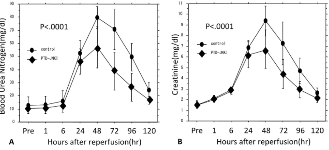
Renal function
In both groups, serum BUN concentrations began to increase starting six hours after
ischemia/reperfusion, and reached a peak on post-operative day 2 (Figure 2A). However, BUN
concentrations were lower in the PTD-JNKI group than in the control group during the entire
post-operative period (P<.0001). The change in serum creatinine concentrations was similar to
that of BUN concentrations (Figure 2B). Serum creatinine concentrations were also lower in the
PTD-JNKI group than in the control group (P<.0001).
Renal Blood Flow
In both groups, renal tissue blood flow, as determined by Doppler flow meter, was
decreased between clamping and unclamping, and then began to increase gradually (Figure 3A).
On post-operative day five, renal tissue blood flow exceeded the pre-ischemic level. There was
no statistically significant difference between groups in renal tissue blood flow (P=0.8461).
Vascular Resistance (Pulsatility Index)
In the control group, PI decreased immediately after reperfusion and reached
minimum values in 30 minutes (Figure 3B). PI then increased and reached maximum values on
post-operative day five. In the PTD-JNKI group, PI also decreased immediately after
reperfusion, but maintained a steady value from 15 minutes until 60 minutes of ischemia (Figure
3B). The PI increased on post-operative day five and reached the pre-ischemic level. The PI in
the PTD-JNKI group was lower than in the control group (P<0.05).
TNF-α Measurement
In both groups, the levels of serum TNF-α one hour after ischemia/reperfusion were
higher than that before ischemia/reperfusion. This difference was not statistically significant
(P=0.1929).
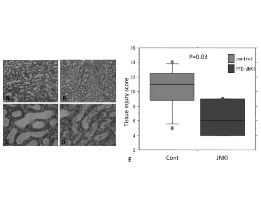
Histological Examination
On post-operative day five, renal tissue samples from the control group demonstrated severe
dilatation and loss of brush borders in proximal tubules, eosinophilic casts in distal tubules, mild
interstitial edema, neutrophil infiltration and interstitial hemorrhage. The severity of injury was
attenuated in all of these parameters in the PTD-JNKI group (Injury score: 10.8±3.13 vs
6.33±2.25; P=.03).
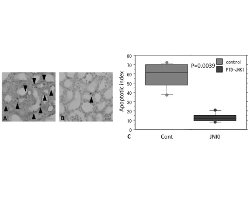
Apoptotic Index Using TUNEL Stain
On post-operative day five, fewer positively stained cells were identified in the
PTD-JNKI group than in the control group. Thus, AI was significantly reduced in the
PTD-JNKI group (AI: 58.3±13.5 vs 12.8±4.75; P=.0039).
Discussion
The present study revealed that PTD-JNKI was induced into renal endothelial cells by
perfusion from the renal artery in the porcine model. Furthermore, PTD-JNKI induced into renal
cells attenuated ischemia/reperfusion injury and reduced apoptotic tubule cells. As a result, this
study suggests that induction of PTD-JNKI into renal endothelial cells may improve renal
transplantation results from marginal donors.
The authors established the renal ischemia/reperfusion injury model in dogs in
previous reports7,9. Similar techniques were used for the present porcine model to confirm
whether PTD-JNKI attenuates renal ischemia/reperfusion injury, and whether it may
consequently improve outcomes of renal transplantation. Because serum BUN and creatinine
levels were slightly elevated when kidneys were exposed to a 60-minute warm ischemia in a
pilot study, it was decided to expose the kidneys to a 90-minute warm ischemia. In addition, the
contralateral kidney was removed in order to simplify the model.
JNK phosphorylates are not only regulatory sites in the N-terminus of the transcription
factor c-Jun but also in other transcription factors such as Elk-1 and p53. Cell apoptosis is
induced by the activation of JNK5. JNK activation is mediated by reactive oxygen species in
tumor necrosis factor-α induced apoptosis6. Many results have been reported that the
inactivation of JNK pathways attenuates ischemia/reperfusion injury in heart12,13, brain14,15,
islets16-18, liver19 and kidney20. Thus, a JNK inhibitor was chosen in the present study to attempt
to attenuate renal ischemia/reperfusion injury.
Previously, intracellular transport of materials was thought to be limited to those of
small size and molecular weight. Recently, however, some articles have reported that large
molecular weight materials can be effectively introduced intracellularly using a mechanism
called PTD10,11. PTD was first reported in 1988 as a part of TAT protein of human
immunodeficiency virus21. The Drosophila melanogaster homeobox protein Antennapedia22,23
and herpes simplex virus protein VP2224 are also well known examples of PTD. These PTDs
have many amino acids, such as arginine and lysine, with a positive electric charge. The
mechanism of introduction of PTD into cells was previously unclear. However, some reports
have recently described that the mechanism of induction is due to endocytosis. Positively
charged PTD adheres to the negatively charged cell membrane lipid bilayer strongly25.
Subsequent transport is performed by endosomes26,27.
In the present study, a peptide consisting of 11 arginine was used as a PTD because
this PTD was reported to be more efficient for transduction into cells28. Induction into renal
endothelial cells using this PTD was confirmed by fluorescence microscopy. PTD-JNKI was
induced into cells at room temperature, in order to replicate the clinical environment. Further
examination in varying environments is necessary.
Renal transplantation is the only curative treatment for end stage renal failure.
However, donor shortage remains a major problem in transplantation of many organs29,30.
Therefore, kidneys must sometimes be used from marginal donors, such as aged organs or those
with prolonged ischemic time. Ischemia/reperfusion injury is one of the factors affecting the
outcomes of renal transplantation, especially from non-heart-beating donors2. This type of
injury needs to be attenuated to improve the outcomes of transplantation from such donors. In
the authors’ previous communications, they reported the effects of a cytokine-suppressive agent
FR167653 or a synthetic protease inhibitor gabexate mesilate to attenuate ischemia/reperfusion
injury on the outcomes of renal or pancreatic transplantation in animal models7-9.
In ischemia/reperfusion injury, disturbance of micro-circulation is largely due to
endothelial damage, leading to an increase in vascular permeability31. It then causes leukocyte
plugging, vasoconstriction, and hemoconcentration32,33. In short, leukocytes localize and adhere
to adhesion receptors of the endothelium, such as intercellular adhesion molecule-1 (ICAM-1),
resulting in immobilization and diapedesis of leukocytes. ICAM-1 expression is then increased
by IL-1 and TNF-α34. In the present study, renal tissue blood flow decreased during ischemic
time, gradually increased after unclamping, and exceeded the pre-ischemic level on
post-operative day five. Although there was no significant difference in renal tissue blood flow
between groups, PI, reflecting peripheral vascular resistance, was lower in the PTD-JNKI group
than in the control group throughout the experimental period especially during the renal injury
period. The reason why tissue blood flow did not increase in the PTD-JNKI group despite the
low peripheral vascular resistance remains unknown. The decrease in the vascular resistance
may have increased tissue oxygen supply, resulting in the improvement of BUN and creatinine
concentrations.
ICAM-1 is intimately involved in acute renal failure caused by ischemia/reperfusion
injury. TNF-α accelerates the expression of ICAM-1. As serum TNF-α was reported to increase
one hour after ischemia/reperfusion in an animal model35, we examined the concentrations of
serum TNF-α one hour after ischemia/reperfusion. In this study, there was no significant
difference in the serum TNF-α concentration between the control and PTD-JNKI groups.
Measuring TNF-α at a later point or in the tissue samples instead of serum may have produced a
different result.
In clinical renal transplantation, characteristic histological findings in acute tubular
necrosis are dilatation of the proximal tubules, degeneration of the tubular epithelium,
interstitial edema, cellular infiltration, and casts in the distal tubules36. The authors previously
reported that loss of brush borders reflected acute tubular necrosis well9. In the present study,
several additional parameters were evaluated (loss of brush borders, detachment of tubular cells,
whole tubular necrosis, interstitial hemorrhage). Using these parameters, the extent of injury in
the control group was greater than in the PTD-JNKI group. The most noticeable differences
observed were dilatation of the proximal tubules, loss of brush borders in the proximal tubules,
and eosinophilic casts in the distal tubules.
The control group also contained many more apoptotic tubular cells as compared to
the PTD-JNKI group. Apoptosis is the principal mechanism leading to organ damage in renal
ischemia/reperfusion injury37.
In conclusion, induction of a cell permeable JNK inhibitor peptide attenuates renal
ischemia/reperfusion injury in pigs. This method may improve the results of renal
transplantation in humans and expand donor availability.
References
1. Charles SM, David AG, Andrew CN. The use of expanded criteria cadaver and live donor
kidneys for transplantation. Urol Clin North Am 2001;28:687-707.
2. Tanabe K, Oshima T, Tokumoto T, Ishikawa N, Kanematsu A, Shinmura H et al. Long-term
renal function in on-heart-beating donor kidney transplantation: a single-center experience.
Transplantation 1998;66:1708-13.
3. Takada M, Nadeau KC, Shaw GD, Marquette KA, Tilney NL. The cytokine-adhesion
molecule cascade in ischemia/reperfusion injury of the rat kidney. Inhibition by a soluble
P-selectin ligand. J Clin Invest 1997;99:2682-90.
4. Davis RJ. Signal transduction by the JNK group of MAP kinases. Cell 2000; 103:239-52.
5. Bogoyevitch MA, Boehm I, Oakley A, Ketterman AJ, Barr RK. Targeting the JNK MAPK
cascade for inhibition: basic science and therapeutic potential. Biochim Biophys Acta
2004;1697:89-101.
6. Shen HM, Liu ZG. JNK signaling pathway is a key modulator in cell death mediated by
reactive oxygen and nitrogen species. Free Radic Biol Med 2006;40:928-39.
7. Kitada H, Sugitani A, Yamamoto H, Otomo N, Okabe Y, Inoue S et al. Attenuation of renal
ischemia-reperfusion injury by FR167653 in dogs. Surgery 2002;131;654-62.
8. Yamamoto H, Sugitani A, Kitada H, Arima T, Nishiyama Ki, Motoyama K et al. Effect of
FR167653 on pancreatic ischemia-reperfusion injury in dogs. Surgery 2001;129:309-17.
9. Inoue S, Sugitani A, Yamamoto H, Kitada H, Motoyama K, Okabe Y et al. Effect of synthetic
protease inhibitor gabexate mesilate on the attenuation of ischemia/reperfusion injury in canine
kidney autotransplantation. Surgery 2005;137:216-24.
10. Schwarze, SR, Ho A, Vocero-Akbani A, Dowdy SF. In vivo protein transduction: delivery
of a biologically active protein into tha mouse. Science 1999;285:1569-72.
11. Wadia JS, Dowdy SF. Protein transduction technology. Curr Opin Biotechnol 2002;13:52-6.
12. Ferrandi C, Ballerio R, Gaillard P, Giachetti C, Carboni S, Vitte PA et al. Inhibition of c-Jun
N-terminal kinase decreases cardiomyocyte apoptosis and infarct size after myocardial ischemia
and reperfusion in anaesthetized rats. Br J Pharmacol 2004;142:953-60.
13. Milano G, Morel S, Bonny C, Samaja M, von Segesser LK, Nicod P et al. A peptide
inhibitor of c-Jun NH2- terminal kinase reduces myocardial ischemia-reperfusion injury and
infarct size in vivo. Am J Physiol Heart Circ Physiol 2007;292:1828-35.
14. Borsello T, Clarke PG, Hirt L, Vercelli A, Repici M, SchordetetDF et al. A peptide inhibitor
of c-Jun N-terminal kinase protects against excitotoxicity and cerebral ischemia. Nat Med
2003;9:1180-6.
15. Hirt L, Badaut J, Thevenet J, Granziera C, Regli L, Maurer F et al. D-JNKI1, a
cell-penetrating c-Jun N-terminal kinase inhibitor, protects against cell death in severe cerebral
ischemia. Stroke 2004;35:1738-43.
16. Noguchi H, Matsushita M, Okitsu T, Moriwaki A, Tomizawa K, Kang S et al. A new
cell-permeable peptide allows successful allogeneic islet transplantation in mice. Nat Med
2004;10(3):305-9.
17. Noguchi H, Nakai Y, Matsumoto S, Kawaguchi M, Ueda M, Okitsu T et al. Cell permeable
peptide of JNK inhibitor prevents islet apoptosis immediately after isolation and improves islet
graft function. Am J Transplant 2005;5:1848-55.
18. Abdelli S, Abderrahmani A, Hering BJ, Bechmann JS, Bonny C. The c-Jun N-terminal
kinase JNK participates in cytokine- and isolation stress-induced rat pancreatic islet apoptosis.
Diabetologia 2007;50:1660-9.
19. Uehara T, Bennett B, Sakata ST, Satoh Y, Bilter GK, Westwick JK et al. JNK mediates
hepatic ischemia reperfusion injury. J Hepatol 2005;42:850-9.
20. Wang Y, Ji HX, Xing SH, Pei DS, Guan QH. SP600125, a selective JNK inhibitor, protects
ischemic renal injury via suppressing the extrinsic pathways of apoptosis. Life Sci
2007;80:2067-75.
21. Frankel AD, Pabo CO. (1988). Cellular uptake of the tat protein from human
immunodeficiency virus. Cell 1988;55:1189-93.
22. Derossi D, Joliot AH, Chassaing G, Prochiantz A. The third helix of the
Antennapedia homeodomain translocates through biological membranes. J Biol Chem
1994;269:10444-50.
23. Derossi D, Calvet S, Trembleau A, Brunissen A, Chassaing G, Prochiantz A. Cell
internalization of the third helix of the Antennapedia homeodomain is
receptor-independent. J Biol Chem 1996;271:18188-93.
24. Elliott G, O’Hare P. Intercellular trafficking and protein delivery by a
herpesvirus structural protein. Cell 1997;88:223-33.
25. Mi Z, Mai J, Lu X, Robbins PD. Characterization of a class of cationic peptides able to
facilitate efficient protein transduction in vitro and in vivo. Mol Ther 2000;2:339-47.
26. Lundberg M, WikstrAm S, Johansson M. Cell surface adherence and endocytosis of protein
transduction domains. Mol Ther 2003;8:143-50.
27. Jean PR, Kamran M, Eric V, Corinne R, Birgit V, Mike JG et al. Cell-penetrating peptides A
reevaluation of the mechanism of cellular uptake. J Biol Chem 2003;278:585-90.
28. Matsushita M, Tomizawa K, Moriwaki A, Li ST, Terada H, Matsui H. A high-efficiency
protein transduction system demonstrating the role of PKA in long-lasting long-term
potentiation. J Neurosci 2001;21:6000-7.
29. Audard V, Matignon M, Dahan K, Lang P, Grimbert P. Renal transplantation from extended
criteria cadaveric donors: problems and perspectives overview. Transpl Int 2008;21(1):11-7.
30. Collini A, De Bartolomeis C, Ruggieri G, Barni R, Bernini M, Carmellini M. Long-term
outcome of renal transplantation from marginal donors. Transplant Proc 2006;38:3398-9.
31. Sutton TA, Mang HE, Campos SB, Sandoval RM, Yoder MC, Molitoris BA. Injury of the
renal microvascular endothelium alters barrier function after ischemia. Am J Physiol Renal
Physiol 2003;285:191-8.
32. Menger MD, Richter S, Yamauchi J, Vollmar B. Role of microcirculation in hepatic
ischemia/reperfusion injury. Hepatogastroenterology 1999;46:1452-7.
33. Mashiach E, Sela S, Winaver J, Shasha SM, Kristal B. Renal ischemia-reperfusion injury:
contribution of nitric oxide and renal blood flow. Nephron 1998;80:458-67.
34. Springer TA. Traffic signals for lymphocyte recirculation and leukocyte emigration: The
multistep paradigm. Cell 1994;76:301-14.
35. Kelly KJ, Williams WW Jr, Colvin RB, Meehan SM, Springer TA, Gutierrez-Ramos JC et al.
Intercellular adhesion molecule-1-deficient mice are protected against ischemic renal injury. J
Clin Invest 1996;97:1056-63.
36. Tisher CC, Brenner BM. Renal pathology with clinical and functional correlations.
Philadelphia: Lippincott Williams & Wilkins; 1989.
37. Daemen MA, de Vries B, Buurman WA. Apoptosis and inflammation in renal reperfusion i
njury. Transplantation 2002;73:1693-700.
Figures
Figure 1. Induction of PTD-JNKI into renal endothelial cells was observed by fluorescence
microscopy. A. Arterial endothelial cells. B. Venous endothelial cells (Original magnification
×400).
0 10 20 30 40 50 60 70 80 90
PTD-JNKI control
Blood Urea Nitrogen(mg/dl)
Hours after reperfusion(hr)
24 72 96 120
1 48
Pre 6
P<.0001
A
0 1 2 3 4 5 6 7 8 9 10 11
PTD-JNKI control
P<.0001
Hours after reperfusion(hr)
24 72 96 120
1 48
Pre 6
Creatinine(mg/dl)
B
Figure 2. Renal function. A. Serum BUN concentrations were lower in the PTD-JNKI group. B.
Serum creatinine concentrations were lower in the PTD-JNKI group. Values are mean±SD; n =
six animals per group.
0 2 4 6 8 10 12 14 16 18 20
PTD-JNKI control
Renal tissue blood flow(ml/min/ g) 100
Time after reperfusion Pre
ischemia
15 30 45 60 120 (min) (hr) A
.2 .4 .6 .8 1 1.2 1.4 1.6
PTD-JNKI control
Time after reperfusion
Pre 15 30 45 60 120
(min) (hr) P<0.05
Pulsatilityindex
B
*
*
Figure 3. Renal blood flow. A. Renal tissue blood flow (RTBF) measured with a Doppler
flow-meter. There is no significant difference between the control group and PTD-JNKI group
(P=0.8461). B. Pulsatility index (PI) measured by echo Doppler. PI was lower in the PTD-JNKI
group than in the controls throughout the study period. * P<0.05 versus control group by
one-way ANOVA. Values are mean±SD; n = six animals per group.
n.s.
TNFα(pg/ml)
-10 0 10 20 30 40 50 60 70 80 90
Pre 1hr
PTD-JNKI control
Figure 4. Serum TNF-α concentrations before and one hour after ischemia/reperfusion. The
concentrations tended to increase, but there is no difference between the control and PTD-JNKI
group. Values are mean±SD; n = six animals per group.
2 4 6 8 10 12 14 16
PTD-JNKI control
A B
C D
E
Tissue injury score
Cont JNKI
P=0.03
Figure 5. Representative microscopic findings at postoperative day five. A&C. Control group:
severe dilatation of the proximal tubules and loss of brush borders, mild interstitial edema,
neutrophil infiltration and interstitial hemorrhage. B&D. PTD-JNKI group: severity of injury
was attenuated in all parameters. (A&B. Original magnification×100; C&D. Original
magnification×400) E. Injury score: 10.8±3.13 vs 6.33±2.25; P=.03 Values are mean±SD; n =
six animals per group.
0 10 20 30 40 50 60 70 80
PTD-JNKI control
Apoptotic index
Cont JNKI
P=0.0039
A B
C
Figure 6. TUNEL stain at postoperative day five. A. In the control group, many apoptotic cells
were confirmed in renal tubule cells (arrow). B. In the PTD-JNKI group, the number of
apoptotic cells was remarkably decreased (Original magnification×400). C. Apoptotic index
(AI). AI was the ratio of TUNEL positive cells to 1000 renal tubular epithelial cells in a clearly
labeled area at ×400 magnification. (AI: 58.3±13.5 vs 12.8±4.75; P=.0039) Values are
mean±SD; n = six animals per group.