伊勢湾海底堆積物中に観察された珪藻類休眠期細胞の種同定
石井健一郎
1・澤山茂樹
1・中村 亨
2・石川 輝
3*・今井一郎
4 1京都大学大学院農学研究科応用生物科学専攻(〒606-8502 京都市左京区北白川追分町) 2三重大学附属練習船勢水丸(〒514-8507 三重県津市栗真町屋町1577) 3三重大学大学院生物資源学研究科生物圏生命科学専攻(〒514-8507 三重県津市栗真町屋町1577) 4北海道大学大学院水産科学研究院海洋生物資源科学部門海洋生物学分野(〒041-8611 北海道函館市港町3-1-1)Ken-Ichiro Ishii1, Shigeki Sawayama1, Toru Nakamura2, Akira Ishikawa3* and Ichiro Imai4: Species identification on diatom resting stage cells in the sediments of Ise Bay, Japan. Jpn. J. Phycol. (Sôrui) 62: 79-87, July 10, 2014
Resting stage cells of 25 diatom species, including one variety, from the sediment of Ise Bay were identified using light microscope. Nineteen species could be identified based on the morphology of these resting stage cells. The rest of six species, including one variety, were unable to be identified based on the morphology of the resting stage cells because there was no diagnostic morphological information of the resting stage cells. Five resting stage cells were identified from the germinated/rejuvenated cells as Chaetoceros eibenii, Ch. laciniosus, Ch. didymus var. anglicus, Actinocyclus cf. octonarius and Coscinodiscus cf. radiatus.
Key Index Words: diatom, germinated/rejuvenated cell, Ise Bay, resting stage cell
1Laboratory of Marine Environmental Microbiology, Division of Applied Biosciences, Graduate School of Agriculture, Kyoto University, Kyoto 606-8502, Japan
2Training Ship of Mie University, Kurima-Machiya, Tsu, Mie 514-8507, Japan
3Graduate School of Bioresources, Mie University, Kurima-Machiya, Tsu, Mie 514-8507, Japan
4Plankton Laboratory, Division of Marine Bioresource and Environmental Science, Graduate School of Fisheries Sciences, Hokkaido University, Hakodate, Hokkaido 041-8611, Japan
* Author for correspondence: [email protected]
緒言
沿岸海域に生息する植物プランクトン群集は,様々な海洋環 境の変化に応じて種交替を繰り返している。なかでも珪藻類は, 海洋生態系において一次生産者として,また魚介類の餌生物と して重要な役割を担う。一方で,ある種の珪藻は赤潮の原因種 としてノリの色落ちを引き起こすことから,水産学上において も重要な生物分類群である(Nishikawa
et al. 2007
)。 珪藻類は栄養塩の枯渇など増殖に不適な環境下で,耐久性を 有する休眠期細胞を形成することが知られている(Hargraves
& French1975
,1983
,Garrison 1984
,板倉2000
など)。この休眠期細胞は,増殖環境の好転によって再び発芽・復活 することで栄養細胞となり,水柱で増殖するためのいわば
「種(タネ)」としての役割を果たす(
Hargraves & French
1983
,McQuoid & Hobson 1996
,Itakura
et al. 1997
など)。したがって,海底堆積物中の休眠期細胞は
seed population
として認識される。珪藻類の生態を包括的に理解するために は,このような水柱に観察されない時期も含めて把握する必 要がある。
珪藻類の休眠期細胞は形態学的に休眠胞子(
resting spore
)と 休 眠 細 胞(
resting cell
) と に 分 け ら れ る(McQuoid &
Hobson 1996
)。前者は栄養細胞の被殻の中に栄養細胞とは 形態が全く異なる新たな被殻が形成され,栄養細胞よりも厚 い珪酸質の被殻構造を有する。この休眠胞子が再び増殖を開 始することを発芽(germination
)と呼称する。これまでに130
種以上の珪藻の休眠胞子が確認されているが(McQuoid
& Hobson 1996
),それらは珪藻類全体ではごく一部にすぎ ず未報告の休眠胞子が多数存在することが予測されている。 一方,休眠細胞は通常の細胞分裂と同様の過程を通じて形成 され,被殻内部の細胞質に栄養細胞よりも色の濃い色素体の 存在が確認されているものの(今井ら1990
,板倉ら1992
,McQuoid & Hobson 1996
,板倉2000
,Ishii
et al. 2012
),被殻の形態は栄養細胞と区別することが不可能である。この
細胞が再び増殖を開始することを復活(
rejuvenation
)と呼称する。これまで,海産珪藻の休眠期細胞が再増殖すること
を発芽と呼称し(例えば,
McQuoid & Hobson 1996)
,淡水珪藻の休眠期細胞が再び増殖することを復活と呼称する傾向 にあったが(例えば
Sicko-Goad
et al. 1989
),これらは明 確に定義されてこなかった。本研究では休眠胞子と休眠細胞 の再増殖を区別する必要があると考え,休眠胞子の再増殖を 発芽(germination
),休眠細胞の再増殖を復活(rejuvenation
) と定義する。 これら海底堆積物中の珪藻類休眠期細胞の密度を正確に把 握する方法として,光学顕微鏡を用いた直接検鏡による各種 休眠期細胞の種同定及び計数が挙げられる。これまで,珪藻 類休眠期細胞を直接検鏡によって種同定し,計数を行った研 究は極めて少ない。Pitcher (1990)
は南アフリカのケープ半島沖の湧昇域において,水柱における
Chaetoceros
属の栄養 細胞と休眠胞子を直接検鏡によって計数し,湧昇域における 両者の動態を明らかにした。この研究では休眠胞子の種同定 に走査型電子顕微鏡(SEM
)が用いられたが,それらの計 数結果は珪藻類の休眠期細胞トータルの値として示されてお り,種レベルでの全体的な計数は行われていない。SEM
を 用いた形態観察は,被殻構造の細部を観察して種同定するに は適しているが,それらを計数することはほぼ不可能である。 その理由として,休眠胞子を種同定するには,適切な角度か ら細胞を観察する必要があり,金属イオン蒸着により固定さ れた状態のSEM
による観察では,全ての細胞を正確に種同 定し,計数することが不可能となることが挙げられる。さら に,海底堆積物中の休眠期細胞を種同定する際には,多くの 堆積物粒子を除きながら観察する必要があるため,倒立顕微 鏡による直接検鏡が欠かせない。すなわち,海底堆積物中の 休眠期細胞密度を把握するには,光学顕微鏡下で種同定する ための形態学的情報が必須となる。 これまで海底堆積物中における珪藻類休眠期細胞の密度 は,最確数法(MPN
法)を用いて見積もられてきた(Imai
et al. 1984
)。このMPN
法は,未知なる形態の生物密度を 把握するには有効な方法であるが,検出限界が200 cells g
-1wet sediment
と密度の低い細胞を検知できないという欠点が あった(今井ら1990
,Itakura
et al. 1999
,板倉2000
)。一方で, 多くの種類の珪藻類が休眠期細胞を形成することが近年明らかになっている(
McQuoid & Hobson 1996
)。さらに最も新しい研究として,
Chaetoceros
属休眠胞子18
種の詳細な 形態とそれらの光学顕微鏡での種同定基準が提案されてい る(Ishii
et al. 2011
)。また,その他の属においても新たに6
種の休眠期細胞の存在が明らかにされ,それぞれの光学顕微 鏡での種同定基準が明らかにされている(Ishii
et al. 2012
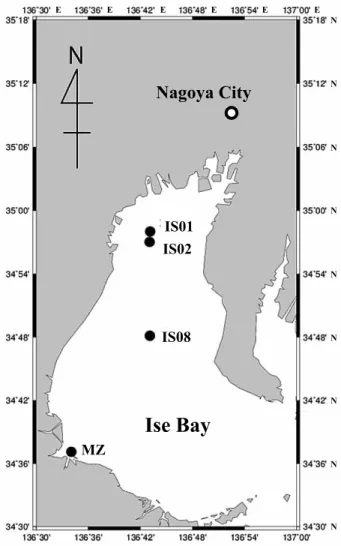
)。 このような近年の珪藻類の休眠期細胞に関する形態学的な研 究の進展により,実際の海底堆積物中にどのような種の休眠 期細胞がどれほど存在しているかを直接的に調査できる可能 性が開かれた。そこで本研究では,これまで明らかにされて きた珪藻類休眠期細胞の形態学的情報を駆使して,まず現場 海底堆積物中の休眠期細胞を光学顕微鏡下で種同定すること が可能かどうかを検証した。材料と方法
海底堆積物の採取 海底堆積物試料は,2011
年5
月21
日から24
日にかけて 行われた三重大学附属練習船勢水丸1105
航海の際に伊勢湾 より採取した。採取点は,IS1
(34
°58.11
ʼN
,136
°43.78
ʼE
),IS2
(34
°57.07
ʼN
,136
°43.49
ʼE)
,IS8
(34
°48.72
ʼN
,136
°43.70
ʼE
),MZ
(34
°37.71
ʼN
,136
°34.34
ʼE
)であり(Fig.
1
),水深はそれぞれ23
,26
,35
,11 m
であった。採泥には 重錘型柱状採泥器(チューブ内径3.4 cm
)を用い,各採取点 において柱状堆積物試料を3
本ずつ採取した。各採取点では, 試料の上部3 cm
を切り取り,プラスチック製容器に収容した。 その後,直ちに冷暗所(7
℃)に移動し,2
ヶ月以上保存する ことで栄養細胞の生残する可能性を排除した。海底堆積物試 料をそのまま観察すると,鉱物粒子等の影響で観察が極めて 困難であるため,試料1 g (wet sediment)
を100 µm
及び20
µm
目の篩を用いてサイズ分画を行った。そして,20 µm
目 の篩上の残渣物を10 mL
の濾過滅菌海水に懸濁し,その中の 珪藻類休眠期細胞を対象に種同定を行った。 種の同定 珪 藻 類 休 眠 期 細 胞 の 種 の 同 定 は 倒 立 顕 微 鏡(Eclipse
TE200, Nikon, Tokyo
)を用いて,Table 1
に示した文献に基づいて行った。観察の過程で,種同定が不可能と判断された 休眠期細胞については発芽・復活実験に供し,得られた栄養 細胞の形態に基づいて種の同定を行った。また,取り扱った 全ての休眠期細胞については,種同定の正誤を判定するため に発芽・復活実験に供し,発芽・復活した細胞の種同定を休 眠期細胞の種同定と併せて行った。発芽・復活実験は,キャ ピラリー操作によって単離した当該細胞を,培養液で充たし
たチャンバースライド(
Matsunami Glass, Osaka
)に収容して培養した(培養温度
20
°C
,光条件50 µmol photons m
-2Ishii
et al. (2011)
に記載されていない種の休眠胞子については,
Table 1
に示された論文に記載された形態情報をもとに種同定を行った。
Table 1
に示された論文の中で,休眠胞子の形態情報が記載され,今回の種同定に主に用いられた論文は,
Rines & Hargraves (1988)
,Jensen & Moestrup (1998)
及びKooistra
et al. (2010)
である。また,休眠細胞は栄養細胞と 同じ形態をしているため,詳しい形態学的情報がTable 1
で示 された論文に記載されていないことがある。そこで,今回の 種同定では栄養細胞の情報を網羅的に記載している,Tomas
(1997)
及びHoppenrath
et al. (2009)
を中心に,各種栄養細 胞に関する既報論文の形態情報をもとに種同定を行った。結果と考察
伊勢湾から得られた海底堆積物中には,10
属24
種1
変種 の休眠期細胞が確認された(Table 1
)。以下に最も種数が多 かったChaetoceros
属の結果を,続いて他属の結果を示す。 なお,調査地点間での休眠期細胞の種構成に大きな変化が無 かったため,全調査地点の結果をまとめてTable 1
に示した。Chaetoceros
属休眠胞子14
種と1
変 種のChaetoceros
属 休眠 胞 子,Chaetoceros
affinis Lauder
,Ch. constrictus Gran
,Ch. contortus
Schütt
,Ch. curvisetus Cleve
,Ch. debilis Cleve
,Ch.
diadema (Ehrenberg) Gran
,Ch. didymus var. didymus
Ehrenberg
,Ch. didymus var. anglicus (Grunow) Gran
,Ch.
distans Ehrenberg
,Ch. eibenii Grunow
,Ch. laciniosus
Table 1. List of diatom species of resting stage cells identified fromsediments of Ise Bay based on the morphological characteristics.
Species Identification
by morphology
Formation of
resting stage cell Referencesb Actinocyclus normanii Possible RC 1
Actinoc. cf. octonariusa Impossible RC
Actinoptychus senarius Possible RC 2
Chaetoceros affinis Possible RS 3, 4, 5, 6, 7, 8 Ch. constrictus Possible RS 3, 4, 5, 9, 10
Ch. contortus Possible RS 8, 7, 11, 12
Ch. curvisetus Possible RS 4, 5, 6, 8, 9, 12, 13, 14 Ch. debilis Possible RS 4, 5, 7, 8, 9, 13, 15 Ch. diadema Possible RS 4, 5, 6, 7, 8, 9, 10, 12, 13, 14, 15, 16, 17, 18, 19 Ch. didymus var. didymus Possible RS 4, 5, 6, 7, 8, 9, 13, 14, 15, 18, 20, 21, 22, 23 Ch. didymus var. anglicus Impossible RS 24
Ch. distans Possible RS 3, 5, 8 Ch. eibenii Impossible RS 21, 25 Ch. laciniosus Impossible RS 4, 5, 7, 9, Ch. lorenzianus Possible RS 3, 4, 5, 7, 8, 9, 12, 14 Ch. pseudocurvisetus Possible RS 4, 5, 8, 9, 26, 27, 28 Ch. radicans Possible RS 4, 5, 6, 7, 8, 9, 11, 15, 29 Ch. vanheurckii Possible RS 3, 4, 5, 8, 15, 18 Coscinodiscus cf. radiatusa Impossible RC
Detonula pumila Possible RS 2
Ditylum brightwellii Possible RS 4, 30, 31
Leptocylindrus danicus Possible RS 4, 19, 30, 32, 33, 34, 35
Paralia sulcata Possible RC 4
Stephanopyxis turris Possible RS 4, 36, 18, 31
Skeletonema sp. Impossible RC
a resting stage cells are unkown
b 1, Sicko-Goad et al. (1989); 2, Ishii et al. (2012); 3, Gran & Yendo (1914); 4, Cupp (1943); 5, Stockwell & Hargraves (1986); 6, Pitcher (1990); 7, Jensen & Moestrup (1998); 8, Ishii et al. (2011); 9, Rines & Hargraves (1988); 10, Riaux-Gobin & Desclas-Gros (1992); 11, Rines & Hargraves (1990); 12, Kooistra et al. (2010); 13, Hargraves (1979); 14, Pitcher (1986); 15, Garrison (1981); 16, Hargraves (1972); 17, Hargraves & French (1975); 18, Hollibaugh et al. (1981); 19, French & Hargraves (1985); 20, Karsten (1905); 21, von Stosch et al. (1973); 22, Round et al. (1990); 23, McQuoid & Hobson (1995); 24, Okuno (1956); 25, Ikari (1925); 26, Kuwata & Takahashi (1990); 27, Kuwata et al. (1993); 28, Oku & Kamatani (1995); 29, Stockwell & Hargraves (1988); 30, Hargraves (1976); 31, Hargraves (1984); 32, Gran (1912); 33, Davis et al. (1980); 34, French & Hargraves (1986); 35, Ishizaka et al. (1987); 36, von Stosch & Drebes (1964)
RC: resting cell, RS: resting spore
sec
-1,24
時間明期)。その後,培養状況が良好な種については,培養株として確立し維持した。種同定をする上で最も困難が
予測された
Chaetoceros
属休眠胞子については,Ishii
et al.
(2011)
の方法に従った。すなわち本属の種同定の際には,まず穿孔列(
single ring of puncta
)の観察により細胞の上下方向を決定することが必要となる。今回の実験では,穿孔列を 倒立顕微鏡下で観察可能かを検証した。また,穿孔列の確認
が困難な場合は,正立顕微鏡(
Eclipse W600, Nikon, Tokyo
)を用いて油浸対物レンズ(総合倍率
1,000
倍)による通常観察及び微分干渉法による観察を行った。穿孔列の観察によっ て上下方向が定まった本属休眠胞子は,細胞表面に存在す る各種形態学的特徴の組み合わせにより種同定した。また,
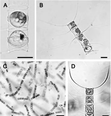
Fig. 2. Optical photomicrographs of Chaetoceros affinis. A: resting spores of Ch. affinis. B: germinated cells 24 hours after the incubation of the resting spore shown in A. C, D: cultured cells established from resting spore shown in A. Scale bars indicate 20 µm.
Schütt
,Ch. lorenzianus Grunow
,Ch. pseudocurvisetus
Mangin
,Ch. radicans Schütt
,及びCh. vanheurckii Gran
の存在が確認された(
Figs 2–4
)。Fig. 2A
は発芽前のCh.
affinis
の休眠期細胞であり,全ての種がFig. 2B
のように発 芽した栄養細胞からの種同定が行われ,休眠胞子からの種同 定に誤りがないことが確認された。また,この方法による単 離培養方法で,多くの種が継続的に培養され,培養株を用い た種同定も併せて行うことができた(Fig. 2C, D
)。なお,本 種の休眠胞子は本来,細胞被殻表面に無数の小棘(spine
)が 存在するが(Ishii
et al. 2011
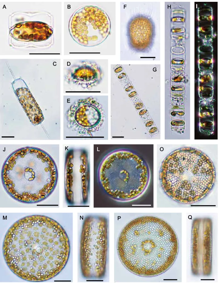
),細胞サイズによってはそれらFig. 3. Optical photomicrographs of Chaetoceros resting spores in sediments of Ise Bay. A-D: resting spores of Ch. constrictus focused on cytoplasm (A, B) using differential interference contrast (B), focused on single ring of puncta (C, D) using differential interference contrast (D). White arrows indicate a single ring of puncta. E, F: resting spore of Ch. lorenzianus, focused on single ring of puncta (arrow). G: resting spores of Ch. contortus, H: resting spores of Ch. debilis. I: resting spore of Ch. diadema. J, K: resting spores of Ch. didymus var. didymus, using differential interference contrast (K). L: resting spore of Ch. distans. Scale bars indicate 20 µm except for L.
が観察することが困難な場合があった(
Fig. 2A
)。このよう な本来観察されるべき同定形質の一部が確認されない場合に ついては,さらに高倍率での顕微鏡観察によりそれらの同定 形質の確認が行われた。Chaetoceros
属休眠胞子の同定に必要な穿孔列は,倒立 顕微鏡下で全ての種において観察できた。Fig. 3A–D
はCh.
constiructus
の休眠胞子の顕微鏡写真である。本属の休眠胞子の後成殻マントル縁辺には穿孔列(
single ring of puncta
)と呼ばれる穴状の構造が一列に並んでいる(
Fig. 3C, D
矢 印)。この構造を有する被殻を後成殻(secondary valve
),そ の上の被殻を初成殻(primary valve
)とし,これにより細胞 の上下方向が決定される。この構造は,細胞輪郭に焦点を合 わせた状態では観察することはできないが(Fig. 3A, B
),焦 点深度を被殻表面よりもやや深い位置で合わせることにより, 倒立顕微鏡でも観察可能であった(Fig. 3C
)。しかし,倒立 顕微鏡を用いた通常の観察では,穿孔列が明確には観察し Fig. 4. Optical photomicrographs of Chaetoceros resting spores and vegetative cells isolated from the resting spores in sediments of Ise Bay. A: resting spore of Ch. vanheurckii. B: resting spore of Ch. pseudocurvisetus. C: resting spore of Ch. radicans. D–G: resting spore of Ch. eibenii, girdle view (D) and valve view (E), germinated cell (F) 24 hours after the incubation of the resting spore shown in D and E, and cultured cells (G) established from the germinated cell shown in F. H, I: resting spore of Ch. didymus var. anglicus in frustule of vegetative cell (H) and higher magnification (I) showing the sheath (arrow). J, K: resting spores of Ch. laciniosus (J) and germinated cells (K) 24 hours after the incubation of the resting spore shown in J. Scale bars indicate 20 µm except for B.Fig. 5. Optical photomicrographs of resting stage cells observed in sediments of Ise Bay. A, B: resting spore of Detonula pumila, girdle view (A) and valve view (B). C: resting spore of Ditylum brightwellii. D, E: resting spore of Leptocylindrus danicus, girdle view (D) and valve view (E). F: resting spore of Stephanopyxis turris. G– I: resting cells of Skeletonema sp., and higher magnification (H), using differential interference contrast (I). J–L: resting cell of Actinocyclus normanii, valve view (J), girdle view (K) and using differential interference contrast (L). M, N: resting cell of Actinocyclus cf. octonarius, valve view (M) and girdle view (N). O: resting cell of Actinoptychus senarius. P, Q: resting cell of Coscinodiscus cf. radiatus, valve view (P) and girdle view (Q). Scale bars indicate 20 µm.
にくい傾向があった。そのような場合は正立顕微鏡による油 浸対物レンズと微分干渉法を用いることによって,より鮮明
に穿孔列が観察できた(
Fig. 3D
)。また,被殻内部の細胞質の観察においても微分干渉法を用いることで,より鮮明な内
部構造を観察することができた(
Fig. 3B
)。Fig. 3E, F
ではCh. lorenzianus
の休眠胞子の顕微鏡写真を示しており,Ch.
constrictus
と同様に焦点深度を適切な位置へ合わせることで 穿孔列が観察できた(Fig. 3F
)。同様にして,今回観察され た本属休眠胞子の全ての種において,この構造が観察され種 同定の際の同定形質に用いられた。 今回観察したChaetoceros
属休眠胞子の中で,既知の形態 学的情報から種の同定ができた種は12
種であった(Table 1
,Figs 2A, 3A–L, 4A–C
)。これらは既報の本属休眠胞子の形態学的情報(
Table 1
)とIshii
et al. (2011)
で提案された本属休眠胞子の同定基準に基づいて種同定することができた。し
かし,残り
2
種と1
変種,Ch. eibenii
,Ch. laciniosus
,及びCh. didymus var. anglicus
は既報の形態学的情報(Table 1
)では種同定が不可能であった。
Ch. eibenii
は発芽実験に供して栄養細胞の形態を根拠に種同定した(
Fig. 4D–G
)。本種の休眠胞子は増大胞子から直接形成することが知られているが
(井狩
1925
,von Stosch
et al. 1973
),これまで詳しい形態学的情報に乏しかった。今回観察された本種の休眠胞子は,細 胞サイズは
50~90 µm
と他のChaetoceros
属休眠胞子よりも 格段に大きかった(Fig. 4D, E
)。被殻の構造は本属休眠胞子 の形態と同様に初成殻と後成殻から成り,細胞表面は滑らか であった。長軸面(Apical plane
)から観察すると初成殻は 緩やかなアーチ状の形態を呈するのに対し,後成殻はより発 達したアーチ状の外形であった(Fig. 4D
)。被殻面(valvar
plane
)から観察すると,他のChaetoceros
属と同様の楕円 形であることが確認された(Fig. 4E
)。本種は,発芽実験に より得られた栄養細胞の形態から種同定が行われた(Fig. 4F,
G
)。また,Ch. didymus var. anglicus
は休眠胞子の周りに栄養細胞の被殻が残存していたため,その形態学的特徴から種
同定することができた(
Fig. 4H, I
)。本種の休眠胞子は初成殻が大きく発達したアーチ状であるのに対し,後成殻は緩や
かなアーチ状であった(
Fig. 4I
)。両殻とも表面は滑らかであったが,後成殻のマントル縁辺からドーム状のシース(
sheath
)が伸びていた(
Fig. 4I
矢印)。Ch. laciniosus
の休眠胞子(Fig.
4J
)の種同定は,発芽実験により増殖した栄養細胞の形態から種同定を行った(
Fig. 4K
)。本休眠胞子の初成殻表面は滑らかなアーチ状をしており,後成殻には小さな棘のような構 造を有していたが,倒立顕微鏡でそれらを観察することは極
めて困難であった(
Fig. 4J
)。この構造はJensen & Moestrup
(1998)
で本種の休眠胞子として記載されている毛状の構造に 該当する可能性がある。また,本種の休眠胞子は2
つの細胞 が対になった状態で多く観察される傾向があった。今後,詳 細な形態観察を行い,本種休眠胞子の同定基準を明らかにす る必要がある。 その他の珪藻類休眠期細胞Chaetoceros
属以外の珪藻類休眠期細胞は,休眠胞子と 休眠細胞を含め,9
属10
種が確認された(Fig. 5
)。休眠胞子は,
Detonula pumila (Castracane) Schütt
,Ditylum
brightwellii (West) Grunow
,Leptocylindrus danicus
Cleve
,Stephanopyxis turris (Greville) Ralfs
の4
種が確認され,これらは全て既報の形態学的情報により種の同定が
可能であった(
Table 1
,Fig. 5A–F
)。また,Skeletonema
属の休眠細胞が確認されたが(
Fig. 5G–I
),Skeletonema
属は近年の分類基準の細分化により光学顕微鏡による種同定
が不可能となったため(
Sarno
et al. 2005
,Zingone
et al.
2005
,Sarno
et al. 2007
,山田2013
),属レベルの同定に留 まった。また,今回確認されたSkeletonema sp.
の休眠細胞は, 連鎖群体を形成した状態で観察されることが多く,その連鎖 群体中における休眠細胞の形態が通常の栄養細胞を委縮させ たような形態をしていた(Fig. 5G–I
)。今後さらに本属休眠 細胞の詳細な形態観察が求められる。その他の休眠細胞は,Actinocyclus normanii (Gregory) Hustedt
,Actinocyclus cf.
octonarius Ehrenberg
,Actinoptychus senarius (Ehrenberg)
Ehrenberg
,Coscinodiscus cf. radiatus Ehrenberg
, 及 びParalia sulcata (Ehrenberg) Cleve
の5
種が確認された(Fig.
5J–Q
)。Actinoc. cf. octonarius
(Fig. 5J–L
) 及 びCo. cf.
radiatus
(Fig. 5P, Q
)の2
種は休眠細胞に関する報告が無い (Table 1
)。本研究の発芽実験により栄養細胞としての増殖が 確認されたことで,両種に休眠ステージが存在することが明 らかになった。しかし,これら2
種の正確な種同定にはSEM
を用いた被殻の詳細な観察が必要になるため(Tomas 1997
), 今回は種名を確定するには至らなかった。 過去の研究により,休眠細胞の色素体が栄養細胞と比較し て暗色を呈し,それらの分布に局在傾向のあることが示され ている(今井ら1990
,板倉ら1992
,Ishii
et al. 2012
)。今回 観察された休眠細胞に関しても同様の傾向が示された。特に,Actinocyclus
属2
種 やActinop. senarius
,Co. cf. radiatus
などの円盤型の中心目珪藻については,色素体が中心よりも 縁辺部に多く分布している特徴が観察された(Fig. 5J–Q
)。 伊勢湾の海底堆積物中から,10
属24
種1
変種の休眠期細 胞が光学顕微鏡観察により確認された。これら休眠期細胞の ほとんどが,形態学的情報に基づく種の同定が可能であるこ とが明らかとなった。したがって,本研究で用いられたサイ ズ分画による20 µm
以上のサイズを有する海底堆積物中の珪 藻類各種の休眠期細胞密度を把握する方法として,光学顕微 鏡を用いた直接検鏡による計数が可能であることが示された。 これまでMPN
法を用いて見積もられてきた海底堆積物中の 珪藻類休眠期細胞の密度が,これにより今後はより正確に把 握することができると考えられる。ただし,堆積物中の休眠期 細胞を計数するためには,倒立顕微鏡を用いて休眠期細胞以 外の鉱物粒子などを除きながら観察する必要がある。しかし, 今回の顕微鏡観察では,倒立顕微鏡で種同定が不可能な種が 存在し,それらは正立顕微鏡の油浸対物レンズを用いることで種同定が行われた。今後の現場海底堆積物中の休眠期細胞 密度を計測するにあたり,どの種がどれほどの分解能の顕微 鏡を用いれば種同定することが可能なのかを明確に定義する 必要があると考えられる。本研究において最も種数の多かっ た
Chaetoceros
属の中で,2
種1
変種が既知の形態学的情報 (Table 1
)だけでは種の同定が不可能であった。Chaetoceros
属は沿岸海域において最も繁栄している属の1
つであり,し ばしば赤潮を形成するなど,重要な分類群である。今後は, 本属休眠胞子の詳細な形態学的情報をさらに充実・整理して いく予定である。謝辞
本研究を進めるにあたり,サンプル採取及び分析に御助力 頂いた三重大学大学院生物資源学研究科生物海洋学研究室 の学生諸氏に心から感謝申し上げる。尚,本研究の一部は,JSPS
科研費22580197
の助成により行われた。引用文献
Cupp, E. E. 1943. Marine Plankton Diatoms of the West Coast of North America. University of California Press, Berkeley.
Davis, C. O., Hollibaugh, J. T., Seibert, D. L. R., Thomas, W. H. & Harrison, P. J. 1980. Formation of resting spores by Leptocylindrus danicus (Bacillariophyceae) in a controlled experimental ecosystem. J. Phycol. 16: 296–302.
French, F. W. & Hargraves, P. E. 1985. Spore formation in the life cycles of the diatoms Chaetoceros diadema and Leptocylindrus danicus. J. Phycol. 21: 477–483.
French, F. W. & Hargraves, P. E. 1986. Population dynamics of the spore-forming diatom Leptocylindrus danicus, in Narragansett Bay, Rhode Island. J. Phycol. 22: 411–420.
Garrison, D. L. 1981. Monterey Bay phytoplankton. II. Resting spore cycles in coastal diatom populations. J. Plankton Res. 3: 137–156.
Garrison, D. L. 1984. Plankton diatoms. In: Steidinger, K. A. & Walker, L. M (eds.) Marine plankton life cycle strategies. pp.1–7. CRC Press, Floloda.
Gran, H. H. 1912. Pelagic plant life. In: Murray, J. & Hjort, J. (eds.) The depths of the ocean. pp. 307–386. Macmillan & Co., Ltd., London. Gran, H. H. & Yendo, K. 1914. Japanese diatoms. I. On Chaetoceros.
Videnskapsselskapets Skrifter. I. Mat.-Naturu. Klasse. 1913. No. 8. Utgit for Friftjof Nansens Fond, Christiania.
Hargraves, P. E. 1972. Studies on marine plankton diatom. I. Chaetoceros diadema (Ehr.) Gran: life cycle, structural morphology, and regional distribution. Phycologia 11: 247–257.
Hargraves, P. E. 1976. Studies on marine plankton diatoms.Ⅱ. Resting spore morpho1ogy. J. Phycol. 12: 118–128.
Hargraves, P. E. 1979. Studies on marine plankton diatoms IV. Morphology of Chaetoceros resting spores. Beihefte zur Nova Hedwigia 64: 99–120. Hargraves, P. E. 1984. Resting spore formation in the marine diatom
Ditylum brightwellii (West) Grun. In: Mann, D. G. (ed.) Proceedings of the Seventh International Diatom Symposium. pp. 33–46. O. Koeltz Science Publishers, Koenigstein.
Hargraves, P. E. & French, F. W. 1975. Observations on the survival of diatom resting spores. Beihefte zur Nova Hedwigia 53: 229–238. Hargraves, P. E. & French, F. W. 1983. Diatom resting spores: significance
and strategies. In: Fryxell, G. A. (ed.) Survival Strategies of the Algae. pp. 49–68. Cambridge University Press, New York.
Hollibaugh, J. T., Seibert, D. L. R. & Thomas, W. H. 1981. Observations
on the survival and germination of resting spores of three Chaetoceros (Bacillariophyceae) species. J. Phycol. 17: 1–9.
Hoppenrath, M, Elbrächter, M & Drebes, G. 2009 Marine Phytoplankton. Selected microphytoplankton species from the North Sea around Helgoland and Sylt. Kleine Senckenberg-Reihe, Band 49, 264 p. 井 狩 二 郞1925.Chaetoceros Eibenii, Grunニ 就 テ. 植 物 学 雑 誌39:
52–59.
Imai, I., Itoh, K. & Anraku, M. 1984. Extinction dilution method for enumeration of dormant cells of red tide organisms in marine sediments. Bull. Plankton Soc. Jpn 31: 123–124.
今井一郎・板倉茂・伊藤克彦1990.播磨灘及び北部広島湾の海底泥中に
おける珪藻類の休眠期細胞の分布.沿岸海洋研究ノート28: 75–84.
Ishii, K-I., Iwataki, M., Matsuoka, K. & Imai, I. 2011. Proposal of identification criteria for resting spores of Chaetoceros species (Bacillariophyceae) from a temperate coastal sea. Phycologia 50: 351– 362.
Ishii, K-I. Ishikawa, A. & Imai, I. 2012. Newly identified resting stage cells of diatoms from sediments collected in Ago Bay, central part of Japan. Plankton Benthos Res. 7: 1–7.
Ishizaka, J., Kaichi, M. & Takahashi, M. 1987. Resting spore formation of Leptocylindrus danicus (Bacillariophyceae) during short time-scale upwelling and its significance as predicted by a simple model. Ecol. Res. 2: 229–242. 板倉茂2000.沿岸性浮遊珪藻の休眠期細胞に関する生理生態学的研究. 瀬戸内水研報, No.2: 67–130. 板 倉 茂・ 今 井 一 郎・ 伊 藤 克 彦1992. 海 底 泥 中 か ら 見 出 さ れ た 珪 藻 Skeletonema costatum休眠細胞の形態と復活過程.日本プランクトン 学会報 38: 135–145.
Itakura, S., Imai, I. & Itoh, K. 1997. “Seed bank” of coastal planktonic diatoms in bottom sediments of Hiroshima Bay, Seto Inland Sea, Japan. Mar. Biol. 128: 497–508.
Itakura, S., Nagasaki, K., Yamaguchi, M. & Imai, I. 1999. Abundance and spatial distribution of viable resting stages of planktonic diatoms in bottom sediments of the Seto Inland Sea, Japan, pp. 213–226. In: Mayama, S., Idei, M. & Koizumi, S. (eds.) Proceedings of the 14th International Diatom Symposium. Koeltz Scientific Books, Koenigstein. Jensen, K. G. & Moestrup, Ø. 1998. The genus Chaetoceros
(Bacillariophyceae) in inner Danish coastal waters. Opera Bot. 133: 5–68.
Karsten, G. 1905. Das Phytoplankton des Antarktischen Meeres nach dem Material der Deutschen Tiefsee-Expedition 1898–1899. Deutschen Tiefsee Expedition 2: 1–136.
Kooistra, W. H., Sarno, D., Hernández-Becerril, D. U., Assmy, P., Prisco, C. D. & Montresor, M. 2010. Comparative molecular and morphological phylogenetic analyses of taxa in the Chaetocerotaceae (Bacillariophyta). Phycologia 49: 471–500.
Kuwata, A. & Takahashi, M. 1990. Life-form population responses of a marine planktonic diatom, Chaetoceros pseudocurvisetus, to oligotrophication in regionally upwelled water. Mar. Biol. 107: 503– 512.
Kuwata, A., Hama, T. & Takahashi, M. 1993. Ecophysiological characterization of two life forms, resting spores and resting cells, of a marine planktonic diatom, Chaetoceros pseudocurvisetus, formed under nutrient depletion. Mar. Ecol. Prog. Ser. 102: 245–255.
McQuoid, M. R. & Hobson, L. A. 1995. Importance of resting stages in diatom seasonal succession. J. Phycol. 31: 44–50.
McQuoid, M. R. & Hobson, L. A. 1996. Diatom resting stages. J. Phycol. 32: 889–902.
Nishikawa, T., Hori, Y., Tanida, K. & Imai, I. 2007. Population dynamics of the harmful diatom Eucampia zodiacus Ehrenberg causing bleachings of Porphyra thalli in aquaculture in Harima-Nada, the Seto Inland Sea, Japan. Harmful Algae 6: 763–773.
Oku, O. & Kamatani, A. 1995. Resting spore formation and phosphorus composition of the marine planktonic diatom Chaetoceros pseudocurvisetus under various nutrient conditions. Mar. Biol. 123: 393–399.
Okuno, H. 1956. Electron-microscopical study on fine structures of diatom frustules. XIV. Observation on the genus Chaetoceros. Bot. Mag. Tokyo 69: 186–192.
Pitcher, G. C. 1986. Sedimentary flux and the formation of resting spores of selected Chaetoceros species at two sites in the Southern Benguela system. Afr. J. Mar. Sci. 4: 231–244.
Pitcher, G. C. 1990. Phytoplankton seed populations of the Cape Peninsula upwelling plume, with particular reference to resting spores of Chaetoceros (Bacillariophyceae) and their role in seeding upwelling waters. Estuar. Coast. Shelf S. 31: 283–301.
Riaux-Gobin, C. & Descolas-Gros, C. 1992. Assemblage de microphytes enkystés á la surface de sédiments profonds, sur le talus continental du golfe du Lion. Comptes Rendus de I’académie de Scienesi Paris 314: 545–552.
Rines, J. E. B. & Hargraves, P. E. 1988. The Chaetoceros Ehrenberg (Bacillariophyceae) flora of Narragansett Bay, Rhode Island, U.S.A. Bibliotheca Phycologica 79: 1–196.
Rines, J. E. B. & Hargraves, P. E. 1990. Morphology and taxonomy of Chaetoceros compressus Lauder var. hirtisetus var. nova, with preliminary consideration of closely related taxa. Diatom Res. 5: 113– 127.
Round, F. E., Crawford, R. M. & Mann, D. G. 1990. The Diatoms. Biology and Morphology of the Genera. Cambridge University Press, Cambridge.
Sarno, D., Kooistra, W. H., Medlin L. K., Percopo, I. & Zingone, A. 2005. Diversity in the genus Skeletonema (Bacillariophyceae). II. An assessment of the taxonomy of S. costatum-like species with the description of four new species. J. Phycol. 41: 151–176.
Sarno, D., Kooistra, W. H., Balzano, S. Hargraves, P. E., & Zingone, A.
2007. Diversity in the genus Skeletonema (Bacillariophyceae). III. Phylogenetic position and morphological variability of Skeletonema costatum and Skeletonema grevillei, with the description of Skeletonema ardens sp. nov. J. Phycol. 43: 156–170.
Sicko-Goad, L., Stoermer, E. F. & Kociolek, J. P. 1989. Diatom resting cell rejuvenation and formation: time course, species records and distribution. J. Plankton Res. 11: 375–389.
Stockwell, D. A. & Hargraves, P. E. 1986. Morphological variability within resting spores of the marine diatom genus Chaetoceros Ehrenberg. In: Ricard, M. (ed.) Proceedings of the Eighth International Diatom Symposium. pp. 81–95. Koeltz Scientific Books, Koenigstein.
Stockwell, D. A. & Hargraves, P. E. 1988. Distribution and size variation of resting spores in Chaetoceros radicans and Chaetoceros debilis within the diatomaceous muds of the Guaymas Basin and the Guaymas Basin Slope, Gulf of California. In: Round, F. E. (ed.) Proceedings of the Ninth International Diatom Symposium. pp. 219–231. Koeltz Scientific Books, Koenigstein, Germany.
Tomas, C. R. 1997. Identifying marine phytoplankton. Academic Press, San Diego, California.
von Stosch, H. A. & Drebes, G. 1964. Entwicklungsgeschichtliche Untersuchungen an zentrischen Diatomeen. Ⅳ. Die Planktondiatomee Stephanopyxis turris-ihre Behandlung und Entwicklungsgeschichte. Helgol. Wiss. Meeresunters.11: 209–257.
v o n S t o s c h , H . A . , T h e i l , G . & K o w a l l i k , K . V. 1 9 7 3 . Entwicklungsgeschichtliche Untersuchungen an zentrischen Diatomeen.V. Bau und Lebenszyklus von Chaetoceros didjmum, mit Beobachtungen über einige andere Arten der Gattung. Helgol. Wiss. Meeresunters. 25: 384–445.
山田真知子2013.珪藻Skeletonema属の最近の分類と生理生態特性(総
説).日本プランクトン学会報 60: 18–28.
Zingone, A., Percopo, I., Sims, P. A. & Sarno, D. 2005. Diversity in the genus Skeletonema (Bacillariophyceae). I. A reexamination of the type material of S. costatum with the description of S. grevillei sp. nov. J. Phycol. 41: 140–150.