〈
Case Report
〉
Mycobacterium heckeshornense
-induced deep abscess
in the gluteus maximus muscle:
a case report and review of the literature
Hirotoshi Kikuchi1,*, Kurumi Asako1, Hajime Kono 1, Miwa Asahara2, Takashi Tanaka2, Go Kamosida3, Takane Ueda3, Shigeru Nagakawa3, Tsuneyuki Ubagai3,
Yuko Kazumi4 and Yasuo Ono1, 3
1 Department of Internal Medicine, Teikyo University School of Medicine,
Tokyo, Japan
2 Department of Central Clinical Laboratory, Teikyo University Hospital,
Tokyo, Japan
3 Department of Microbiology and Immunology,
Teikyo University School of Medicine, Tokyo, Japan
4 The Research Institute of Tuberculosis, Japan Anti-Tuberculosis Association,
Tokyo, Japan
(Received for publication October 29, 2018)
Mycobacterium heckeshornense is rarely isolated from a clinical sample. We
report a case of a patient with polymyositis in whom a deep abscess formed in the gluteus maximus muscle and in which M. heckeshornense was identified by DNA sequencing. Combination therapy of levofloxacin and clarithromycin for 2 years improved inflammatory findings and 5 years have passed without recurrence of the gluteal abscess. Identification of the bacterial strain in nontuberculous mycobacterium (NTM) infection is important to determine the treatment plan and accumulation of data on drug sensitivity is required to establish a therapeutic strategy for rare pathogen such as M. heckeshornense.
Introduction
The incidence of nontuberculous mycobacterium (NTM) infection has shown a tendency to increase in Japan. The rates for individual bacterial strains are 60% for Mycobacterium avium, 25% for M. intracellulare, and 8% for M. kansasii, while other strains including M. abscessus are
rare1). In contrast, the incidence of NTM infection in Europe is highest for M. avium complex
(MAC), followed by M. xenopi2,3).
Herein, we report a case of a patient with polymyositis in whom a deep abscess formed in the gluteus maximus muscle and in which M. heckeshornense is isolated.
Case Report
A 62-year-old man developed myalgia at the proximal muscle 10 years ago. An elevated blood creatine kinase (CK) level and positive anti-Jo-1 antibody were found and he was diag-nosed with polymyositis based on the results of muscle biopsy. Since the disease redeveloped when the dose of adrenal cortical steroids was decreased, maintenance treatment was provided with prednisolone (12 mg/day), cyclosporine (150 mg/day) and methotrexate (10 mg/week). The patient became aware of an uncomfortable feeling in his left buttock 4 months ago and swelling in the same region 2 months ago.
At his first visit to our hospital, he had a temperature of 36.0°C with no myalgia, arthralgia, or obvious trauma. Swelling was confirmed from the left buttock to the external side of the left femoral region. A blood test showed a white blood cell count of 15,800/μL (stab cells 2.0%, seg-ment cells 94.0%, and lymphocytes 4.0%), a C-reactive protein level of 1.90 mg/dL, and an eryth-rocyte sedimentation rate of 40 mm/h. The levels of CK (69 IU/L), IgG (1,270 mg/dL), IgA (163 mg/dL), IgM (142 mg/dL), antinuclear antibody (titer <40), and anti-Jo-1 antibody (142.5 IU/L, positive) were similar to those in a previous examination, suggesting stable polymyositis.
Magnetic resonance imaging of the area from the buttock to the femoral region suggested fluid accumulation in a deep area to the side of the left femoral muscle (Fig. 1). Puncture was per-formed under ultrasound guidance. A standard plate count of bacteria was negative in purulent as-pirate however the concentrating method for tubercle bacillus was 1+. Chest X-ray and chest computed tomography performed in the same period gave no clear findings other than pulmonary fibrosis associated with polymyositis, and acid fast bacterium was negative in gastric fluid and blood culture. Puncture was performed twice for the abscess and growth of NTM was found in both aspirates on the 15th day. In a DNA-DNA hybridization (DDH) test (DDH Mycobacteria, Kyokuto Pharmaceutical Industrial Co., Ltd, Japan), the bacterium was identified as M. xenopi.
M. xenopi is rare in Japan and the number of reports on infection of tissues other than the lungs
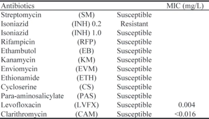
is small 4). In addition, a drug susceptibility test for the detected bacterium showed sensitivity to
ethambutol (Table 1), which is inconsistent with M. xenopi. Furthermore, DNA sequencing of 16S rRNA and rpoB gene of the bacterium suggested concordance rates with M. heckeshornense of 100% and 99%, respectively, while those with M. xenopi were 95%. Thus, the NTM was identified as M. heckeshornense. After drainage of the abscess, the patient was treated with concomitant 500 mg/day levofloxacin and 400 mg/day clarithromycin for 2 years, and there has been no
redevel-opment of the condition for 5 years without administration of these antibacterial agents.
Discussion
There are several methods for identification of NTM bacterial strains. In 2011, Morimoto et
al. suggested that M. heckeshornense, which cannot be identified using the DDH kit, may be
in-correctly identified as M. xenopi5). In addition, two case reports showed that a NTM identified as
M. xenopi by the DDH kit was found to be M. heckeshornense, a slow acid fast bacterium 6).
Figure 1.
A. Magnetic resonance imaging (sagittal view) of the area from the buttock to the femoral region. Arrow: deep abscess in the gluteus maximus muscle. B. MRI findings after 10 months from treatment are shown. Abscess has disappeared.
Table 1. Results drug susceptibility testing in our case
Drug susceptibility tests were determined using a Vite Spectrum-SR kit. (Kyokuto Pharmaceutical Industrial Co., Ltd, Japan). There are no criteria of CLSI (Clinical and Laboratory Standards Institute) in M.
Table 2. Summary of documented Mycobacterium heckeshornense case r eports
These reports indicate a problem with the method used to identify bacterial strains.
At first, we used a DDH kit, with which 18 bacterial strains can be identified at one time by hybridization using DNA of a type strain and DNA of a suspected strain. However, the NTM was sensitive to ethambutol (Table 1) and the infection site was not in the lungs, both of which differ from the typical characteristics of M. xenopi. Therefore, we performed DNA sequencing for 16S rRNA and rpoB gene of the bacterium and identified the strain as M. heckeshornense.
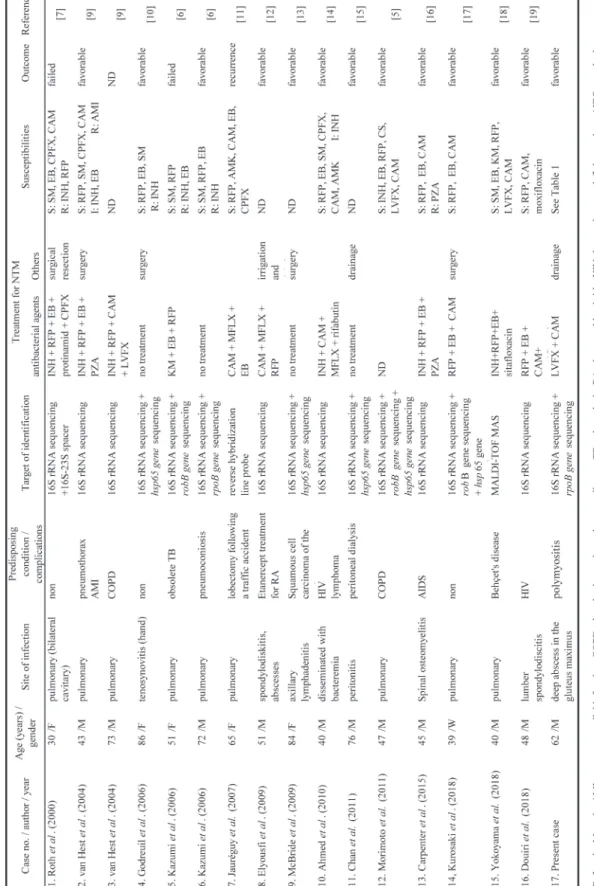
M. heckeshornense was first reported by Roth et al.7) and has been confirmed in only 17
cases (Table 2)5∼7, 9∼19), including our case, because DNA sequencing is required for
identifica-tion. In recent years, Yokoyama et al.19) diagnosed M. heckeshornense using matrix-assisted laser
desorption ionization-time of flight mass spectrometry (MALDI-TOF MS). They had also con-firmed in the DNA sequence and had reported that MALDI-TOF MS was useful in the diagnosis of M. heckeshornense.
A half of these cases (47.1%) had infection at sites other than the lungs. The outcomes were favorable in most cases (improvement: 81.2%, redevelopment: 18.8%) and improvement oc-curred without administration of antibacterial agents in some cases. Pulmonary infection with M.
xenopi is often unresponsive to drug treatment alone, and thus surgery is required in many
cases8), however the pathogenicity of M. heckeshornense is unclear. van Hest et al. suggested that
M. heckeshornense detected from lung tissues with normal immunity was pathogenic9), but a
therapeutic strategy has not been established because of the small number of reported cases. In our case, multiple immunosuppressants were used to control the disease activity of poly-myositis. Initially we were considering treatment with three drugs such as rifampicin, levofloxa-cin and clarithromylevofloxa-cin, but because the patient refused to increase the amount of steroids associ-ated with rifampicin administration, we treassoci-ated with two agents with reference to the case report of M. xenopi. Since the possibility that the combination of levofloxacin and clarithromycin in-duces drug resistance of M. heckeshornense cannot be denied either, studies on the resistance mechanism of M. heckeshornense by clarithromycin are desired.
We conclude that identification of the bacterial strain in NTM infection is important to deter-mine the treatment plan. Accumulation of data on drug sensitivity is required to establish a thera-peutic strategy for rare pathogen such as M. heckeshornense.
Conflict of interests
We declare that we have no conflict of interest. Acknowledgments
We thank the microbiology technologist (The Research Institute of Tuberculosis, Japan Anti-Tuberculosis Association) for performing 16S rRNA, rpoB, and hsp65 gene sequence analysis for the identification of M. heckeshornense. This study was supported by the Private University
Research Branding Project.
References
1) Kajiki A: Non-tuberculous mycobacteriosis. What has been coming out. Kekkaku 2011; 86: 113– 25.
2) Lamden K, Watson JM, Knerer G, Ryan MJ, Jenkins PA: Opportunist mycobacteria in England and Wales: 1982 to 1994. Commun Dis Rep CDR Rev. 1996; 6: R147–51.
3) Dailloux M, Abalain ML, Laurain C, et al.: Respiratory infections associated with nontuberculous mycobacteria in non-HIV patients. Eur Respir J. 2006; 28: 1211–5.
4) Matsui Y, Tamura A, Nagayama N, et al.: Review of pulmonary Mycobacterium xenopi infection cases: 11 cases of our own and 18 other cases reported in Japan. Kekkaku 2010; 85: 647–53. 5) Morimoto K, Kazumi Y, Maeda S, et al.: Mycobacterium heckeshornense lung infection that was
diagnosed as Mycobacterium xenopi disease by DNA-DNA hybridization (DDH). Intern Med. 2011; 50: 1251–3.
6) Kazumi Y, Sugawara I, Wada M, Kimura K, Itono H: Microbiologically identified isolates of My-cobacterium heckeshornense in two patients. Kekkaku 2006; 81: 603–7.
7) Roth A, Reischl U, Schönfeld N, et al.: Mycobacterium heckeshornense sp. nov., a new patho-genic slowly growing Mycobacterium sp. Causing cavitary lung disease in an immunocompetent patient. J Clin Microbiol. 2000; 38: 4102–7.
8) Lang-Lazdunski L, Offredo C, Le Pimpec-Barthes F, Danel CDujon, A, Riquet M: Pulmonary re-section for Mycobacterium xenopi pulmonary infection. Ann Thorac Surg. 2001; 72: 1877–82. 9) van Hest R, van der Zanden A, Boeree M, et al.: Mycobacterium heckeshornense infection in an
immunocompetent patient and identification by 16S rRNA sequence analysis of culture material and a histopathology tissue specimen. J Clin Microbiol. 2004; 42: 4386–9.
10) Godreuil S, Marchandin H, Terru D, et al.: Mycobacterium heckeshornense tenosynovitis. Scand J Infect Dis. 2006; 38: 1098–101.
11) Jauréguy F, Ioos V, Marzouk P, et al.: Mycobacterium heckeshornense: an emerging pathogen re-sponsible for a recurrent lung infection. J Infect. 2007; 54: e33–5.
12) Elyousfi AA, Leiter JR, Goytan MJ, Robinson DB: Mycobacterium heckeshornense lumbar spon-dylodiskitis in a patient with rheumatoid arthritis receiving etanercept treatment. J Rheumatol. 2009; 36: 2130–1.
13) McBride SJ, Taylor SL, Pandey SK, Holland DJ: First case of Mycobacterium heckeshornense lymphadenitis. J Clin Microbiol. 2009; 47: 268–70.
14) Ahmed RA, Miedzinski LJ, Shandro C: Mycobacterium heckeshornense infection in HIV-infected patient. Emerg Infect Dis. 2010; 16: 1801–3.
15) Chan WW, Murray MC, Tang P, Romney MG: Mycobacterium heckeshornense peritonitis in a peritoneal dialysis patient: a case report and review of the literature. Clin Microbiol Infect. 2011; 17: 1262–4.
16) Carpenter RJ, Graf PC.: Pott s disease? AIDS-associated Mycobacterium heckeshornense spinal osteomyelitis and diskitis. J Clin Microbiol. 2015; 53 :716–8.
17) Kurosaki F, Yoshimoto T, Nakayama M, Bando M, Hagiwara K: Pulmonary Mycobacterium heckeshornense infection in a healthy woman. J Infect Chemother. 2018; 24: 483–6.
18) Yokoyama A, Kage H, Ohama Y, et al.: Mycobacterium heckeshornense lung infection diagnosed by matrix-assisted laser desorption ionization-time of flight mass spectrometry (MALDI-TOF
MS). J Infect Chemother. 2018; 24: 763–5.
19) Douiri N, Lefebvre N, Hansmann Y, Partisani M, Schramm F: Relapsing Pott disease caused by Mycobacterium heckeshornense in a well-controlled HIV-infected patient. Med Mal Infect. 2018; 48: 157–8.