Successfully Rescued Left Main Coronary Trunk Dissection during Percutaneous Coronary Intervention by Blood Transmission to the Left Anterior Descending Branch Via a Microcatheter
5
0
0
全文
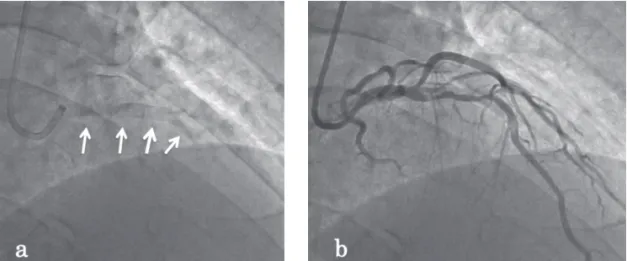
(2) J Jpn Coron Assoc 2018; 24: 190-194. Fig. 1 (a) Severe calcification is visible on the non-contrast imaging. (b) Contrast imaging of the same site reveals stenosis of the LAD. Calcification is easily visible even on the non-contrast imaging and extends as far as the LMT.. Fig. 2 The lesion in the LAD was dilated with a 2.25-mm balloon, but the dilation was not sufficient for the indentation to disappear.. GWs for balloon dilation. Although the IVUS device could be. Fig. 3 IVUS images of the LMT (A), LAD just proximal to the lesion (B), and lesion in segment 6 (C). Severe circumferential calcification extends from the LMT to the LAD.. passed through with some difficulty, adequate dilation was not achieved. Severe circumferential calcification of the vessel existed, with an estimated 3.5 mm diameter at the lesion (Fig. 3). A. (ECG) showed ST elevation, and the patient became disturbed. larger balloon catheter, measuring 2.5×6 mm, was then dilated at. (Fig. 5). Dissection, a common complication of PCI, was sus-. a pressure <20 atm, at which point the blood pressure at the tip. pected. However, no obvious dissection was visible on the coro-. of the guiding catheter suddenly could not be measured. The bal-. nary angiography. A balloon catheter was inserted again, but the. loon catheter was withdrawn, and coronary angiography was. tip did not advance beyond the LMT. A FINECROSS (Terumo,. performed, revealing that the LMT was occluded by the guiding. Japan) microcatheter was inserted via the right radial artery,. catheter tip and was not contrasted as far as the LAD. The left. which was advanced into the LAD. Contrast enhancement of the. circumflex (LCX) artery and D1 were visualized late (Fig. 4).. LAD via the microcatheter showed, fortunately, no occurrence. Dilation was attempted again using two smaller balloon cathe-. of dissection on the distal side (Fig. 6). To avoid shock induced. ters, measuring 2.5×6 mm and 2.0×12 mm, but neither could be. by LMT occlusion, a 20 ml syringe was used to repeatedly inject. advanced, and the patient suddenly went into shock. Systolic. arterial blood collected from the sheath introducer in the right. blood pressure dropped to 70 mmHg, the electrocardiogram. radial artery into the LAD via the microcatheter. Each adminis-. ─ 191 ─. 0622-Sugiura(早).indd 191. 2018/12/11 10:08.
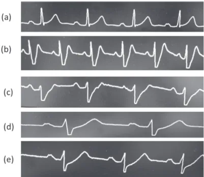
(3) J Jpn Coron Assoc 2018; 24: 190-194. Fig. 4 Contrast angiography when the LMT dissection occurred. Neither the LMT nor the LAD was visualized, and visualization of the LCX, right lateral branch, and part of the first diagonal branch was delayed.. Fig. 5 Changes over time in ECG lead II monitor waveforms during PCI. (a) Start of PCI; (b), (c), (d) between LMT occlusion and the injection of arterial blood, tachycardia develops, ST-T fluctuates, and heart rate gradually slows down; (e) during arterial blood injection via the microcatheter.. tinuing with PCI, but the balloon catheter would probably not pass through the lesion and that, even if it did, the lesion was so severely calcified that adequate dilation would be impossible. Thus, we converted to coronary artery bypass grafting (CABG). As our hospital is not equipped to perform CABG, the patient was transported to an affiliated hospital. During transportation, a two-way stopcock attached to the sheath was connected to the microcatheter, and arterial blood infusion was continued by manual pumping with a 20 ml syringe. CABG was performed at the affiliated hospital approximately 2 hours later. The chest was opened, and arterial blood infusion was continued via a pump oxygenator until the patient was on a cardiopulmonary pump. The surgery was successful. Percutaneous cardiopulmonary supFig. 6 Contrast angiography of the FINECROSS microcatheter. Contrast angiography after the microcatheter was advanced into the LAD, and the guidewires were withdrawn showing no arterial dissection of the LAD distal to the lesion; therefore, LAD is well visualized.. port (PCPS) remained in use temporarily because of the risk for postoperative heart failure, but was discontinued on Day 4. The patient’s subsequent course until discharge was uneventful. Cardiac rehabilitation was implemented after discharge. Echocardiography showed hypokinesis of the anterior wall, and the ejection fraction was 45%, indicating a poor cardiac function.. tration of approximately 20 ml of blood required a mean time of. However, the patient continued to visit our hospital as an outpa-. 80 seconds. When approximately 40–50 ml had been injected in. tient, with no chest symptoms, no need for readmission, no neu-. 2–3 administrations, the elevated ST segment began to normal-. rological deficit, and no heart failure exacerbation.. ize, and by the time 2–3 further administrations had been per-. III. Discussion. formed, resulting in continuous injection of over 100 ml of arterial blood, the ST segment had returned to baseline. The systolic. A variety of complications may occur during PCI, and techni-. blood pressure also stabilized at around 90 mmHg, and prema-. cal problems during catheter insertion alone include vascular in-. ture ventricular contractions (PVC) and ventricular tachycardia. jury, coronary vasospasm, thrombosis, and coronary artery dis-. (VT) resolved as well (Fig. 5). At this point, we considered con-. section, which may cause coronary occlusion, shock, and even. ─ 192 ─. 0622-Sugiura(早).indd 192. 2018/12/11 10:08.
(4) J Jpn Coron Assoc 2018; 24: 190-194. Fig. 7 Changes over time in 12-lead ECG: before PCI, immediately after PCI, and immediately before CABG.. death. For severe calcification and LMT lesions, CABG via open. with two GWs, without using a Rotablator, but dissection meant. chest surgery is frequently indicated rather than PCI.. that the opportunity to use it was lost. According to a study by. In the present case, although calcification was comparatively. Boyle et al., coronary artery dissection caused by a guide cathe-. severe in the LAD, the stenotic lesion was less than 10-mm. ter is relatively uncommon, with an incidence of 0.008%–0.02%. long ; thus, PCI was performed. However, the patient went into. on catheterization and 0.06%–0.07% on PCI3); however, when it. shock when a balloon catheter was dilated during several at-. does occur, it may become serious4, 5). Potential factors of LMT. tempts to insert catheters of varying sizes. CABG is the gold. dissection include the presence of an LMT lesion6); strong cathe-. standard for LMT and multiple branch lesions, and numerous. ter backup and angle of advancement, deep engagement, or. clinical research studies comparing PCI and CABG have con-. rough catheter manipulation7); and excessive contrast medium. cluded that CABG is superior to PCI1). Approximately 1,240. injection pressure8). In the present case, however, dissection. hospitals in Japan perform PCI, but CABG can be performed in. during PCI was unlikely to have been caused by the guiding. only half of these hospitals2). Therefore, it is important that car-. catheter as there was no stenotic lesion in the LMT, deep en-. diologists should not be wedded to PCI and should be aware of. gagement was not performed, it coincided with balloon dilation,. the complication risks and limitations of this procedure. More-. and there was no dissection at the distal side of LAD on contrast. over, they should be open to other surgical options and be pre-. angiography with the microcatheter (Fig. 5). Although thrombot-. pared to make immediate decisions in the case of complications.. ic occlusion was possible, it was not considered here because the. As a strategy for calcified lesions, after abandoning PCI in this. balloon catheter could not pass through after guidewire reinser-. case, we learned that the use of a Rotational Atherectomy Sys-. tion. We cannot deny the possibility that intramural hematoma. tem (Rotablator, Boston Scientific, USA) may be indicated for. caused vascular occlusion. Using IVUS, intramural hematoma. severely calcified circumferential lesions around the bifurcation. was confirmed after 6.7% of PCIs, and the mechanism is report-. with the LAD, and that the use of a scoring device was helpful.. ed to be dissection into the media where blood accumulated be-. On the basis of our hospital’s criteria, we carried out dilation. cause of a lack of re-entry9). However, we were unable to con-. ─ 193 ─. 0622-Sugiura(早).indd 193. 2018/12/11 10:08.
(5) J Jpn Coron Assoc 2018; 24: 190-194. firm this in our case, as IVUS did not pass the obstruction. The patient was able to recover from shock and had stable hemodynamics during the 2 hour interventions owing to the tip cir-. References 1)Serruys PW, Morice MC, Kappetein AP, et al; SYNTAX investigators: Percutaneous coronary intervention versus coronary-artery by-. culation by autologous arterial blood injection via the microcath-. pass grafting for severe coronary artery disease. N Engl J Med 2009;. eter. Physiologically, resting coronary artery blood flow is. 360: 961-962. normally 40–80 ml/min, which is approximately 4% of the car-. 2)Nishigaki K, Yamazaki T, Fujiwara H; Japanese Coronary Interven-. diac output. In this case, arterial blood was suctioned from the. tion Study (JCIS) Group: Assessment of coronary intervention in Ja-. sheath using a 20 ml syringe and pumped manually with one. pan from the Japanese Coronary Intervention Study (JCIS) group:. hand to supply blood at an estimated rate of 15 ml/min. Although. Comparison between 1997 and 2000. Circ J 2004; 68: 181-185. the amount was insufficient, over 100 ml of blood was injected. 3)Boyle AJ, Chan M, Dib J, et al: Catheter-induced coronary artery dissection: risk factors, prevention and management. J Invasive Cardiol. within a period of less than 10 minutes, until the ST elevation on the ECG returned to baseline. Patients with chronic complete coronary artery occlusion who do not develop myocardial infarc-. 2006; 18: 500-503 4)Guss SB, Zir LM, Garrison HB, et al: Coronary occlusion during coronary angiography.Circulation 1975; 52: 1063-1068. tion because of small amounts of perfusion via collateral circula-. 5)Ozdol C, Oral D, Tutar E: Catheter-induced left main coronary artery. tion are commonly encountered. Although it is uncertain whether. dissection resulting in abrupt closure and cardiac arrest. Successful. this mechanism is related to the present case, it is likely that the. stenting during resuscitation.J Invasive Cardiol 2007; 19: E93-95. continuous administration of arterial blood for over 2 hours was. 6)Curtis MJ, Traboulsi M, Knudtson ML, et al: Left main coronary ar-. effective in preventing myocardial ischemia. We show the chang-. tery dissection during cardiac catheterization. Can J Cardiol 1992;. es over time before PCI, immediately after PCI, and immediately before CABG of each 12 lead ECG (Fig. 7).. 18: 725-728 7)Dunning DW, Kahn JK, Hawkins ET, et al: Iatrogenic coronary artery dissections extending into and involving the aortic root. Catheter Cardiovasc Interv 2000; 51: 387-393. IV. Conclusion. 8)Jain D, Kurowski V, Katus HA, et al: Catheter-induced dissection of. We treated a patient who went into shock after LMT dissec-. the left main coronary artery, the nemesis of an invasive cardiologist:. tion during cardiac catheterization. Tip circulation by injecting. a case report and review of the literature. Z Kardiol 2002; 91: 840-. arterial blood via a microcatheter to prevent myocardial ischemia. 845. during the time required for conversion to CABG might be an. 9)Maehara A, Mintz GS, Bui AB, et al: Incidence, morphology, angio-. effective method for ischemia prevention after an unsuccessful coronary artery reperfusion.. graphic findings, and outcomes of intramural hematomas after percutaneous coronary interventions: an intravascular ultrasound study. Circulation 2002; 105: 2037-2042. Conflict of interest Authors have no conflict of interest. ─ 194 ─. 0622-Sugiura(早).indd 194. 2018/12/11 10:08.
(6)
図
関連したドキュメント
Standard domino tableaux have already been considered by many authors [33], [6], [34], [8], [1], but, to the best of our knowledge, the expression of the
The input specification of the process of generating db schema of one appli- cation system, supported by IIS*Case, is the union of sets of form types of a chosen application system
That is, sequential parts within a given cluster in the Gauss Map are mapped to sequential members of the corresponding branch in the left-half of the Stern-Brocot Tree.. Right
H ernández , Positive and free boundary solutions to singular nonlinear elliptic problems with absorption; An overview and open problems, in: Proceedings of the Variational
Keywords: Convex order ; Fréchet distribution ; Median ; Mittag-Leffler distribution ; Mittag- Leffler function ; Stable distribution ; Stochastic order.. AMS MSC 2010: Primary 60E05
В данной работе приводится алгоритм решения обратной динамической задачи сейсмики в частотной области для горизонтально-слоистой среды
Inside this class, we identify a new subclass of Liouvillian integrable systems, under suitable conditions such Liouvillian integrable systems can have at most one limit cycle, and
Starting from a dualisable, strongly irregular algebra M, we may use the general theory of P lonka sums to produce a version of Theorem 2.3 that preserves the type of M ∞
