Study of Drilling Properties in Bone Biomodel
著者
Muramoto Yuta
学位授与機関
Tohoku University
学位授与番号
11301甲第19387号
Doctoral Thesis
Study of Drilling Properties in Bone Biomodel
A Dissertation Submitted for the Degree of Doctor of Philosophy (Biomedical Engineering)
Graduate School of Biomedical Engineering
TOHOKU UNIVERSITY
by
Yuta MURAMOTO
(ID No. B7WD1004)
TOHOKU UNIVERSITY
Graduate School of Biomedical Engineering
Study of Drilling Properties in Bone Biomodel
(骨モデルにおける切削特性に関する研究)
A Dissertation Submitted for the Degree of Doctor of Philosophy (Biomedical Engineering)
Department of Biomedical Engineering
by
Yuta MURAMOTO
February, 2020
修了年度 2020 年度 課程 博士課程後期3 年の課程 英文Abstract
Title: Study of Drilling Properties in Bone Biomodel Author: Yuta MURAMOTO
Supervisor: Makoto OHTA
Drilling of bone is a fundamental surgical skill in orthopedics, dentistry, and neurosurgery. Bone biomodels are indispensable for surgical training and mechanical tests of medical devices, having their merits in the ease of handling and consistency of material properties. However, a bone biomodel produced under the standard specification is reported to show different drilling properties compared to those of cortical bone. Toward development of bone biomodels that cover drilling properties of bone, this thesis finds out the relationship among mechanical and drilling properties, and tactile feedback during drilling. To do so, acrylic composite materials including ceramic additives were fabricated, and the effects of additives on drilling properties were studied. Assuming that the alternation of drilling properties is related to the changes of mechanical properties dominant on drilling, mechanical tests were performed. Besides, tactile feedback was obtained through manual drilling by surgeons. The experimental results suggest that additives can alter both drilling and mechanical properties. This effect becomes larger along the increase in additive amount up to 40 wt%. Acrylic composite materials exhibit the good similarity to bone in perceptual feedback during drilling. These results are considered to be attributed to changes of thrust force during drilling, which were brought by the changes of hardness and elasticity of acrylic resin due to additives.
和文アブストラクト 論文題目: 骨モデルにおける切削特性に関する研究 提出者氏名: 村元 雄太 指導教員: 太田 信 骨切削は整形外科,歯科,脳外科における基本手技である.骨切削の訓練や切削に 関連する医療機器の力学試験のために骨モデルは必要不可欠であり,生体骨に比べ て扱いの容易さや品質の恒常性に特徴がある.規格に沿って製造された骨モデルも 存在するが,生体骨と異なる切削特性を示すことが指摘されている.本研究では,皮 質骨の切削特性を再現する骨モデル材料の開発指針獲得のため,骨モデル材料に用 いられるアクリル樹脂にセラミック系添加物を混合させた複合材料を作製し,これ を用いた切削試験を行うことによって,添加物が切削特性に与える影響を解析した. また切削特性の違いは,切削に寄与する力学特性の違いに基づくと考え,切削に影響 すると考えられる力学特性を解析した.更に,切削特性に付随する切削感覚との関連 を解明するため,医師による複合材料切削時の感覚調査と切削特性の測定を行った. 試験の結果より,セラミック系添加物を混合することにより複合材料の切削およ び力学特性が変化することがわかった.混合量40wt%まで調べると,混合比が高くな るに従い各特性は大きくなった.また複合材料は既存の骨モデルよりも皮質骨に近 い切削感覚を示すとの評価を受けた.これらの結果は、添加物が複合材料の硬さや弾 性を変化させ,医師が切削時のスラスト力を変化させたことによる可能性がある.
ii
Table of Contents
Abstract ... i
Table of contents ... ii
Chapter 1: Bibliography Synthesis ... 1
1.1. Introduction ... 2
1.2. Biomodel ... 3
1.2.1.The role of biomodel to fulfill medical resources ... 3
1.2.2. Definition and appplications of biomodel ... 4
1.2.3. Biomodel in development of medical devices ... 6
1.2.4. Biomodel as a standard test material ... 8
1.2.5. Biomodel in surgical training ... 11
1.3. Bone ... 13
1.3.1. Physiological functions ... 13
1.3.2. Biomechanical aspects ... 15
1.3.3. Bone among engineering materials ... 17
1.4. Drilling ... 18
1.4.1. Environments surrounding drilling in industry ... 18
1.4.2. Surgical drilling for operations ... 19
1.4.3. Quantitative aspects in drilling ... 24
1.4.3.1. Geometry of a drill bit ... 24
1.4.3.1. Drilling mechanics in bone ... 26
1.4.3.1. Drilling of bone ... 28
1.4.3.1. Drilling of bone biomodels ... 31
1.5. Research scopes and objectives ... 32
1.6. Organization of the thesis ... 33
Chapter 2: Characterization of drilling in bone and SawbonesⓇ test materials ... 35
2.1. Introduction... 36
2.2. Test materials ... 36
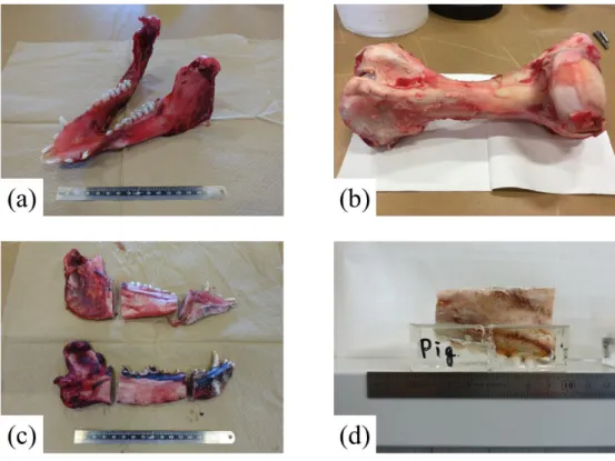
2.2.1 Bone ... 36
2.2.2 SawbonesⓇ test materials ... 37
2.2.3 Comparison of general properties ... 38
2.3. Drilling test methods ... 39
2.3.1 Experimental apparatus ... 39
2.3.2 Test measurements ... 41
iii
2.4. Analysis methods ... 46
2.4.1 Processing of acquired data during drilling tests ... 46
2.4.2 Observation by optical microscope ... 47
2.4.3 Observation by Scanning Electron Microscopy (SEM) ... 47
2.5. Results and discussions ... 48
2.5.1 Drilling properties ... 48
2.5.2 Observation of cutting chips... 65
2.6. Conclusions... 67
Chapter 3: Characterization of drilling in acrylic resin as a matrix of composite materials ... 69
3.1. Introduction... 70
3.2. PMMA as a matrix of composite materials ... 70
3.4.1 PMMA ... 70
3.4.2 Fabrication methods ... 71
3.3. Experimental methods ... 72
3.4.1 Drilling tests ... 72
3.4.2 Dynamic Mechanical Analysis (DMA) measurements ... 73
3.4. Results and discussions ... 73
3.4.1 Characterization of cutting chips ... 73
3.4.2 Drilling properties related to chip formation ... 75
3.4.3 Effects of machining conditions on drilling properties ... 77
3.4.4 Thermal effects on mechanical properties and drilling ... 79
3.5. Conclusions... 80
Chapter 4: Fabrication of composite materials and their drilling properties ... 81
4.1. Introduction... 82
4.2. Materials ... 82
4.2.1. Ceramic additives ... 82
4.2.2. Fabrication of composite materials ... 84
4.3. Experimental methods ... 86
4.3.1. Drilling under constant load ... 86
4.3.2. Drilling under constant feed rate ... 86
4.4. Results and discussions ... 86
4.4.1. The effects of additives under constant load drilling ... 86
4.4.2. The effects of additives under constant feed rate drilling... 90
4.5. Conclusions... 92
Chapter 5: Relationship between drilling and mechanical properties ... 95
5.1. Introduction... 96
iv
5.2.1. Specimens ... 96
5.2.2. Bending tests ... 96
5.2.3. Fracture toughness tests ... 97
5.2.4. Microindentation tests ... 98
5.2.5. DMA measurements ... 99
5.3. Results and discussions ... 99
5.3.1. Bending test results ... 99
5.3.2. Fracture toughness tests ... 101
5.3.3. Microindentation tests ... 102
5.3.4. DMA measurements results... 105
5.4. Conclusions... 107
Chapter 6: Characterization of manual drilling by surgeons ... 109
6.1. Introduction... 110
6.2. Materials ... 110
6.3. Experimental and analytical methods ... 110
6.4. Results and discussions ... 112
6.5. Conclusions... 117
Chapter 7: Concluding Remarks ... 119
References ... 124
Chapter 1: Bibliography Synthesis
This chapter presents the state of the art in drilling of bone biomodels, by reviewing the literatures concerning biomodel, bone, and drilling, followed by the description of the objective of this study and the research approach toward improvement of bone biomodels.
Chapter 1: Bibliography Synthesis
-2-
1.1. Introduction
Drilling of bone is one of the common surgical steps. Surgical treatment results are largely influenced by the surgical skills of surgeons and the performance of medical devices. Then, it is important especially in Japan, the most super-aged society in the world, that surgical training of doctors and development of high-performance medical devices should be carried out efficiently.
Biomodels can function to meet the rapidly increasing demands for medical resources. Bone biomodel is one of the biomodels that replicate human tissue, and known for its usage in surgical training for doctors or mechanical tests of medical devices.
A number of bone biomodels are currently available in the market, but the reproducibility of drilling of bone has little been paid attention and barely been in the research scope up until today. Therefore, conventional bone biomodels cannot fully reproduce the unique drilling behavior of natural bones. Besides, evaluation items among physical and mechanical properties of biomodels toward the replication of drilling of bone is uncertain.
Replication of drilling includes both drilling properties such as thrust force and torque, and tactile feedback during drilling. These aspects should be influenced by physical and mechanical properties of work materials. In order to improve the conventional bone biomodels, it is necessary to fabricate the alternative materials based on understanding of the correlation among mechanical and drilling properties, and tactile feedback during drilling.
Then, the objective of this study is to find out the relationship between mechanical and drilling properties, and also drilling properties and tactile feedback during drilling for the development of bone biomodels that cover drilling characteristics of natural bone.
To accomplish this objective, the present study adopts the fabrication of composite materials to look into the effects of additives on mechanical and drilling properties, and tactile feedback during drilling. Contrary to the conventional approach that relied on the tactile feedback of doctors, where there were a lot of trials and errors for improvement, quantitative characterization of mechanical and drilling properties from the standpoint of engineering are applied to drilling of bone biomodels as well as natural bones and conventional bone substitute materials.
In this chapter, backgrounds and literature studies focusing on biomodel, bone, and drilling are summarized, including the current limitations and challenges in the development of bone biomodels. The research objectives and approaches to achieve the objectives are also described as well as the outline of this thesis.
Chapter 1: Bibliography Synthesis
-3-
1.2. Biomodel
1.2.1. The role of biomodel to fulfill medical resources
In 2007, Japan has entered the “super-aged society”, which is defined by the World Health Organization (WHO). The WHO defines the “aging rate” as the proportion of a society’s population for those aged 65 or older. If a society has the aging rate more than 7%, the society is an “aging society”. If the rate exceeds 14%, it is an “aged society”, and a “super-aged society” in case that the rate surpasses 21%. Fig. 1-1 shows a demographic change in Japan from 1990 to 2065 as illustrated in the statement on the social welfare renovation by the Ministry of Health, Labour and Welfare [1]. Although it has been stressed for a long time since the Japan faced the super-aged society, the aging rate at the year of 2016 in Japan is about 27.3% as reported in the White Paper on Aging Society [2]. Moreover, according to the 15th estimated population reported in 2017 by the National Institute of Population and Social Security Research [3], the elderly over 65 years old can account for about 30% of the whole Japanese population in 2025, and almost 40% in 2065.
Chapter 1: Bibliography Synthesis
-4-
The advancement of the super-aged society holds problematic potentialities especially in medical and welfare support systems. A larger number of orthopaedic surgeries has been performed along the drastic increase in the proportion of the elderly. For example, the number of total joint replacement of hip, knee, and shoulder operated from April 2017 to March 2018 reached 220,000, which is 28.3% larger than that of the previous year period [4]. This trend is not only the case in Japan, but also in other developed countries. In the U.K., the number of joint replacement operations for hips, knees, ankles, shoulders, and elbows performed in 2018 statically came up to 240,163, marking 9.5% increase compared to 2017 as reported in the annual reports by the National Joint Registry (NJR) [5,6]. Also in the U.S., the number of primary total knee arthroplasty (TKA) performed from 2012 to 2017 was 650,674 according to the 5th annual report in 2018 by the American Joint Replacement Registry [7], and the number of such operation is estimated to increase by 3.48 million per year by 2030 [8].
As just described, the number of orthopedic operations is undoubtedly keep increasing all over the world in the future. To meet this rapidly increasing demands for medical care, the society is required to rapidly fulfill medical resources such as a sufficient number of skillful doctors, and good-quality medical devices. To do so, the joint effort has been made between the industry, government, and academia for enhancement of medical educational system, research and development of high-performance medical devices, and establishment of appropriate testing standards of medical devices or test materials for facilitation of evaluation procedure.
Biomodel is an attractive material that steadily supports the progress of medical technology in all the aspects mentioned above; enhancement of educational system, development of medical devices, and establishment of test standards. The use of biomodels will be detailed in the next sessions.
1.2.2. Definition and applications of biomodel
According to Lohfeld et al. [9], “A biomodel is an entity that replicates the geometry or morphology of a biological structure, which can be realized in either a computer-based form or a solid physical form.” Based on this definition, there are two kinds of biomodels available; a computer-based
biomodel and a physical biomodel. Computer-based biomodel covers not only a virtual biomodel, but
also a computational biomodel [9]. A virtual biomodel shall be created for the purpose of visualization of biological structures, such as a skeletal model based on 3D computer-based images generated from computed tomography (CT) scans, normally used for preoperative planning. A computational
Chapter 1: Bibliography Synthesis
-5-
analysis on a biological structure, often used for determination of stress and strain distributions in reality [10].
A physical model is a biomodel in a solid physical form that can be fabricated by engineering technologies such as computerized numerical control (CNC) milling, injection molding, or rapid prototyping (RP) technologies [11,12]. Fig. 1-2 shows several examples of a physical model [13–15] mainly replicating the geometry or morphology of biological structures respectively. As far as engineering technology permits, all the parts of soft and hard physiological tissue can be generated. Recently, 3D printing technology is also applied in development of biomodel widely from blood vessels to bones [16–20].
Fig. 1-2 Examples of physical biomodel. (a) A Prosthetic Restoration Jaw Model [13], (b) A Drilling
Chapter 1: Bibliography Synthesis
-6-
Biomodel is used for a wide range of applications. Representative application is, for example, preoperative planning [21–23], surgical training [16,24–26], and mechanical tests as laboratory study for evaluation of medical devices [27–30]. The efficacy of preoperative explanation of surgical procedure using biomodel is also regarded as helpful to obtain informed consent about operation [11]. For each application, the intended role of biomodel is different. That is to say, the desired ability that biomodel has to exhibit should be well determined. For the purpose of preoperative explanation to patients, the realistic appearance of target tissue with its anatomical accuracy should be of its high priority, rather than its similarity of tactile feedback or physical properties. This application is relevant to the definition of biomodel in [9]. On the other hand, tactile feedback as well as anatomical structure should have high priority for surgical training, while physical and mechanical responses related to target function should be reproduced for the mechanical tests of medical devices. In these applications, the functional behavior of living tissue is more requested rather than the geometry or morphology of the biological structure over the conventional definition of biomodel. The functional characteristics of biomodel is the recently emerging aspect that should be replicated in the use of physical models, which used to be out of the scope of its use.
1.2.3. Biomodel in development of medical devices
In Japan, sales and production of medical devices are regulated by the Pharmaceuticals and Medical Devices Law (PMDL). Under the PMDL, the Pharmaceuticals and Medical Devices Agency (PMDA) is in charge of evaluation of medical devices in terms of quality, efficacy, and safety taking into account the current scientific and technological standards. In the U.S., these services are under the jurisdiction of the Food and Drug Administration (FDA). Fig. 1-3 shows the overview of PMDA’s reviews and related services during product development procedure consisted of several stages; research and development, non-clinical tests, clinical trials, filing of application, approval, and marketing [31]. The PMDA provides various services at each stage of the procedure, such as consultation in relation to regulatory submission, compliance assessments focusing on Good Laboratory Practice (GLP), Good Clinical Practice (GCP), and Good Post-marketing Study Practice (GPSP) to ensure the submitted data shall be in accordance with the ethical and scientific standards, and inspections in terms of Good Manufacturing Practice (GMP), Quality Management System (QMS), and Good Gene, Cellular, and Tissue-based Products Manufacturing Practice (GCTP) in order to ensure the quality management of the manufacturing facility for the pending products.
Chapter 1: Bibliography Synthesis
-7-
As illustrated in Fig. 1-3, one of the characteristics in development of medical devices before distribution to a customer in comparison of other industrial products is the presence of clinical trials to obtain the approval. Since the intended purpose of medical devices is to help health care providers diagnose, prevent, and treat sickness or disease of their patients, with often influencing patients’ anatomical structure or physiological function, clinical trials have been regarded as indispensable in Japan. What is essential in clinical trial is to see whether an emerging device has the capability to fulfill the intended purpose, balancing the benefits and the risks on patients’ body. Therefore, newly developed medical devices are tested using living tissue either in vivo or in vitro.
Chapter 1: Bibliography Synthesis
-8-
It is unsurprising that performing clinical trial is not easy. As developers of medical device, there are several steps to overcome, in order to obtain the approval. In addition to a number of administrative works, developers have to recreate the intended usage environments (e.g. a number of intact vascular systems surrounded by human soft tissue and bones), and prepare a sufficient number of test specimens and subjects. No matter what kind of living tissue is required, obtaining live specimens of acceptable sample size and quality, from the limited stock, within a reasonable time period, before environmental deterioration like dehydration or biological decay affects and alters the specimens [32,33], is a complicated and complex work. Besides, individual variance in material properties also make it difficult to obtain statically reliable data due to animal species, gender, anatomic location, food history, and the presence of disease [34,35]
Then, the use of inanimate biomodel can play a role as alternative materials to living tissue, somehow to mitigate these limitations and predict the biomechanical testing results. Compared to living tissue from cadavers or animals, the use of biomodel has two major advantages; ease of handling (biomodel does not require any special storage methods, licenses, or approvals from ethical committee and do not carry the risk of infection), and reproducibility (biomodel can provide statically reliable testing outcomes owing to the consistence of material composition, density, and geometry). Thanks to those advantages, the use of biomodel attracts more and more attentions over the world.
1.2.4. Biomodel as a standard test material
Another difficulty for evaluation of medical devices lies in the authorities’ side. In Japan, PMDA is in charge of fixing up the testing methodology and requirements in the evaluation items for each medical device, but designing valid testing system could take time, especially in case that genuinely required evaluation items related to the accomplish of the intended function is not clear. The term “medical device” covers a wide range of devices used for health care, in various medical specialties. Nowadays, numerous types of medical devices have been invented, and thus evaluation methods are diverse. Therefore, PMDA is responsible to study the intended purpose of each medical device, and determine the most appropriate evaluation methodology to see if the medical devices can exhibit the desired function, taking into account the possibility of securement of a sufficient number of subjects, guarantee of long-run capability of products, uncertainty of surgical outcome depending on surgeons, and unique usage environments (particularly implantable devices such as artificial joint or artificial heart). In this regard, it is academically expected to provide a better understanding of the
Chapter 1: Bibliography Synthesis
-9-
relationship between the required properties of medical devices and the capacity of the medical devices to fulfill the intended function, for facilitation of the development of medical devices by helping to establish evaluation items composed of the bare minimum requirements.
Standards development is one of the services of PMDA as illustrated in the Fig. 1-3. In this service, testing methodology valid for evaluation of medical devices are established as standards. Standards are reviewed and protected by the International Organization for Standardization (ISO) and ASTM International (ASTM; American Society for Testing and Materials) over the world, and by the Japanese Industrial Standards (JIS) in Japan. Hundreds of standards have been already defined for medical devices, for example, about the implants for surgery [36–43]. By following the ASTM standard designated such as F543 [40], metallic bone screws for bone plates in orthopedics were evaluated by researchers [44–46], and comparable among the screws. Specification by the standards covers not only terminology and testing methodology, but also materials both for medical devices and test materials as well as their fabrication methods. The use of bone biomodel, which is made of polyurethane foam, is regulated by the specification F1839 [47], for determination of the axial pullout strength of metallic bone screws [40].
SawbonesⓇ is one of bone biomodels commercially available around the world (Fig. 1-4) [48].
Among the products of Sawbones🄬, solid rigid polyurethane foam is defined as a standard test material (Fig. 1-5) [47]. Taking into account the specification, researchers have studied its static physical and mechanical properties [49–51]. However, as Hausmann described, obtaining clinically relevant data is limited for cases of biomechanical testing, due to the essential difference in mechanical properties compared to those of genuine bone, whereas the use of Sawbones🄬 as a test material for comparable study between a series of identical devices can be agreeable [52]. Nevertheless, it is also true that there is still lack of quantitative information available regarding machining characteristics, such as cutting forces and cutting temperatures during drilling, whose information are essential for the assessment of orthopedic or other specialties dealing bone and prosthesis. Among limited literatures, Cseke reported that there is a large difference in machining characteristics between natural bones and Sawbones🄬 test materials [53]. Therefore, only a comparable study between bone drills, bars, and prosthesis can be possible using the current bone biomodels. In order to improve the use of bone biomodel to evaluate the machining functions of medical devices, enhanced biomodel that can reproduce the machining properties of natural bone should be developed.
Chapter 1: Bibliography Synthesis
-10-
current technology for fabrication of biomodel permits only comparable studies among the same type of medical devices such as screws or pins. To take a further step, biomodel that equips with the realistic properties of natural bone, related to the assessment of intended purpose for each medical device is necessary.
Fig. 1-4 SawbonesⓇ biomechanical test materials [48]
Fig. 1-5 Standard specification for rigid polyurethane foam regulated by the ASTM international
Chapter 1: Bibliography Synthesis
-11-
1.2.5. Biomodel in surgical training
Surgical outcomes can be largely influenced by a user of medical devices or instruments, even though highly-performant products have been newly developed. In case that surgeons lack their knowledge and/or operational capability of surgical devices, medical accidents can possibly occur. In orthopedic surgery, surgeons would manually cut and remove patients’ skins and other tissues using surgical knife and electric scalpel in order to expose the target inner organs. Here, surgeons are required to have a good command of surgical instruments to properly conduct operations as initially planned.
Regarding this point, there are certain surgical skills that are complicated and take time for mastery, such as clipping of aneurysms [54] or drilling of bone [55]. Drilling of bone is one of a series of surgical steps in dentistry or orthopedics, and often performed during dental implant surgery or artificial joint replacement. Therefore, dentists and surgeons are required to acquire the skill of drilling. In this regard, the surgeons are expected to accurately and steadily handle the surgical tool with controlling their level of force along the progress of drilling displacement depending on the bone structure and the individual difference of bone’s characteristics, in order to avoid severe risks to their patients.
Medical and dental students can learn the basic knowledge about frequently occurring diseases and disorders, and their treatment methods. They have a chance to practice surgeries using actual instruments on dummies, and work as doctor-in-training in hospital under senior doctor at various specialties. In this manner, the students learn to conduct surgeries by following a proper procedure. In fact, however, the students do not have many chances to train themselves on living tissue while in school, because of the difficulty in handling as well as the limited accessibility. Besides, during internship, the students often only observe the surgeries and have less chance to give treatment [56]. The progress in medical technology has extended the surgical knowledge, and as a result increased the students’ burden. The industry, government, and academia have been somehow responding to this trend by developing educational materials and increasing learning opportunities [57,58].
Basically surgical training for amateur doctors are performed in operating rooms in a hospital, but the currently increasing medical demands in our society require a number of experienced doctors and therefore amateur doctors are expected to do supplementary training away from hospital. Workshop using cadavers or animal models has been traditionally performed since long years, but nowadays clinical practice is becoming more and more difficult, because of the less tolerance of our
Chapter 1: Bibliography Synthesis
-12-
society against the use of cadavers or animals in medical research and education [59]. As alternative training methods, the advancement in technology brought computer-based training systems [60–62] and virtual reality (VR) simulators [63,64]. Although these emerging training systems show high efficiency in educational performance and have great advantages, the technology still has some room for improvement and also not many hospitals can introduce the systems because of its initial and maintenance cost [58].
For the purpose of surgical training, the use of biomodel has high potential. Similarly, as the use in mechanical tests of medical devices, inanimate biomodels possess strong advantages in ease of obtaining, conserving, and handling as well as reliability for repeated times of use over a long-duration. In a wide field of specialties, biomodel for surgical training has been developed. For biomodels of bone, there are many sorts of biomodels already available. One of the bone biomodels, made of acrylic resin and wood flour (Exsurg🄬, Tecno Cast Co., Ltd.) (Fig. 1-6) [65], has relatively better reputation
among the existing models, but the reputation is by no means based on quantitative evaluation. Widely as for the evaluation of biomodels, there has been no specific criteria to determine a good biomodel except the perceptual feedback by doctors. Therefore, development of biomodel has been a series of trials and errors without concrete direction for improvement.
To address this situation, researchers aware of the gap between human tissue and conventional biomodels have been working on reproducing more realistic biomodels [66–69]. Recently, a bionic humanoid was invented in Japan in a framework of research and development program driven by the Japanese government [70–72]. The invented humanoid consisted of artificial living tissues and equipped with a series of sensors that enabled tactile force measurements of operators. Using this model, surgical training can be possible with quantitatively monitoring mechanical parameters and simultaneously assessing the surgical skill of the operator. However, the development of human bone tissue that can reproduce the realistic drilling haptics has no yet been its scope and no quantitative research can be found for the assessment of the tactile perception during drilling based on mechanical criteria.
Chapter 1: Bibliography Synthesis
-13-
Fig. 1-6 An example of mandibular biomodels for surgical training [65]
1.3. Bone
1.3.1. Physiological functions
Bone is one of human hard tissues, accounting for around 18% of the weight of our human body. Bone constitutes the skeletal system and six main functions are displayed in Table 1-1. In a microscopic level of view, bone, or osseous tissue, contains an abundant extracellular matrix that surrounds widely isolated cells. Those extracellular matrixes are composed of about 25% water, 25% collagen fibers, and 50% crystallized mineral salts [73]. The richest mineral salt is calcium phosphate, and it combines with another mineral salt, calcium hydroxide [Ca(OH)2], to form crystals of
hydroxyapatite [Ca10(PO4) 6(OH)2]. Those crystals combine with other mineral salts, such as calcium
carbonate, and ions such as magnesium, fluoride, potassium, and sulfate. Those mineral salts are generally embedded on the collagen fibers of the extracellular matrix, and the crystalizing process, which hardens the bone tissue, is called calcification.
Bone is not completely solid but has many small spaces between its cells and extracellular matrix components. Several spaces work as vascular channels which provide nutrients to bone cells. Other spaces function as storage area for red bone marrow. Bone is categorized as compact (or cortical) and spongy (or cancellous) bone, according to the size and distribution of the spaces. About 80% of the skeletal system is cortical bone and 20% is cancellous bone.
Fig. 1-7 shows an overview of both compact and spongy bones [73]. Compact bone, also called cortical bone, has strong and dense form of bone tissue. Cortical bone tissue forms the outer layer of
Chapter 1: Bibliography Synthesis
-14-
every bone and provides protection and support and endures the stresses induced by weight and movement. Blood vessels, lymphatic vessels, and nerves from the periosteum run through cortical bone inside perforating canal, often referred to as Volkmann’s canals, and they connect with those of medullary cavity, periosteum, and central or Haversian canals. The central canals run longitudinally through the bone. Cortical bone tissue is composed of several repeating units called osteons or Haversian systems. Each osteon has a Haversian canal surrounded by several parts of lamellae, lacunae, osteocytes, and canaliculi. Osteons in cortical bone tissue are arranged in a parallel way to the lines of stress. The distribution of osteons is not settled and osteons remodel their structure according to the physical needs of the skeleton.
On the other hand, spongy bone, also called cancellous bone, tissue is light and has porous structure literally similar to sponge. Cancellous bone tissue forms interior part of bone, normally surrounded by cortical bone for protection. The bone tissue is composed of trabeculae, lamellae distributed in a random lattice of thin columns. Distribution of trabeculae appears to be arranged randomly, but in fact they are optimized precisely along lines of stress; the distribution helps bones endure and propagate stresses without breaking. The empty spaces between the trabeculae are often filled with red bone marrow.
Chapter 1: Bibliography Synthesis
-15-
Fig. 1-7 Osteons (Haversian systems) in compact bone and trabeculae in spongy bone [73]
1.3.2. Biomechanical aspects
Biomechanics of bone have been widely studied since the middle of 20th century. The previous
studies have shown the macroscale to microscale of mechanical properties of bone about tensile, compressive, and shear strength and elasticity or fracture and fatigue behavior with categorizing the anatomical location in both cortical and cancellous bone [33,74–81]. Particularly, Currey found that the stiffness of bone increases drastically with the mineral density of bone [74], while Bonfield et al. found the anisotropy of stiffness of bone using ultrasonic measurement technique [76]. It was the early days of the study of bone from the standpoint of material engineering.
The recent progress in measurement technique and the further research interests about bone in these three decades brought deeper understanding of bone [82]. Most famously, nanoindentation technique enabled researchers to measure the mechanical properties of bone precisely at nano-scale. Focusing on the effects of anisotropy of bone with distinguishing at the scale of osteons [83–88], the anisotropy in mechanical properties at the level of osteons was confirmed as the longitudinal moduli is higher than the transverse moduli [87,88]. General mechanical properties of bone such as tensile strength and elastic modulus are listed in Table 1-2 [32,49,75,83,89–91]. Human cortical bone shows
Chapter 1: Bibliography Synthesis
-16-
elastic modulus from 17.6 to 23.5 GPa in longitudinal direction, which is slightly higher in comparison with that of traverse direction as an anisotropic material. As shown in Table 1-2, cortical bone is superior to cancellous bone in mechanical properties. There is also difference in mechanical properties depending on animal types.
Moreover, research interests have been expanded to dominant factors on mechanical properties such as not only the effects of mineral contents [85,92] or experimental parameters as represented by strain rate [93,94], but also specimen size, wet or dry conditions [95,96], and conservation methods [97–100]. Taken together, bone specimen is recommended to preserve frozen rather than in chemical liquids in order to maintain its mechanical properties.
Numerical simulation by finite element method (FEM) also supports the biomechanical aspects of bone, as firstly introduced, for example, by Richmond et al. [101]. Currently, combining with scanning images at high resolution using CT, stress distribution of bone in a realistic geometry can be obtained [102,103]. The advance in computer processor made it possible to analyze numerically as far as the machining of bone [104,105].
Chapter 1: Bibliography Synthesis
-17-
1.3.3. Bone among engineering materials
Bone can be regarded as a composite material consisting of organic (collagen) and inorganic (hydroxyapatite) tissues, having unique characteristics among engineering materials widely applied in industry. Fig. 1-8 shows the comparison of stress-strain curves for four representative engineering materials with different elasticities, such as steel, glass, bone, and rubber [106]. It indicates that bone exhibits higher stiffness rather than that of rubber, but lower than steel and glass, locating the curve of bone between those of rubber and glass. To the authors’ knowledge, there is no such alternative materials that can show the similar mechanical response to that of bone.
Fig. 1-8 Stress-strain curves of representative engineering materials [106]
On the other hand, development of new materials such as engineering plastics and fine ceramics have been undertaken in last decades in order to expand the area of use from plastics and ceramics respectively. In the fields of ceramics, the development of fine ceramics has enthusiastically progressed thanks to the advance in technique that allowed the accomplishment of highly refined materials and the control of resultant composition and geometry. In the early 20th century, fine ceramics
Chapter 1: Bibliography Synthesis
-18-
gradually came into practical realization as represented by the vehicle engines and semiconductors. Besides, the application for medical purpose caught attention at the late 20th century, though the
primary use started even the late 18th century in dentistry and the late 19th in orthopedics for bone
filling [107]. After that, the improvement in toughness and strength of fine ceramics, such as alumina (Al2O3) and zirconia in particular, led to the use into implantable devices. Eventually since 1990, a
tremendous number of joint replacement, using alumina components and zirconia femoral heads, has been implanted across the world [107]. In addition to these “bioinert” ceramics consisted of alumina and zirconia, “bioactive” ceramics, mainly from hydroxyapatite (HAP) or tricalcium phosphate (TCP) because of the similarity of their compositions to the mineral part of bone, is known for the clinical use.
Reinforced plastics was also keenly developed since 1960s along the rapid industrial growth of petroleum chemistry for various applications. Glass or carbon fiber reinforced plastics (GFRP or CFRP) are the two representative products, having such characteristics in specific strength and specific stiffness, where epoxy resin and polyester have been mostly chosen for the majority of the matrix in previous studies [108]. CFRP is particularly applied to the constructional materials in aircrafts or vehicles as well as sports goods due to its advantage.
These fiber-reinforced plastics (FRPs) are categorized as composite materials. Not only for the improvement of mechanical properties of a matrix, but also for the adjustment of target characteristics, composite materials are fabricated. Exsurg🄬 [65], one of the bone biomodel presented at the section 2.5, is also a composite material, which consists of acrylic resin as a matrix and wood flour as fillers. According to the inventors’ patent, the inclusion of wood flour was intended for the adjustment of tactile perception during drilling to give the similarity to that of natural bone [109]. Like this case, polymeric (sometimes composite) materials are often used to replace human tissue.
1.4. Drilling
1.4.1. Environments surrounding drilling in industry
Drilling is one of the machining techniques for material removal similarly to milling or grinding, which is usually performed as finishing process in order to obtain the desired geometry of engineering materials. A drill bit is used to make a hole of circular cross-section in any solid materials such as wood, metal, ceramics, plastics, and composites. Rotated at a certain rate of revolutions per minute,
Chapter 1: Bibliography Synthesis
-19-
the drill bit is pressed against a work piece at a voluntary feed rate. The force conveyed through the drill bit makes the cutting chips from the drilled hole along the penetration of drill bit. Although the remarkable advancement in engineering technology brought the various machining methodology, the drilling process continuously stays as an indispensable technique for material removal in manufacturing.
Development of machining tool for drilling dates back to the time of ancient Egypt, but it is only since the late 18th century in England that the machining tool was renovated during the industrial
revolution. Basic knowledge about cutting theory of our time was widely obtained in the middle of 20th century [110–114]. After that, machining tools became numerically controlled by a computer,
making automatic machining possible for mass production with high precise machinability and productive efficiency. Cutting tools have been developed in response to the appearance of new materials such as strengthened alloy, reinforced plastics, and fine ceramics. A tremendous number of research works on drilling have been done along the increasing demands of improvement in machinability and due to the complexity of processing mechanism of emerging materials.
Contrary to the simple purpose of drilling, making a hole, drilling is known as a complex mechanical phenomenon because of various factors affecting the resultant outcome. Focusing on FRPs, both GFRP and CFRP show low machinability due to additives. In both cases, enhanced strength of the composite materials makes cutting tools difficult to penetrate and subsequently the used tools show short useful life, resulting in the increase in manufacturing cost. A number of researchers have been struggling to address the low machinability of FRPs [108,115–117]. In this regard, most of literatures are focusing on the accomplishment of good finish surface or drilled hole quality, by optimizing machining parameters such as spindle speed, feed rate and geometry of a cutting tool [118–125].
To investigate drilling outcomes, the correlation between those machining parameters and drilling responses such as thrust force, torque, temperature rise, and cutting chips morphology is often characterized for the analysis of drilling. However, it is barely quantitatively mentioned the relationship between drilling characteristics and mechanical properties of work materials, such as surface roughness, hardness, and strength, which are known dominant [114]
1.4.2. Surgical drilling for operations
Drilling performs a practical role for medical purpose. Surgical drilling on natural bone is often performed in several specialties such as dentistry, orthopedics, and neurosurgery. This section
Chapter 1: Bibliography Synthesis
-20-
summarizes case examples of bone drilling in operations and risks on human body carried by drilling. Previous findings on drilling of bone are also summarized.
Fig. 1-9 shows an example of typical surgical process in dental implant surgery [126]. Drilling is performed on maxillary or mandibular bone to make a pilot hole for dental prosthesis. After determination of drilling site, several kinds of drill bits are used with gradually increasing the diameter of drill bits before implantation of dental prosthesis. In orthopedics, bone pins, screws, and plates can be inserted as traumatic injury treatments wherever the anatomical location is (e.g. distal femur and proximal humerus as shown in Fig. 1-10 [127]). According to the surgical procedure manual for the insertion of NCB🄬 bone screws as illustrated in Fig. 1-11, drilling shall be performed to make guide holes for bone screws [128]. The insertion of bone screws and plates is a fundamental procedure also for joint replacement and spine surgery. In neurosurgery, surgeons deal with diseases or disorders related to the nervous system including the brain, spinal cord, peripheral nervous system, and cerebrovascular system. Hence, neurosurgical treatments on the brain or cerebrovascular system accompany craniotomy procedure to reach the inner system through the skull, which makes drilling a mandatory step. As stated so far, various types of surgical operations require drilling technique.
It is obvious that drilling technique is an essential and still fundamental skill for surgeons, but drilling carries a large risk on patients’ body:
- Firstly, there is a possibility that failure of implanted devices or bone fractures can accidentally occur in case of inappropriate fastening. As shown in Fig. 1-12, Natali et al. reported breakage of drill bit left inside bone tissue resulting from overloaded drilling [129]. Motoyoshi et al. suggested recommended values of fixation torque for tightening an orthodontic mini-implant to avoid failure of implant devices [130].
- Second risk is the accidental damages to the surrounding tissue because of the error in position of drilling site or in drilling depth. Especially when drill bits are penetrated deeper than needed, serious damage can occur in bone tissue as well as in nerves or vascular channels adjacent to bone tissue, which may bring medical accidents such as excessive bleeding, paralysis, or abnormality of sensation.
- Thirdly, bone necrosis, often referred as osteonecrosis, can be caused due to either exposure of excess force or high temperature attributed to drilling. Since osteonecrosis is regarded as a serious risk on mother body, due to not only delaying the regeneration of bone cells but also facilitating bone fracture, the effects of drilling on temperature elevation have been
Chapter 1: Bibliography Synthesis
-21-
extensively studied. After it was turned out as early as 1984 by Eriksson et al. that bone temperature must not be more than 47℃ for 1 min to avoid osteonecrosis [131–133], various aspects such as machining parameters [134–139], machining tools [140,141], and cooling methods [136,142–144] were the major scope of research topics. There are still rooms left for further research about the effects of application of surface coating or textile on drill bits and the improvement of numerical modelling of temperature rise during bone drilling [132,133,145].
Considering these risks, surgical education as well as mechanical tests of medical devices are important since surgical outcomes are strongly dependent on operators’ command of medical devices. The advancement of information technology also offers significant benefits on surgeons, such as robot-assisted drilling systems [146–149] or remote controlled robots for surgery [150,151].
Chapter 1: Bibliography Synthesis
-22-
Chapter 1: Bibliography Synthesis
-23-
Chapter 1: Bibliography Synthesis
-24-
Fig. 1-12 An example of failure of drill bit left in situ [129]
1.4.3. Quantitative aspects in drilling
1.4.3.1. Geometry of a drill bit
Above all, a drill bit is an indispensable tool for drilling procedure. Fig. 1-13 illustrates a sketch of a drill bit, consisted of a shank, flutes, and cutting edges [152]. The shank is used to connect to a piece to a chuck of a hand-drill or a machining system. Material removal takes place by the cutting edge. Cutting chips and debris are extracted along the flutes in response to penetration of a drill bit. The cutting edges function to produce a series of slices as the drill bit progresses.
The cutting face can be divided into several parts, as shown in Fig. 1-13 (b). The chisel edge contributes seldom to cutting but largely to the axial thrust force of the drill bit. This is because of a relatively slow rotating velocity in the center of the drill bit and the rake angle nearly zero, meaning the cutting edge almost perpendicular to the work surface.
The point angle is the angle on the tip of the drill bit formed by both cutting chips (Fig. 1-14). Optimal point angles in the orthopedics for bone surface is recommended such as 90° and 118° by the literature [152]. Hillery et al. reported that there seems no significant difference in temperature elevation in bovine and cadaveric bone in vitro in their experiments investigating the effects of point angles between 70°, 80°, and 90° [135]. Similarly, Augustin et al. found trivial effects on drilling temperature using 2-fluted drill bits with 80°, 100°, and 120° [153]. Therefore, point angle has little effect on the increase in temperature during drilling.
The helix angle is the angle between the longitudinal axis of the drill bit and a tangent to the leading edge of the land. Surgical twist drill bits are often slow-spiral, which means the helix angle is relatively small. This small helix angle was assumed ideal for the drilling of bone [154].
Chapter 1: Bibliography Synthesis
-25-
Fig. 1-13 Sketch of a drill bit. (a) Overview, (b) Point geometry, and (c) Relief and helix angles
[152]
Chapter 1: Bibliography Synthesis
-26-
1.4.3.2. Mechanism of drilling in bone
Drilling requires the mechanical input for the rotational motion (rpm) and torque (N・m), where torque means the moment of lateral force that is required for material removal by drill bits. These input are often exerted by the hand-drill or the machining system. Axial thrust force (N) is also loaded vertically to work material for material removal, which is applied manually by the operator or automatically by the machining device under numerical control (NC). The moving velocity of the drill bit through the work material is defined as the feed rate (mm/s).
In industrial manufacturing, a constant feed rate is normally applied under NC systems whereas in the clinical circumstances, a quasi-constant axial thrust force is applied to the hand-drill by the surgical operators. This distinction of the two drilling system is considered to be of crucial importance especially for the studies on surgical drilling of cortical bone.
The literature seems to suggest that drill bit diameter is an important variable that determines the magnitude of thrust force, in addition to the bone quality of drilling site as another contributing factor. Allotta et al. found the linear relationship between drill bit diameter and axial thrust force to produce a given feed rate [155]. Hobkirk et al. found the mean values applied during oral surgery between 4 and 19 N [156], and likewise Natali measured a maximum of between 10 and 20 N in case of 2.5-mm diameter drilling [129]. On the other hand, as much as 110-N mean thrust force was applied on drilling 3.2-mm diameter holes in cortical bone [157]. Altogether, previous studies focusing on dentistry generally applies axial thrust force no larger than 25 N [137], while orthopedic studies has a range between 20 and as much as 120 N [137,152,158,159].
An idealized illustration of the oblique cutting mechanism is shown in Fig. 1-15. The removal of bone at the cutting face takes place by the cutting edges that remove a certain thickness, t, with each rotation as they spiral through the bone, following a helical path. The work material being cut is associated with a unique cutting force, and this determines the optimal rake angle, which is around 25 to 35° for cortical bone [154]. Moreover, the unique anisotropy of bone gives a complexity to drilling characteristics because the cutting resistance vector is continuously changing along the rotation of the drill bit [53]. This dependency of the cutting process on the osteon direction in cortical bone was firstly demonstrated by Jacobs et al., as the cutting forces were greatest when cutting perpendicularly to the longitudinal direction of osteon [154]. Based on this work related to the orthogonal cutting in bone, it was established that a rake angle of 45° was recommended because of the markedly reduced cutting force, regardless of the osteon direction.
Chapter 1: Bibliography Synthesis
-27-
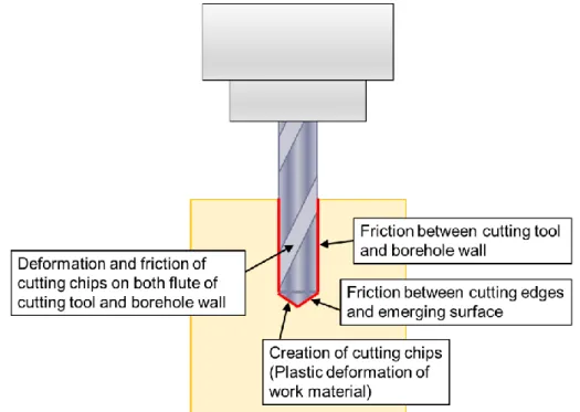
Heat generation inevitably occurs during drilling. According to Fig. 1-15 [152], the primary heat sources pertain to shear deformation of work material (1), friction between the cutting chips from work material and the rake face of cutting tool (2), and friction between cutting edge and under surface of work material touching the relief face of the cutting tool (3). Secondary, indirect heat sources are driven purely by friction involving cutting chips, especially between bone chips and flutes, bone chips and drilled wall of work material when travelling the flute. In case of drilling of metals, almost 60 to 70% of the total heat are transferred to cutting chips [160]. In case of drilling of bone, approximately 60% of the heat energy generated during drilling can be converted to bone chips [152], transferring the rests to the surrounding tissues as well as the drill bit itself. Numerical and mathematical models have been currently developed for the analysis of heat generation and transfer during drilling of bone [104,105,161–164].
Thermal conductivity is a thermodynamic parameter that determines an ability of materials to conduct heat. Cortical bone has a relatively poor heat conductivity as a composite material. Recently, Feldmann et al. determined the thermal conductivity of cortical bone to be 0.64 W/mK for bovine, 0.68 W/mK for human [165], whereas that of surgical-grade stainless steel, often used to drill bits, is around 16.3 W/mK. Specific heat is also known as another material property that influences the temperature rise during drilling [165].
Fig. 1-15 Mechanism of material removal by a drill bit at the rake angle of oblique cutting, with regions
of primary heat generation indicated; (1) shear deformation of the bone, (2) friction between the bone chip and cutting tool, and (3) friction between the tangential surface and cutting tool. (b) Rake and relief angles [152].
Chapter 1: Bibliography Synthesis
-28-
A relief angle is designed in cutting tools to relieve thermal dissipation and mechanical damages due to the friction between cutting tools and the emerging surface of work materials. Chacon et al. reported the significant effects of relief angle on the scale of the temperature elevation during drilling of bone [166].
1.4.3.3. Characterization of drilling in bone
Drilling behavior is the outcome deriving from variables such as cutting tools, machining conditions, and mechanical, thermal, and chemical properties of work materials. Drilling behavior can be characterized by cutting forces (thrust force and torque) and resultant temperatures. Under constant thrust force drilling, drilling time and feed rate are another key properties describing drilling. Drilling behavior can also be characterized by the cutting tool life observed in wear of cutting edge, quality of drilled holes seen in surface roughness and dimension accuracy, and cutting chips generated during drilling. Since these aspects cannot be directly converted from the mechanical properties of materials, drilling tests shall be carried out for the characterization of drilling behavior.
Cutting forces
Thrust force and torque have been reported as the mechanical outputs in drilling to describe drilling characteristics of bone since as early as 1970s [154,167]. Until now, a large number of researchers have worked to find out the haptic aspects on drilling of bone [133,152,168–170]. Wang
et al. reported the effects of rotation speed, feed rate and drill bit diameter on thrust force and torque
under constant feed rate drilling, describing the decreased force and torque along the increase in rotation speed, and increased force and torque in case of increase in feed rate and drill bit diameter [171]. This trend is in a good agreement with other researchers [133,167,170]. Tuijthof et al. compared the thrust force between different surgical machining tools such as twist drill bits, round burrs, and kirschner wire in bones of pig and goat [172].
Cutting temperature
Temperature elevation during drilling can be measured mainly by two major methods; thermal images obtained from infrared camera, or thermocouples placed in bone [173]. Both methods have advantage and drawback. In case of using the infrared camera, what can be obtained is thermal images on the very surface of work piece during drilling, thus temperature rise inside the drilled hole can be more accessible in case of using thermocouples. However, as cortical bone usually has a thickness of less than 5 mm in radial direction, placing thermocouples in cortical bone is not always appropriate
Chapter 1: Bibliography Synthesis
-29-
due to the limitation of spaces. It is important to distinguish the measurement purpose and select the suitable method.
Recently, a lot of researchers carry out experimental analysis of bone drilling as a validation method of their numerical models [174–177]. Lugnmani et al. particularly established a numerical model to predict thrust force and torque that were in a good accordance with obtained experimental results [176]. Feldmann et al. even established a numerical model that predicts temperature elevation during bone drilling [145].
Cutting chips
Chip formation is another important factor in the characterization of drilling in bone. The morphology of cutting chips indicates fracture behavior of work specimens. There are certain literatures focusing on bone chips formation during drilling [178–180]. Apparently, bone often exhibits crack-typed cutting chips in drilling possibly due to its brittleness, which was different from those of synthetic materials, as flow-typed chips was observed for cutting epoxy-based bone biomodels [180]. In addition to the nature of bone, the progress of machining process accompanies temperature increase in bone, which consequently change the fracture mode of bone chips alternating the morphology of cutting chips from flow-typed to crack-typed [178,179]
Wear of cutting tools
A repeated contact of the cutting edge on the emerging surface of work pieces causes wear and dulling of cutting edges, which subsequently requires the application of a higher thrust force for the progress of drill bit. Wear of cutting tools can cause defective cutting usually with higher elevation of temperature, and the initiation of vibration due to an increase in surface roughness of the cutting edges. Observation of cutting edges using optical microscope or Scanning Electron Microscope (SEM) is the imaging methodology to analyze wear of cutting edges. Literatures report that abrasive wear as well as plastic deformation can occur to alter the geometry of the chisel and cutting edges, and the rake face of the drill bit [181,182]. Allan et al. investigated the effects of various magnitude of wear on maximum temperature elevation in cortical bone in vitro. Three types of drill bits (1.5 mm diameter, 2-fluted Leibinger) were compared which were fresh, used in the drilling of 600 holes in porcine mandibular bone, and provided from operating theatres after the use for several months with measuring maximum temperatures during drilling tests [181]. They revealed that 600 holes was statistically sufficient to cause significant temperature rise compared to fresh drill bits with showing the images of three drill bits as shown in the Fig. 1-16.
Chapter 1: Bibliography Synthesis
-30-
There seems a large number of information available on drilling of bone, but in most cases machining parameters such as rotation speed, feed rate, tool geometry, animal species, and anatomical positions are diverse and those reported results cannot be always comparable. Therefore, in order to study the drilling characteristics of bone toward development of biomodel, it should be necessary to determine the information of target bone and machining conditions respectively.
Fig. 1-16 Images of three drill bitsfrom side view and tip. Upper row: fresh, Middle row: used in the
Chapter 1: Bibliography Synthesis
-31-
1.4.3.4. Drilling of bone biomodels
Drilling characteristics of bone biomodels have not yet been reported until Cseke et al. reported the drilling of bone and SawbonesⓇ test materials in comparison [53]. They described that drilling
properties such as thrust force and torque of Sawbones🄬 test materials under constant feed rate drilling are quite lower than those of porcine and bovine bones [53], implying a discrepancy under surgical training or evaluation of medical devices. No other literatures on drilling of Sawbones🄬 test materials can be found at this moment.
Nonetheless, Tawara et al. pointed out the difference of mechanical properties between human bone and conventional bone biomodels and so manufactured a new bone biomodel made of polyurethane, whose drilling characteristics not yet available experimentally but pull-out strength [183,184]. Tai et al. developed a plaster-based material that showed the same order to bone in thrust force and torque under constant feed rate drilling, still having limitations in replicating cutting chips morphology [20,21]. In addition, there are two patents available for bone biomodels; acrylic-based composite materials with ceramic additives [186], and acrylic-based composite materials with wood fibers [109]. However, drilling characteristics of those models are not available in the patents but describing the accomplishments of good sensory feedback from doctors, thus lacking scientific grounds.
Bone biomodels are often made of plastics, ceramics, or composites of plastics and ceramics. Therefore, there should be the knowledge in those materials currently available that can be applied to drilling of bone biomodel. Drilling of plastics maybe especially applicable that has been studied along the development of FRPs for the components of industrial products [108,113,116–118,187–189]. Conventionally, thermoset polymers such as epoxies and polyesters are on the main scope of research works [116], but the usage of thermoplastic polymers is recently getting more and more attention for FRPs (CFRTP for carbon reinforced thermoplastic polymer, and GFRTP for glass fiber-reinforced thermoplastic polymer), thanks to their recyclability based on the thermoplasticity. Among thermoplastic polymers, polypropylene (PP), polycarbonate (PC), polyvinylchloride (PVC), and nylon (polyamide) are the main matrices often studied and applied to composite materials [188,190–193]. Regardless of the registration of patents as bone model, poly(methylmethacrylate) (PMMA) has not yet been a main focus of literatures. Since polymers show distinct material properties each other and thus different machinability, drilling characteristics of polymers shall be studied respectively for polymer species [188].
Chapter 1: Bibliography Synthesis
-32-
Among limited literatures, Kobayashi studied the three-dimensional drilling characteristics of PMMA [113], although studied machining conditions cannot necessarily be applied to surgical drilling. Apart from drilling, two-dimensional cutting, single point diamond turning (SPDT), on PMMA was reported for the frequent application of PMMA to optical lenses [194], but likewise, the cutting theory in two-dimensions cannot be always applicable in three-dimensional machining.
PMMA is known for its use as bone cement or teeth fixation in medical applications. Acrylic-based composite materials including ceramic additives can be found in literatures [195–197], but those acrylic composite materials are used for fixation of prosthesis or restoration of teeth, not yet intended for drilling. Thus, drilling characteristics of acrylic-based composite materials with ceramic additives have not yet been quantitatively studied.
1.5. Research scopes and objectives
In this chapter, backgrounds and literatures focusing on biomodel, bone, and drilling are studied, including the current limitations and challenges in the development of bone biomodels. After a vast literature review, the research background can be summarized as follows:
1. Bone biomodels are useful particularly in surgical training and for mechanical tests of medical devices,
2. Polyurethane foam from Sawbones🄬 is one of the standard bone biomodels for cancellous bone defined in a test standard, while no biomodels are referred for cortical bone,
3. Bone drilling is one of the fundamental surgical steps in dentistry or orthopedics, but the replication of bone drilling has been out of research scope,
4. Thus, no quantitative evaluation items are currently available for the development of bone biomodels, except for perceptual feedback of surgeons.
Therefore, there have been many trials and errors for the development of bone biomodels. This study will address this situation by quantitatively studying drilling characteristics of bone biomodels. As stated above, contrary to a wide range of options possible for materials and machining conditions, there are only a limited number of previous studies related to drilling characteristics of bone biomodels available, resulting in the lack of understanding in drilling properties of bone biomodels. This study chooses to perform drilling tests under surgical machining conditions, focusing on acrylic composite materials with ceramic additives as well as bones and Sawbones🄬 test materials. Drilling characteristics are dependent on various dominants. This study focuses on mechanical properties of
Chapter 1: Bibliography Synthesis
-33-
work materials, acrylic composite materials. The authors aim to control drilling characteristics by altering the mechanical properties that shall be controlled in response to the material composition. Through controlling the material properties, the objectives of this study are as follows:
1. Apply engineering standpoints into surgical drilling of bone biomodels, by quantitatively measuring drilling properties such as thrust force, torque, and temperature rise during drilling.
2. Understand the drilling characteristics of acrylic resin and acrylic composite materials under surgical drilling conditions toward the use application as bone biomodels.
3. Elucidate the effects of additives on mechanical and drilling properties of the matrix by controlling the composition of composite materials.
4. Elucidate also the effects of drilling properties on tactile feedback of surgeons.
5. Obtain the future direction of the development of bone biomodels through understanding of the relationships between material compositions, mechanical and drilling properties, and perceptual properties during drilling.
1.6. Organization of the thesis
In consideration of research motivations and objectives as stated above, the contents of this thesis are outlined as follows:
Chapter 1: backgrounds and literature reviews on this study focusing on biomodel, bone, and
drilling are summarized, including the use application of bone biomodels, the current knowledge of drilling of natural bone and bone biomodels. The current limitations and challenges in the development of bone biomodels led to the research objectives.
Chapter 2: drilling tests of natural bone and Sawbones🄬 test materials are preliminarily performed to understand the discrepancy of drilling characteristics and the current limitations of bone biomodels. Machining parameters are selected to reproduce realistic surgical drilling.
Chapter 3: drilling of acrylic resin are studied as a matrix toward fabrication of acrylic
composite materials. The effects of cutting parameters and temperature elevation during drilling are investigated.
Chapter 4: Acrylic composite materials are fabricated using ceramic additives, and then their
drilling properties are studied for the replication of drilling of bone.
![Fig. 1-3 Services of PMDA at each stage of product development (Reprinted from [31])](https://thumb-ap.123doks.com/thumbv2/123deta/5915899.1050737/18.892.133.758.442.978/fig-services-pmda-stage-product-development-reprinted.webp)
![Fig. 1-9 Diagrams of surgical sequence using a bur and drill bits in dental implant placement [126]](https://thumb-ap.123doks.com/thumbv2/123deta/5915899.1050737/32.892.148.741.538.1024/diagrams-surgical-sequence-using-drill-dental-implant-placement.webp)
![Fig. 1-11 Procedure manual of the insertion of cortical screws (Reprinted from [128])](https://thumb-ap.123doks.com/thumbv2/123deta/5915899.1050737/34.892.138.740.218.1015/fig-procedure-manual-insertion-cortical-screws-reprinted.webp)
![Fig. 1-12 An example of failure of drill bit left in situ [129]](https://thumb-ap.123doks.com/thumbv2/123deta/5915899.1050737/35.892.237.658.143.413/fig-example-failure-drill-bit-left-situ.webp)
![Fig. 1-14 Comparison of point angles between two drill bits [152]](https://thumb-ap.123doks.com/thumbv2/123deta/5915899.1050737/36.892.260.633.731.1083/fig-comparison-point-angles-drill-bits.webp)