First record of Larsonella pumilus (Teleostei:
Gobiidae) from Japan, with phylogenetic placement of the genus Larsonella
Author NOZOMI HANAHARA, TAKUO HIGASHIJI, CHUYA SHINZATO, RYO KOYANAGI, KEN MAEDA
journal or
publication title
Zootaxa
volume 4695
number 4
page range 367‑377
year 2019‑11‑11
Publisher Magnolia Press
Rights (C) 2019 Magnolia Press.
Author's flag publisher
URL http://id.nii.ac.jp/1394/00001098/
doi: info:doi/10.11646/zootaxa.4695.4.4
ZOOTAXA
ISSN 1175-5326 (print edition) ISSN 1175-5334 (online edition) Zootaxa 4695 (4): 367–377https://www.mapress.com/j/zt/
Copyright © 2019 Magnolia Press
Article
https://doi.org/10.11646/zootaxa.4695.4.4
http://zoobank.org/urn:lsid:zoobank.org:pub:38DB2B6D-FA80-40FF-925B-F402787FF7F6
First record of Larsonella pumilus (Teleostei: Gobiidae) from Japan, with phylogenetic placement of the genus Larsonella
NOZOMI HANAHARA1, TAKUO HIGASHIJI2, CHUYA SHINZATO3, RYO KOYANAGI4 & KEN MAEDA4
1Okinawa Churashima Foundation Research Center, 888 Ishikawa, Motobu-cho, Okinawa 905-0206, Japan.
E-mail: [email protected]
2Okinawa Churaumi Aquarium, 424 Ishikawa, Motobu-cho, Okinawa 905-0206, Japan.
3Atmosphere and Ocean Research Institute, The University of Tokyo, Chiba 277-8564, Japan
4Okinawa Institute of Science and Technology Graduate University (OIST), 1919-1 Tancha, Onna, Okinawa 904-0495, Japan
Abstract
During a survey of deep-sea fauna, using a Remotely Operated Vehicle, a single specimen (21.6 mm in standard length) of Larsonella pumilus (Larson & Hoese, 1980) was collected at a depth of 214 m off the coast of Okinawa Island, Japan.
It represents the first record of this species from Japan. The collection site was far deeper than previous reports for this species. This suggests that the main habitat of L. pumilus is deeper than previously recognized and it may explain the paucity of records of this species. As the previously available morphological description of L. pumilus was based on only a single specimen (holotype), this new specimen is described herein. Its morphology corresponds closely to the original description of the holotype, except that faint melanophores are arranged radially around the eyes and scattered on the trunk and the fins. Mitochondrial genome sequences of L. pumilus and 19 related species demonstrate close relationships between L. pumilus and the genus Priolepis. These data also indicate that the genus Priolepis is not monophyletic.
Key words: goby, deep sea, mitochondrial genome, Ryukyu Archipelago
Introduction
The order Gobiiformes (= suborder Gobioidei in Nelson 2006) is a very diverse fish taxon, including more than 2,000 goby species, belonging to more than 300 genera (Nelson et al. 2016). Gobies are distributed worldwide and occur in fresh, brackish, and marine waters. In the sea, they occupy nearshore environments, including rocky and sandy bottoms, mudflats, tidepools, coral reefs, and caves (Patzner et al. 2011). Although the deep sea may seem to be atypical goby habitat, more than ten genera (Antilligobius, Karsten, Lepidogobius, Lesueurigobius, Obliquogobi- us, Palatogobius, Pinnichthys, Priolepis, Sufflogobius, Suruga, Thorogobius, and Varicus) have been reported from
>200 m depth (Eschmeyer & Herald 1983; Goren 1992; Goren & Baranes 1995; Bianchi et al. 1999; Shinohara et al. 2001; Greenfield 2002; Murdy 2002; Mytilineou et al. 2005; Shibukawa & Aonuma 2007; Tornabene et al.
2016; Baldwin et al. 2018; Sauberer et al. 2018). However, information about deep-sea gobies is extremely limited.
Probably due to their small body sizes (usually <10 cm) and their cryptic natures (often dwelling in substrata or abandoned invertebrate exoskeletons), gobies are not easily detected. Most deep-sea gobies recorded were collected by trawling or dredging. We collected a small goby specimen identified as Larsonella pumilus from a depth of 214 m off the coast of Okinawa Island, Japan, using a Remotely Operated Vehicle (ROV) during a survey of deep-sea fauna.
Larsonella pumilus was originally described by Larson & Hoese (1980) as Lubricogobius pumilus. Subse- quently, Randall & Senou (2001) reviewed the genus Lubricogobius and established a new genus Larsonella for this species on the basis of its depressed head, slenderer body, lack of a pelvic frenum, five rows of sensory papillae that radiate perpendicularly from the lower edge of the orbit, a snout longer than the eye, and the presence of scales posteriorly on the body. Currently L. pumilus is the only member of the genus Larsonella. The phylogenetic place- ment of Larsonella has not been studied, but Hoese & Larson (2010) suggested close relationships among Larson-
ella, Lubricogobius, and Priolepis based on their morphological similarity. On the other hand, Thacker (2015) put Larsonella and Lubricogobius into the “Gobiodon lineage” sensu Agorreta et al. (2013) (= the “coral gobies” group of Thacker & Roje 2011) with Gobiodon, Eviota, etc., rather than into the “Priolepis lineage” sensu Agorreta et al.
(2013) (= the “tiny banded gobies” group of Thacker & Roje 2011).
In this paper, we report the first record of Larsonella pumilus from Japan. The morphology of this specimen is described because all morphological information about L. pumilus (see Larson & Hoese 1980 and Randall & Senou 2001) is based only on a single specimen (holotype) and our material had melanophores which are not described for the holotype. We also discuss the phylogenetic placement of Larsonella based on mitochondrial genome sequences of Larsonella pumilus and species in related genera.
Materials and methods
Sampling. A single specimen of Larsonella pumilus was collected with invertebrate specimens (see details in the
“Habitat” section) on a muddy bottom at a depth of 209–220 m off East China Sea coast of Seragaki, Onna Village, Okinawa Island, Japan (26°31′N, 127°52′E), during a survey of deep-sea fauna. Sampling was conducted using a ROV (LEO, KOWA Corporation, Osaka, Japan) on 11 August 2017. The specimen was kept alive in a tank, and fixed in 10% formalin after it died on 22 November 2017, and then preserved in 70% ethanol. A piece of muscle was removed from a damaged part of the left side of the body prior to 10% formalin fixation and was preserved in 99.5% ethanol for mitochondrial DNA analysis.
Morphological observation. Measurements and counts were taken from the right side of the fish, because of damage to the left side. Measurements were made point-to-point to the nearest 0.1 mm, using a vernier caliper or a divider under a stereomicroscope, and were expressed as a percentage of standard length (SL). Measurements and counts follow Nakabo (2002), with the following modifications: body depth was measured at the pelvic- and anal- fin origins; length of first dorsal-fin base was measured from the origin of first dorsal fin to the base of the spine of the second dorsal fin. Scales and cephalic sensory organs were observed after staining with cyanine-blue solution.
Teeth and osteological features were observed using computed tomography (CT) data. In order to observe nonde- structively, the specimen was placed in a plastic pack containing water and scanned with X-ray microcomputed tomography (micro-CT) R_mCT2 (Rigaku Co., Tokyo, Japan) at an X-ray setting of 90 kV and 200 mA. Micro-CT images were reconstructed from CT-slices (0.02 mm interval) using DICOM editing software AZE VirtualPlace (AZE Ltd., Tokyo, Japan). Teeth were also observed with a stereomicroscope. The dorsal-fin pterygiophore formula follows Birdsong et al. (1988). Color in life was described from a photograph taken when it was alive (Fig. 1A).
Symbolic codes used to represent collections and institutions follow Sabaj (2016), except OCF (Okinawa Churashi- ma Foundation Research Center).
Mitochondrial DNA analysis. Total genomic DNA was extracted from muscle of the Larsonella pumilus specimen, and right pectoral fins, right eyes, or muscle piece of 19 related species preserved in 99.5 % ethanol, using Maxwell RSC Blood DNA Kit (Promega, Fitchburg, Wisconsin, USA) or DNeasy Blood & Tissue Kit (Quia- gen, Hilden, Germany). Whole-genome shotgun sequencing libraries were prepared using a KAPA HyperPlus Kit, PCR-free (KAPA Biosystems, Wilmington, Massachusetts, USA). Extracted genomic DNA was enzymatically frag- mented into pieces of 200–1000 bp. After repairing protruding ends and A-tailing, sequencing adaptors were ligated onto both ends of DNA fragments. Shotgun libraries were then sequenced on an Illumina HiSeq 2500 sequencer (Illumina, San Diego, California, USA) in Rapid Run mode version 2 using a HiSeq Rapid Cluster Kit v2 – Paired- End (Illumina) and a HiSeq Rapid SBS Kit v2 (Illumina) or an Illumina HiSeq 4000 sequencer (Illumina) using HiSeq 3000/4000 PE Cluster Kit (Illumina) following manufacturer’s instructions.
Sequencing data from each library were assembled with the IDBA_UD assembler version 1.1.1 (Peng et al.
2012) with different kmer lengths (60, 80, 100). Identification of complete mitochondrial genomes from assembled contigs was performed by 1) comparing them with the complete Stiphodon alcedo mitochondrial genome (acces- sion: AB613000.1) (BLASTN e-value ≤ 1e-100), and by 2) confirming that 100 bp of both head and tail DNA sequences of a contig were identical, indicating that the sequence was circular. Complete mitochondrial genomes were aligned using MAFFT v7.244 (Katoh & Standley 2013) and all positions with gaps were removed using tri- mAl (Capella-Gutierrez et al. 2009). Phylogenetic model selection for aligned whole mitochondrial genomes was performed using ModelTest-NG version 0.1.5 (Darriba et al. 2019). We performed molecular phylogenetic analyses
of aligned mitochondrial genomes using the GTR+I+Gamma model suggested by the above evolutionary model selection. Maximum likelihood (ML) analysis was performed using RAxML version 8.2.3 (Stamatakis 2014) with 100 bootstrap replicates, and Bayesian inference analysis was performed using MrBayes version 3.2.7 (Ronquist et al. 2012) with one million generations. All sequenced raw data are available in the DDBJ Sequence Read Archive under BioProject accession number PRJDB5763. Assembled mitochondrial genome sequences with gene annota- tions are available in the DDBJ under accession numbers: AP019315–AP019347. Accession numbers for each individual are shown in Table 1.
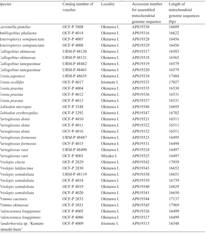
TABLE 1. Accession numbers and lengths of mitochondrial genome sequences analyzed in this study.
Species Catalog number of
voucher
Locality Accession number for assembled mitochondrial genome sequence
Length of mitochondrial genome sequences (bp)
Larsonella pumilus OCF-P 3808 Okinawa I. AP019338 16609
Amblygobius phalaena OCF-P 4014 Okinawa I. AP019316 16622
Asterropteryx semipunctata OCF-P 4007 Okinawa I. AP019328 16456
Asterropteryx semipunctata OCF-P 4008 Okinawa I. AP019329 16456
Callogobius okinawae URM-P 48120 Okinawa I. AP019317 16503
Callogobius okinawae URM-P 48121 Okinawa I. AP019318 16565
Callogobius tanegasimae URM-P 48462 Okinawa I. AP019319 16579
Callogobius tanegasimae URM-P 48463 Okinawa I. AP019320 16579
Eviota japonica URM-P 48635 Okinawa I. AP019334 17484
Eviota ocellifer OCF-P 4017 Iriomote I. AP019333 17027
Eviota prasina OCF-P 4004 Okinawa I. AP019335 16530
Eviota prasina OCF-P 4012 Okinawa I. AP019336 16531
Eviota prasina OCF-P 4013 Okinawa I. AP019337 16531
Gobiodon micropus OCF-P 3248 Okinawa I. AP019346 16695
Gobiodon erythrospilus OCF-P 3292 Okinawa I. AP019347 16702
Parioglossus dotui OCF-P 4010 Okinawa I. AP019321 16511
Parioglossus dotui OCF-P 4011 Okinawa I. AP019322 16511
Parioglossus dotui OCF-P 4016 Okinawa I. AP019332 16511
Parioglossus formosus URM-P 48487 Okinawa I. AP019323 16495
Parioglossus formosus OCF-P 4015 Okinawa I. AP019331 16494
Parioglossus raoi URM-P 48490 Okinawa I. AP019324 16497
Parioglossus raoi OCF-P 4003 Miyako I. AP019325 16497
Priolepis cincta OCF-P 2829 Okinawa I. AP019342 17059
Priolepis latifascima OCF-P 2830 Okinawa I. AP019343 16652
Priolepis semidoliata URM-P 48119 Okinawa I. AP019330 16651
Priolepis semidoliata OCF-P 4018 Okinawa I. AP019339 16739
Priolepis semidoliata OCF-P 4019 Okinawa I. AP019340 16829
Priolepis semidoliata OCF-P 4020 Okinawa I. AP019341 16650
Trimma caesiura OCF-P 2833 Okinawa I. AP019344 17137
Trimma okinawae OCF-P 3851 Okinawa I. AP019345 17969
Valenciennea longipinnis OCF-P 4005 Okinawa I. AP019326 16499
Valenciennea longipinnis OCF-P 4006 Okinawa I. AP019327 16499
Vanderhorstia sp. ‘Komon- yatsushi-haze’
OCF-P 4009 Iriomote I. AP019315 16548
Larsonella pumilus (Larson & Hoese, 1980) [New Japanese name: Yuuna-haze]
(Figs. 1–4; Table 2)
Lubricogobius pumilus Larson & Hoese, 1980: 41 (type locality: Indian Ocean, 3°25’N 47°14.8’E, 37–38 m depth).
Larsonella pumilus (Larson & Hoese, 1980): Randall & Senou 2001: 11.
Material examined. OCF-P 3808, 21.6 mm SL, East China Sea off Seragaki, Onna Village, Okinawa, Japan, 11 August 2017.
FIGURE 1. Larsonella pumilus, OCF-P 3808 (21.6 mm in standard length), in life (A), fresh (B), and after preservation (C).
FIGURE 2. Three-dimensional images of the head of Larsonella pumilus reconstructed from microcomputed tomography data.
A, frontal view, showing the outermost teeth. B, mid-sagittal view of the right side (cut along section x-x’ in A), showing inner teeth without a part of ceratohyal and 5thbranchiostegal ray to observe teeth easily. Red, premaxilla and outermost teeth on the premaxilla; blue, dentary and outermost teeth on the dentary. Triangles, vestiges of missing teeth. Scale bars, 1 mm.
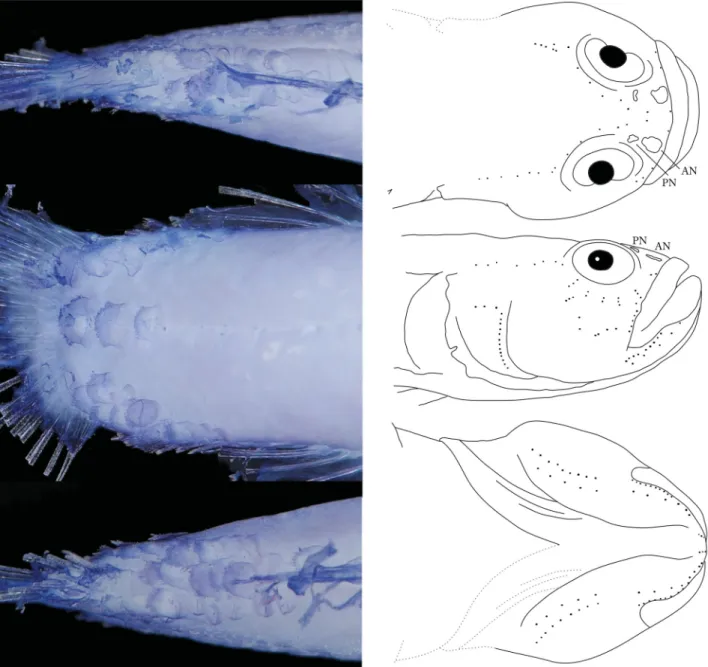
FIGURE 3. Picture of the caudal peduncle, stained with cyanine-blue solution to show scale arrangements (left) and schematic illustrations of cephalic sensory papillae (right) of Larsonella pumilus (OCF-P 3808, 21.6 mm SL) in dorsal (top), lateral (mid- dle), and ventral (bottom) views. AN and PN, anterior and posterior nares, respectively. Dotted lines, the probable undamaged states of the damaged parts.
Description. Counts and measurements are shown in Table 2. Head depressed, trunk nearly cylindrical, and tail compressed. Eyes located dorsolaterally. Mouth strongly oblique with angle to body axis about 60 degrees. Lower jaw protruding beyond upper jaw. Posterior end of upper jaw reaching below middle point between anterior margin of iris and anterior margin of pupil. Canine-like teeth aligned on edges of anterior halves of premaxilla and dentary;
four and three teeth on one side of premaxilla and dentary, respectively; posterior teeth larger (Fig. 2A). An inner row of conical teeth extending from anterior part to more posterior part of dentary than the outer canine-like teeth row (Fig. 2B). Additional small conical teeth observed on inner parts of premaxilla and dentary with a stereomicro- scope, but no such small teeth with micro-CT.
First dorsal fin with six spines. Second dorsal fin with one spine and nine soft rays. First and second dorsal fins connected by a low membrane behind last spine of the first dorsal fin. Anal fin with one spine and eight soft rays.
Caudal fin rounded, with 17 segmented rays. Pectoral fin with 18 soft-rays. Pelvic fin with one spine and five soft rays. Posterior tips of pectoral fins reaching or exceeding position of anus, according to a photograph taken in life
(Fig. 1A), although they are broken in the preserved specimen. Pelvic fins without frenum. Vertebrae 10 + 16 = 26;
dorsal-fin pterygiophore formula 3-22110; epural 1; anal-fin pterygiophores anterior to first haemal spine 2.
Head and body largely naked except for posterior part of caudal peduncle, involving three rows composed of 7–10 ctenoid scales along dorsal midline. A row of three ctenoid scales along lateral midline, and three rows com- posed of 6–8 ctenoid scales along ventral midline (Fig. 3). No sensory canals or associated pores on head. Cephalic sensory papillae patterns illustrated in Fig. 3. Infraorbital area with five transverse rows of sensory papillae.
Color in preservative (Fig. 1C): Background of head and tail white, trunk yellowish white. All fin membranes transparent. Melanophores scattered on dorsal half of trunk and on membranes of first and second dorsal, anal, and pelvic fins. Pectoral fin also with a few melanophores. Three rows of tiny melanophores arched between right and left eyes (Fig. 4A). Infraorbital area with two transverse rows of tiny melanophores (Fig. 4B) and another row of tiny melanophores behind eyes (Fig. 4B). These melanophore rows arranged radially around eyes.
FIGURE 4. Pigmentations around the eyes of Larsonella pumilus (OCF-P 3808, 21.6 mm SL) in dorsal (left) and lateral (right) views. Photos were taken after preservation.
FIGURE 5. Underwater photograph (A) and a dried shell (B and C) of Xenophora chinensis. A, Collecting the shell in which Larsonella pumilus may have taken refuge, using a Remotely Operated Vehicle at a depth of 214 m in the East China Sea off Seragaki, Onna Village, Okinawa, Japan. B (dorsal view) and C (ventral view) are likely the same shell as A. Scale bars, 10 mm.
Color in life (Fig. 1A): Body and all fins yellow or yellowish orange. Arrangement of melanophores same as after preservation, described above (Fig. 1A).
Habitat. We collected ten specimens of five invertebrate species from the muddy bottom at a depth of 209–220 m using the ROV, on 11 August 2017. These included five live comb jellies, Lyrocteis imperatoris, a sea cucumber, Holothuria dura, a starfish, Asterodiscides japonicus, a heart urchin, Pericosmus sp., and an empty shell of Xe- nophora chinensis (Fig. 5). After these animals were put into a tank on the boat, the L. pumilus specimen was found
in the tank. Because Lubricogobius species, close relatives of Larsonella pumilus, often inhabit empty shells, sea urchin tests, tunicate siphons, bottles, etc. (Randall & Senou 2001; Allen & Erdmann 2016), we believe that the L.
pumilus was inside the empty shell of X. chinensis (collected at a depth of 214 m) and was collected with it.
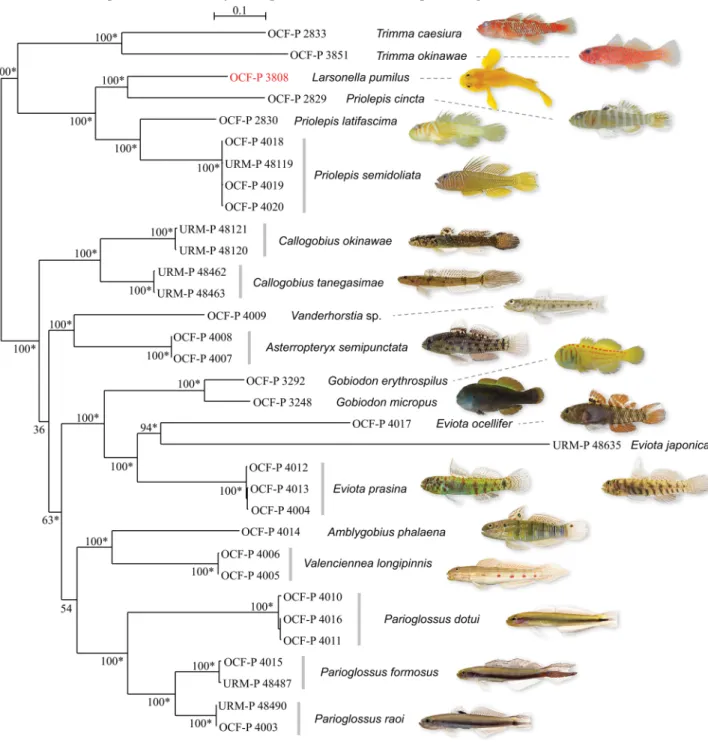
Mitochondrial DNA analysis. We succeeded in assembling the entire mitochondrial genomes of Larsonella pumilus and 19 related species (Table 1). In the phylogenetic tree, using 15559 bp of aligned mitochondrial genomes (Fig. 6), most nodes, including L. pumilus, were supported by high bootstrap values (100%) and bayesian posterior probabilities (1), indicating that L. pumilus was placed in a clade including Priolepis spp. and Trimma spp., while Gobiodon spp. was placed in another clade with Callogobius spp., Vanderhorstia sp., Asterropteryx semipunc- tata, Eviota spp., Amblygobius phalaena, Valenciennea longipinnis, and Parioglossus spp. Larsonella pumilus was paired with Priolepis cincta, and they were placed within the Priolepis lineage.
FIGURE 6. Molecular phylogeny of Larsonella pumilus and 19 related goby species from the Ryukyu Archipelago. Tree topol- ogy and branch lengths were obtained from maximum likelihood analysis of aligned mitochondrial genomes (15559 bp) with 100 bootstraps based on the GTR+I+Gamma model. The scale bar indicates 0.1 substitutions per site. Numbers on major nodes represent ML bootstrap support. Bayesian posterior probabilities are summarized as asterisks for values equal to 1.
TABLE 2. Counts and measurements of Larsonella pumilus. Values of the holotype are based on Larson & Hoese (1980).
D1, first dorsal fin; D2, second dorsal fin; A, anal fin; C, caudal fin; P1, pectoral fin; P2, pelvic fin; PO, preopercular.
Holotype
OCF-P 3808 ZMH 6165
Standard length (mm) 21.6 14.5
Counts
D1 VI VI
D2 I, 9 I, 9
A I, 8 I, 8
C (segmented rays) 17 17
P1 18 17
P2 I, 5 -
Measurements as % of standard length
Head length 31.9 34.5
Head depth at PO margin 20.4 24.1
Head width at PO margin 27.8 28.3
Snout length 7.4 9.7
Eye diameter 8.8 6.9
Upper jaw length 15.3 15.2
Body depth at P2 origin 21.8 -
Body depth at A origin 18.1 18.6
Caudal peduncle depth 13.9 13.8
Caudal peduncle length 19.0 20.7
Predorsal length 39.8 -
Preanal length 65.7 -
Length of D1 base 19.9 -
Length of D2 base 23.6 23.4
Length of A base 19.4 -
Discussion
Identification. The present specimen was identified as Larsonella pumilus (Larson & Hoese, 1980) as its morpho- logical characters correspond almost exactly to those of the holotype described by Larson & Hoese (1980), although some characters, including fin morphology and nare shapes could not be confirmed due to their damaged condition.
The only major difference found was the presence of melanophores. Larson & Hoese (1980) noted “no traces of dark pigment”, while our specimen has melanophores arranged radially around eyes and scattered on trunk and fins.
Because these melanophores were very small and invisible to the naked eye, we suppose that the melanophores were overlooked in the original description. It is also possible that melanophores vary individually or depending on body size and/or sex (we could not determine the sex of our specimen).
Distribution and habitat. There have been few records of this species since the holotype of Lubricogobius pumilus was collected from the Indian Ocean off Somalia using an Agassiz trawl at a depth of 37–38 m (Larson &
Hoese 1980). Other available records from the Indian Ocean are from La Digue in the Seychelles (4°23’S 55°49’E) from dredging at a depth of 30 m (Randall & van Egmond 1994; BPBM 35515 and ROM 66149; as L. pumilis) and from the northwest shelf of Australia (16°49’S to 19°52’S) (Hoese & Larson 2006). This species also appeared in a list of marine and brackish water goby species in the western central Pacific (Larson & Murdy 2001), but detailed locality information was not provided. According to the GBIF database (GBIF Secretariat 2017), L. pumilus was also collected in New Caledonia and Tonga (69 and 67 m in depth, respectively). The specimen reported in the pres- ent study was caught at a depth of 214 m off Okinawa Island. This is the first record of L. pumilus from Japan and the only report substantiated with a voucher from the Pacific Ocean. The depth is far greater than other records (214 m vs. 30–69 m). Thus, the main habitat of L. pumilus may be deeper sea than previously recognized. This may also
explain the paucity of records since this area has been poorly explored. It also suggests that this species is distributed much more widely in the Indo-Pacific.
Phylogenetic placement. Among their hypothesized lineages based on molecular data, morphological charac- ters, and literature review, Thacker (2015) placed the genus Larsonella into the “Gobiodon lineage (sensu Agorreta et al. 2013)” with the genera Bryaninops, Eviota, Gobiodon, etc. However, our results clearly demonstrate that L.
pumilus belongs to the “Priolepis lineage (sensu Agorreta et al. 2013)” with Priolepis spp. and Trimma spp., not to the “Gobiodon lineage” (Fig. 6). Our tree also indicates that the genus Priolepis is not monophyletic, as L. pumilus nested within a clade composed of three species of Priolepis. But we could analyze only three of 35 species in Prio- lepis and no Lubricogobius species were involved in the present study, although L. pumilus is considered to be a close relative of Lubricogobius (Hoese & Larson 2010). Hoese & Larson (2010) doubted the validity of Larsonella based on the fact that a species of Lubricogobius actually has scales on its caudal peduncle, which is one of the ma- jor diagnostic characters to distinguish Larsonella from Lubricogobius. The mitochondrial genome sequence of L.
pumilus included in the present study will help future research to review phylogenetic relationships among species in the genera Priolepis, Lubricogobius, and Larsonella.
Remarks. Larsonella pumilus is mentioned as “Larsonella pumila” in some online databases (e.g. Fricke et al.
2018; Froese & Pauly 2018), although no author spelled it this way. This species was originally described as a new species in Lubricogobius and the specific epithet is considered as a noun as the authors stated “Derivation of name:
Latin – pumilus = dwarf fish” (Larson & Hoese 1980). Therefore, the original spelling of the specific epithet is to be retained with gender ending unchanged, even if gender of the generic name is changed. Although the specific epithet of this species is spelled “pumilis” in figure 536 of Larson & Hoese (1980) and Randall & van Egmond (1994), this is also a misspelling.
Acknowledgements
We are grateful to Akira Kawaguchi (University of the Ryukyus), Taketeru Tomita, and Kei Miyamoto (Okinawa Churashima Foundation Research Center) for microcomputed tomography scans at the Department of Human Bi- ology and Anatomy, Graduate School of Medicine, University of the Ryukyus. We wish to thank Helen K. Larson (NTM), Toshiaki Hirobe (Marine Dream, Onna, Okinawa), and Yoshihiro Fujiwara (JAMSTEC) for valuable in- formation, Steven D. Aird (OIST) for editing the manuscript, Kanako Hisata and members of Marine Genomics Unit (OIST) for their kind support of our study, the staff of the Deep-Sea Animal Section of Okinawa Churaumi Aquarium for sampling and for maintaining the specimen, and Motobu Fisheries Cooperative Association for its kind cooperation. Computations were partially performed on the NIG supercomputer at ROIS National Institute of Genetics. This research was partially funded by an OIST internal grant to the Marine Genomics Unit (Noriyuki Satoh).
References
Agorreta, A., San Mauro, D., Schliewen, U., Van Tassell, J.L., Kovačić, M., Zardoya, R. & Ruber, L. (2013) Molecular phy- logenetics of Gobioidei and phylogenetic placement of European gobies. Molecular Phylogenetics and Evolution, 69, 619–633.
https://doi.org/10.1016/j.ympev.2013.07.017
Allen, G.R. & Erdmann, M.V. (2016) Lubricogobius tunicatus, a new species of goby (Pisces: Gobiidae) from Papua New Guinea and the first record of L. ornatus from the East Indies. Journal of the Ocean Science Foundation, 24, 24–34.
Baldwin, C.C., Tornabene, L. & Robertson, D.R. (2018) Below the Mesophotic. Scientific Reports, 8, 4920.
https://doi.org/10.1038/s41598-018-23067-1
Birdsong, R.S., Murdy, E.O. & Pezold, F.L. (1988) A study of the vertebral column and median fin osteology in gobioid fishes with comments on gobioid relationships. Bulletin of Marine Science, 42, 174–214.
Capella-Gutiérrez, S., Silla-Martínez, J.M. & Gabaldón, T. (2009) trimAl: a tool for automated alignment trimming in large- scale phylogenetic analyses. Bioinformatics, 25, 1972–1973.
https://doi.org/10.1093/bioinformatics/btp348
Bianchi, G., Carpenter, K.E., Roux, J.-P., Molloy, F.J., Boyer, D. & Boyer, H.J. (Eds.) (1999) FAO Species Identification Field Guide for Fishery Purposes. The Living Marine Resources of Namibia. FAO, Rome, 256 pp.
Darriba, D., Posada, D., Kozlov, A.M., Stamatakis, A., Morel, B. & Flouri, T. (2019) ModelTest-NG: a new and scalable tool for
the selection of DNA and protein evolutionary models. Molecular Biology and Evolution, msz189. [published]
https://doi.org/10.1093/molbev/msz189
Eschmeyer, W.N. & Herald, E.S. (1983) A field guide to Pacific Coast fishes of North America from the Gulf of Alaska to Baja California. Houghton Mifflin Company, Boston, 336 pp.
Fricke, R., Eschmeyer, W.N. & van der Laan, R. (Eds.) (2018) Catalog of fishes: genera, species, references. Available from:
http://researcharchive.calacademy.org/research/ichthyology/catalog/fishcatmain.asp (accessed 19 September 2018) Froese, R. & Pauly, D. (Eds.) (2018) FishBase. World Wide Web electronic publication. Version (06/2018). Available from:
http://www.fishbase.org (accessed 19 September 2018)
GBIF Secretariat (2017) GBIF Backbone Taxonomy. Checklist dataset. Available from: https://doi.org/10.15468/39omei (ac- cessed 30 August 2018)
Goren, M. (1992) Obliquogobius turkayi, a new species of gobiid fish from the deep water of the central Red Sea. Senckenber- giana Maritima, 22 (3/6), 265–270.
Goren, M. & Baranes, A. (1995) Priolepis goldshmidtae (Gobiidae), a new species from the deep water of the northern Gulf of Aqaba, Red Sea. Cybium, 19, 343–347.
Greenfield, D.W. (2002) Palatogobius grandoculus: a new deep-water western Caribbean goby (Teleostei: Gobiidae). Copeia, 2002, 716–718.
https://doi.org/10.1643/0045-8511(2002)002[0716:PGANDW]2.0.CO;2
Hoese, D.F. & Larson, H.K. (2006) Gobiidae. In: Beesley P.L. & Wells A. (Eds.), Zoological catalogue of Australia. Vol. 35.
ABRS and CSIRO Publishing, Canberra, pp. 1612–1697.
Hoese, D.F. & Larson, H.K. (2010) Description of two new species of the genus Priolepis from the Indo-Pacific with redescrip- tion of Priolepis profunda and Priolepis psygmophilia. Ichthyological Research, 57, 373–388.
https://doi.org/10.1007/s10228-010-0170-6
Katoh, K. & Standley, D.M. (2013) MAFFT multiple sequence alignment software version 7: Improvements in performance and usability. Molecular Biology and Evolution, 30, 772–780.
https://doi.org/10.1093/molbev/mst010
Larson, H.K. & Hoese, D.F. (1980) Fische des Indischen Ozeans. Ergebnisse der ichthyologischen Untersuchungen während der Expedition des Forschungsschiffes “Meteor” in den Indischen Ozean, Oktober 1964 bis Mai 1965. A. Systematischer Teil, XXIII. Gobiidae. Meteor Forschungsergebnisse, Reihe D, Biologie, 32, 33–43.
Larson, H.K. & Murdy, E.O. (2001) Families Eleotridae, Gobiidae. In: Carpenter, K.E. & Niem, V.H. (Eds.), Species identifica- tion guide for fishery purposes. The living marine resources of the western central Pacific. Vol. 6. Bony fishes part 4 (Lab- ridae to Latimeriidae), estuarine crocodiles, sea turtles, sea snakes and marine mammals. FAO, Rome, pp. 3574–3603.
Murdy, E.O. (2002) Karsten: a new genus of eel goby (Gobiidae: Amblyopinae) with a key to “Trypauchen” group genera.
Copeia, 2002, 787–791.
https://doi.org/10.1643/0045-8511(2002)002[0787:KANGOE]2.0.CO;2
Mytilineou, C., Politou, C.-Y., Papaconstantinou, C., Kavadas, S., D’Onghia, G. & Sion, L. (2005) Deep-water fish fauna in the Eastern Ionian Sea. Belgian Journal of Zoology, 135, 229–233.
Nakabo, T. (2002) Fishes of Japan with pictorial keys to the species. English Edition. Tokai University Press, Tokyo, 1749 pp.
Nelson, J.S. (2006) Fishes of the world, fourth edition. John Wiley & Sons, Inc., Hoboken, 601 pp.
Nelson, J.S., Grande, T.C. & Wilson, M.V.H. (2016) Fishes of the world. 5th Edition. John Wiley & Sons, Inc., Hoboken, 707 pp.
https://doi.org/10.1002/9781119174844
Patzner, R.A., van Tassell, J.L., Kovačić, M. & Kapoor, B.G. (2011) The biology of gobies. Science Publishers, New Hampshire, 685 pp.
https://doi.org/10.1201/b11397
Peng, Y., Leung, H.C., Yiu, S.M. & Chin, F.Y. (2012) IDBA-UD: a de novo assembler for single-cell and metagenomic sequenc- ing data with highly uneven depth. Bioinformatics, 28, 1420–1428.
https://doi.org/10.1093/bioinformatics/bts174
Randall, J.E. & van Egmond, J. (1994) Marine fishes from the Seychelles: 108 new records. Zoologische Verhandelingen Leiden, 297, 43–83.
Randall, J.E. & Senou, H. (2001) Review of the Indo-Pacific gobiid fish genus Lubricogobius, with description of a new species and a new genus for L. pumilus. Ichthyological Research, 48, 3–12.
https://doi.org/10.1007/s10228-001-8111-z
Ronquist, F., Teslenko, M., van der Mark, P., Ayres, D.L., Darling, A., Höhna, S., Larget, B., Liu, L., Suchard M.A. & Huelsen- beck, J.P. (2012) MrBayes 3.2: Efficient Bayesian phylogenetic inference and model choice across a large model space.
Systematic Biology, 61, 539–542.
https://doi.org/10.1093/sysbio/sys029
Sauberer, M., Iwamoto, T. & Ahnelt, H. (2018) Two new deep-water species of the genus Thorogobius (Teleostei: Gobi- idae) from the upper continental slope of the Eastern Central Atlantic. Zootaxa, 4429 (2), 357–371.
https://doi.org/10.11646/zootaxa.4429.2.10
Sabaj, M.H. (2016) Standard symbolic codes for institutional resource collections in herpetology and ichthyology: an Online Reference. Version 6.5. American Society of Ichthyologists and Herpetologists, Washington, D.C. Available from: http://
asih.org/standard-symbolic-codes/about-symbolic-codes (accessed 30 August 2018)
Shibukawa, K. & Aonuma, Y. (2007) Three new species of the deep-dwelling goby genus Obliquogobius (Teleostei: Gobi- idae: Gobiinae) from Japan, with comments on the limits of the genus. Bulletin of the National Science Museum Series A (Zoology) Supplent1, 137–142.
Shinohara, G., Endo, H., Matsuura, K., Machida, Y. & Honda, H. (2001) Annotated checklist of the deepwater fishes from Fishes from Tosa Bay, Japan. National Science Museum Monographs, 20, 283–343.
Stamatakis, A. (2014) RAxML version 8: a tool for phylogenetic analysis and post-analysis of large phylogenies. Bioinformat- ics, 30, 1312–1313.
https://doi.org/10.1093/bioinformatics/btu033
Thacker, C.E. (2015) Biogeography of goby lineages (Gobiiformes: Gobioidei): origin, invasions and extinction throughout the Cenozoic. Journal of Biogeography, 42, 1615–1625.
https://doi.org/10.1111/jbi.12545
Thacker, C.E. & Roje, D.M. (2011) Phylogeny of Gobiidae and identification of gobiid lineages. Systematics and Biodiversity, 9, 329–347.
https://doi.org/10.1080/14772000.2011.629011
Tornabene, L., Van Tassell, J.L., Gilmore, R.G., Robertson, D.R., Young, F. & Baldwin, C.C. (2016) Molecular phylogeny, analysis of character evolution, and submersible collections enable a new classification for a diverse group of gobies (Teleostei: Gobiidae: Nes subgroup), including nine new species and four new genera. Zoological Journal of the Linnean Society, 177, 764–812.
https://doi.org/10.1111/zoj.12394