九州大学学術情報リポジトリ
Kyushu University Institutional Repository
HuC/D expression in small round cell tumors and neuroendocrine tumors: a useful tool for
distinguishing neuroblastoma from childhood small round cell tumors
武本, 淳吉
http://hdl.handle.net/2324/2236082
出版情報:九州大学, 2018, 博士(医学), 課程博士 バージョン:
権利関係:
Original contribution
HuC/D expression in small round cell tumors and neuroendocrine tumors: a useful tool for
distinguishing neuroblastoma from childhood small round cell tumors ☆
Junkichi Takemoto MD
a,b, Masaaki Kuda MD, PhD
c, Kenichi Kohashi MD, PhD
a, Yuichi Yamada MD, PhD
a, Yutaka Koga MD, PhD
a, Izumi Kinoshita MD
a,
Ryota Souzaki MD, PhD
b, Tomoaki Taguchi MD, PhD
b, Yoshinao Oda MD, PhD
a,⁎
aDepartment of Anatomic Pathology, Graduate School of Medical Sciences, Kyushu University, Fukuoka 812-8582, Japan
bDepartment of Pediatric Surgery, Graduate School of Medical Sciences, Kyushu University, Fukuoka 812-8582, Japan
cDepartment of Digestive and General Surgery, Graduate School of Medicine, University of Ryukyus, Okinawa 903-0215, Japan
Received 19 September 2018; revised 30 October 2018; accepted 2 November 2018
Keywords:
Neuroblastoma;
HuC/D;
PHOX2B;
Tyrosine hydroxylase;
Immunohistochemistry;
Differential diagnosis
SummaryThe RNA-binding protein HuC/D displays a neuron-specific expression and is involved in neu- ronal differentiation and the maintenance of the nervous system. Here we investigated the diagnostic value of HuC/D in neuroblastomas. We evaluated 85 neuroblastic tumors: 81 neuroblastomas; 3 ganglioneuro- blastomas, intermixed; 1 ganglioneuroma, maturing; and 101 other tumors consisting of 34 Ewing sarco- mas, 14 nephroblastomas, 11 rhabdomyosarcomas, 15 pulmonary small cell carcinomas, 18 pancreatic neuroendocrine tumors, and 9 pheochromocytomas. Immunohistochemistry for HuC/D, PHOX2B, and ty- rosine hydroxylase was performed. The immunoreactivity for HuC/D was semiquantified using the total score (TS; range, 0-8). HuC/D positivity was defined as a TS≥6. The TS of the neuroblastic tumors (mean TS, 7.94) was significantly higher than those of the other small round cell tumors and neuroendocrine tu- mors (Pb.001) except for the pheochromocytomas (mean TS, 6.89;P= .074). HuC/D was positive in all 85 neuroblastic tumors, 1 (2.9%) Ewing sarcoma, 1 (6.7%) pulmonary small cell carcinoma, and 8 (89%) pheochromocytomas. PHOX2B was positive in all of the neuroblastic tumors and pheochromocyto- mas. Tyrosine hydroxylase was positive in 80 (94%) neuroblastic tumors, 1 (9.1%) rhabdomyosarcoma, and all of the pheochromocytomas. Therefore, HuC/D serves as a highly sensitive diagnostic marker to distin- guish neuroblastomas from other small round cell tumors. The combination of HuC/D and PHOX2B stain- ing may be valuable for the diagnosis of neuroblastic tumors, especially in the assessment of small sections.
HuC/D expression in tumors may be related to catecholamine production or a neural crest–derived cell or- igin.
© 2018 Elsevier Inc. All rights reserved.
☆ Competing interests: The authors declare that there are no conflicts of interest to disclose.
⁎ Corresponding author at: Department of Anatomic Pathology, Pathological Sciences, Graduate School of Medical Sciences, Kyushu University, Maidashi 3-1- 1, Higashi-ku, Fukuoka 812-8582, Japan.
E-mail address:[email protected](Y. Oda).
www.elsevier.com/locate/humpath
https://doi.org/10.1016/j.humpath.2018.11.004 0046-8177/© 2018 Elsevier Inc. All rights reserved.
1. Introduction
Neuroblastomas are small round cell tumors that arise mostly in the adrenal glands and sympathetic nervous system. They are the most common extracranial solid tumor in children. Sponta- neous regression and maturation to a benign ganglioneuroma are the characteristic features of neuroblastomas[1-5], and it is also known that chemotherapy leads to the maturation of neuroblastoma cells. Neuroblastoma cells are typically posi- tive for neural antibodies (eg, neuron-specific enolase, synap- tophysin, chromogranin A, NB84, and S-100 protein)[6,7], but these antibodies are not specific to neuroblastoma[8,9].
Tyrosine hydroxylase (TH) was the most widely used sym- pathoadrenal marker specific for neuroblastoma, but its sensi- tivity was insufficient (84%)[10,11]. The protein known as paired-like homeobox 2b (PHOX2B) has recently attracted at- tention as a sensitive and specific marker for neuroblastoma.
HuC/D, a member of the Hu family, is an RNA-binding protein that displays a neuron-specific expression and is in- volved in neuronal differentiation and the maintenance of the nervous system[12,13]. It has also been reported that HuC/
D is a credible pan-neuronal marker for the enteric nervous system neurons[14,15]. However, there are few published studies about HuC/D expression in neuroblastoma.
We conducted the present study to determine the HuC/D ex- pression along with PHOX2B and TH expressions in neuroblas- tomas, small round cell tumors, and neuroendocrine tumors, and we discuss the potential utility of HuC/D expression as a differ- ential diagnostic tool in neuroblastoma.
2. Materials and methods
2.1. Patients and tissue samplesThis study was approved by the Ethics Committee of Kyu- shu University (no. 29-429). We examined 85 paraffin-embed- ded samples from neuroblastic tumor tissue registered in the Department of Anatomic Pathology, Graduate School of Med- ical Sciences, Kyushu University, between 1996 and 2015.
Clinicopathological data were obtained from the patients' med- ical charts. We also stained (as comparison samples) other small round cell tumors and neuroendocrine tumors, comprising 34 Ewing sarcomas, 14 nephroblastomas, 11 rhabdomyosarcomas (5 alveolar rhabdomyosarcomas and 6 embryonal rhabdomyo- sarcomas), 15 pulmonary small cell carcinomas, 18 pancreatic neuroendocrine tumors, and 9 pheochromocytomas.
One Ewing sarcoma case and 1 pulmonary small cell carci- noma case were excluded from the analyses of PHOX2B and TH expression because of a shortage of tissue samples.
2.2. Histologic review
In all cases, the diagnosis was based on a light microscopic examination with hematoxylin-eosin staining according to the most recent World Health Organization classification[16]. In
addition, immunoperoxidase procedures were performed using the streptavidin-biotin peroxidase method (Histofine;
Nichirei Biosciences, Tokyo, Japan) or the universal immuno- peroxidase polymer method (Envision-kit; Dako Japan, To- kyo, Japan) when necessary. Each sample was from a different patient.
2.3. Immunohistochemistry
The immunohistochemical study was performed using the streptavidin-biotin-peroxidase method. The primary antibod- ies used in this study were anti-HuC/HuD neuronal protein (mouse monoclonal, clone 16A11; A21271; dilution 1:400;
Life Technologies, Carlsbad, CA), anti-PHOX2B antibody (rabbit monoclonal, clone EPR14423; no. ab183742; dilution 1:500; Abcam, Cambridge, MA), and anti-TH antibody (rabbit polyclonal; no. ab112; dilution 1:500; Abcam). Ganglion cells from small intestine were used as a positive control.
Three pathologists (J. T., M. K., and K. K.) evaluated the stained sections according to the following criteria. The inten- sity of HuC/D nuclear staining (intensity score [IS]) was scored from 0 to 3 as follows: 0, none; 1, weak; 2, moderate;
and 3, strong. The extent of staining (proportion score [PS]) was graded as follows: 0, absent; 1, b1%; 2, 1%–9%; 3, 10% to 32%; 4, 33% to 66%; and 5,N66% of tumor cells.
The total score (TS) was calculated by adding the IS and PS (IS + PS), so that the possible scores ranged from 0 to 8 [17]. HuC/D positivity was defined as a TS≥6.
The extent of PHOX2B and TH immunoreactivity was graded according to the percentage of positive tumor cells as follows: 0, absent; 1+, b5%; 2+, 5% to 50%; 3+, 51% to 75%; and 4+, 76% to 100% of tumor cells. Positivity for PHOX2B and TH was defined as grade≥2+[18].
2.4. Statistical analysis
Categorical variables were compared using the Pearsonχ2 est or Fisher exact test as appropriate. Statistical significance was defined as Pb.05. All analyses were performed with the JMP Pro statistical software package (ver. 13.1.0; SAS In- stitute, Cary, NC).
3. Results
3.1. Clinicopathological characteristics
The clinicopathological features of the patients with neuro- blastic tumors and the comparative tumors are summarized in Tables 1and2. In the group of neuroblastic tumor cases, all 85 patients were children (median age, 0 years; range, 0-8 years).
3.2. HuC/D expression
The results of the immunohistochemical analysis are sum- marized inTable 3. In regard to extent of staining, 98% (83/
163 HuC/D expression in neuroblastoma
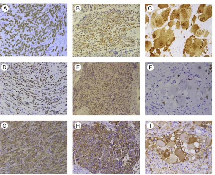
85) of the neuroblastic tumors showed a PS value of 5 (Figure A). In these cases, the percentage of positive tumor cells was almost 100%. Two (2%) tumors, which were the undifferenti- ated subtype, showed a PS of 4 (FigureB). In the ganglioneur- oblastomas and ganglioneuroma, immature and mature ganglion cells were positive for nuclear and cytoplasmic stain- ing, but Schwann cells were negative (FigureC).
In contrast, none of the Ewing sarcomas (Supplementary Fig. 1A), nephroblastomas (Supplementary Fig. 1B), rhabdo- myosarcomas (Supplementary Fig. 1C), pulmonary small cell carcinomas (Supplementary Fig. 1D), and pancreatic neuroen- docrine tumors (Supplementary Fig. 1E) showed a PS of 5 for
HuC/D immunoreactivity. The remaining pheochromocyto- mas had a PS 5 in 8 of the 9 cases (89%; Supplementary Fig. 1F).
From the viewpoint of intensity, 96% (82/85) of the neuro- blastic tumors, 9% (3/34) of the Ewing sarcomas, and 33% (3/
9) of the pheochromocytomas had the IS value of 3. All of the nephroblastomas, rhabdomyosarcomas, pulmonary small cell carcinomas, and pancreatic neuroendocrine tumors had an IS b3.
The TS values were significantly higher in the neuro- blastomas compared with the other small round cell tumors and neuroendocrine tumors (Pb.001), except for the pheo- chromocytomas (P= .074). Regarding the immunohistochem- ical positivity of HuC/D, all 85 (100%) neuroblastomas, 1 (2.9%) of the 34 Ewing sarcomas, none of 14 nephroblasto- mas, and none of the 11 rhabdomyosarcomas were interpreted as positive. The single HuC/D-positive Ewing sarcoma showed PS 3 and IS 3. Among the neuroendocrine tumors, 8 (89%) of the 9 pheochromocytomas, 1 (6.7%) of the 15 pul- monary small cell carcinomas, and none of the 18 pancreatic neuroendocrine tumors were positive for HuC/D.
3.3. PHOX2B and TH expression
The immunohistochemical results for PHOX2B and TH expression are summarized inTable 4. All 85 of the neuroblas- tic tumors (FigureD-F) and all 9 of the pheochromocytomas were positive for PHOX2B. In contrast, PHOX2B expression was completely absent in the Ewing sarcomas, nephroblasto- mas, rhabdomyosarcomas, pulmonary small cell carcinomas, and pancreatic neuroendocrine tumors.
Eighty (94%) of the 85 neuroblastic tumors (FigureG-I), 1 of the 11 rhabdomyosarcomas, 3 of the 18 pancreatic NETs, and all 9 of the pheochromocytomas were positive for TH.
In the Ewing sarcomas, nephroblastomas, pulmonary small cell carcinomas, and pancreatic neuroendocrine tumors, the TH expression was 0 or 1+.
4. Discussion
In this study, we evaluated the immunohistochemical HuC/
D expression profile in neuroblastic tumors, small round cell Table 1 Clinicopathological characteristics of neuroblastic
tumors
Characteristics n
Sex
Male 52
Female 33
Age (y)
b1.5 52
1.5≤,b5 30
5≤ 3
Site
Adrenal gland 45
Retroperitoneum 25
Others 15
Subtype
Neuroblastoma, undifferentiated 2
Neuroblastoma, poorly differentiated 78
Neuroblastoma, differentiating 1
Ganglioneuroblastoma 3
Ganglioneuroma, maturing 1
Stage
1 24
2A 12
2B 1
3 11
4 29
4s 3
Unknown 5
N-myc
Positive 17
Negative 68
Table 2 Clinicopathological characteristics of small round cell tumors and neuroendocrine tumors
Tumor type ES WT RMS SCLC Pancreatic NET PCC
n 34 14 11 15 18 9
Sex, n (%)
Male 13 (38) 8 (57) 3 (27) 12 (80) 5 (28) 2 (22)
Female 21 (62) 6 (43) 8 (73) 3 (20) 13 (72) 7 (78)
Age (y), mean (range) 17 (1-74) 1 (0-10) 2 (0-17) 74 (60-84) 59 (20-79) 56 (21-76)
Abbreviations: ES, Ewing sarcoma; NET, neuroendocrine tumor; PCC, pheochromocytoma; RMS, rhabdomyosarcoma; SCLC, small cell lung cancer (pul- monary small cell carcinoma); WT, Wilms tumor (nephroblastoma).
Figure Examples of HuC/D (A-C), PHOX2B (D-F), and TH (G-I) staining in the neuroblastic tumors. A, D, and G, Adrenal neuroblastoma, poorly differentiated in a 1-year-old boy. A, HuC/D (TS 8 [PS 5 + IS 3]). D, PHOX2B (4+). G, TH (4+). B, E, and H, Intra-abdominal undiffer- entiated neuroblastoma in a 2-year-old girl. B, HuC/D (TS 6 [PS 4 + IS 2]). E, PHOX2B (2+). H, TH (4+). C, F, and I, Adrenal ganglioneuroblas- toma, intermixed in a 4-year-old boy. C, HuC/D (TS 8 [PS 5 + IS 3]). F, PHOX2B (3+). I, TH (4+).
Table 3 Summary of HuC/D immunohistochemical total scoring
Tumor type NB Other small round cell tumors Other neuroendocrine tumors
ES WT RMS SCLC Pan-NET PCC
n 85 34 14 11 15 18 9
HuC/D positivea 85 1 0 0 1 0 8
(%) (100) (2.9) (0) (0) (6.7) (0) (89)
8 82 0 0 0 0 0 3
7 1 0 0 0 0 0 5
6 2 1 0 0 1 0 0
0-5 0 33 14 11 14 18 1
Mean TS 7.94 1.5 0.43 0.82 3.73 1.28 6.89
Abbreviations: ES, Ewing sarcoma; NB, neuroblastoma; Pan-NET, pancreatic neuroendocrine tumor; PCC, pheochromocytoma; RMS, rhabdomyosarcoma;
SCLC, small cell lung cancer (pulmonary small cell carcinoma); WT, Wilms tumor (nephroblastoma).
aHuC/D positivity was defined as the TS≥6.
165 HuC/D expression in neuroblastoma
tumors, and neuroendocrine tumors. The results showed that HuC/D was expressed more diffusely and strongly in the neu- roblastic tumors than in the other tumors, with the exception of the pheochromocytomas.
It is of note that HuC/D was reported to be a useful pan- neuronal marker in both central and enteric nervous system neurons[12-15]. Few studies have addressed the expression of Hu antigens in small round cell tumors. Dalmau et al et al [19], using frozen tumor tissue and anti-Hu IgG (prepared from the sera of patients with paraneoplastic sensory neuropa- thy/encephalomyelitis) for immunohistochemical and Western blot analyses, observed evidence of Hu expression in 4 of 8 neuroblastomas and both of 2 Ewing sarcomas. One rhabdo- myosarcoma was negative for Hu antigen[19]. In another study of neuroblastomas, Gultekin et al [20]found that 39 (78%) of 50 neuroblastomas expressed the Hu antigens. How- ever, HuC/D immunohistochemistry in small round cell tu- mors and neuroendocrine tumors has not been systematically conducted to investigate HuC/D as a diagnostic marker for neuroblastomas.
PHOX2B has been described as a sensitive and highly spe- cific immunohistochemical marker for peripheral neuroblastic tumors[21], including neuroblastoma, with 92% sensitivity and 100% specificity[18]. In our present study, HuC/D ex- pression was detected with diffuse (PS 4-5) staining in all 85 neuroblastic tumor cases and with strong (IS 3) staining in 82 (96%) cases; in addition, we observed positive HuC/D ex- pression in neuroblastic tumors with 100% sensitivity and 98% specificity among small round cell tumors (neuroblastic tumors, nephroblastomas, Ewing sarcomas, and rhabdomyo- sarcomas). The positive expression of PHOX2B and TH showed 100% and 94% sensitivity and 100% and 98% speci- ficity, respectively. In 8 (9%) cases, grade 2+ PHOX2B
expression was detected. Therefore, the evaluation of HuC/D immunoexpression may be a highly sensitive diagnostic tool to distinguish neuroblastomas from other pediatric small round cell tumors.
The histologic definitive diagnosis of neuroblastoma is sometimes challenging because of small specimens with sub- optimal histology, such as specimens of bone marrow, which is the most common site of infiltration in children presenting with metastatic disease at the time of diagnosis and also a fre- quent site of recurrences[22]. Burchill et al[23]stated that for bone marrow disease assessments in children with neuroblas- tomas, the value of immunohistochemistry depends on the quality of the sample and the specificity and sensitivity of the antibodies, and they encouraged the use of at least 2 anti- bodies. Thus, the combination of HuC/D and PHOX2B stain- ing may be especially valuable in the assessment of small sections.
Our study included 2 neuroblastomas of the undifferenti- ated subtype. These 2 cases were both TS 6 (PS 4 and IS 2), which is lower than the TS values of the neuroblastomas of the poorly differentiated subtype, 99% (77/78) of which were TS 8 (PHOX2B expression in these cases was 3+ and 2+, and TH expression was 4+ and 1+, respectively). It was reported that HuC/D may be used as a specific marker in progenitor cell cultures for the identification of“postmitotic”ganglion cells [12]. Wakamatsu and Weston[24]reported that Hu genes dis- played sequential expression during neuronal differentiation, and that HuC and HuD were not detected in apparently undif- ferentiated cell populations. These reports suggest the associa- tion between the differentiation of neuroblastoma cells and HuC/D expression, although the number of undifferentiated subtype cases in the present study was very limited. Further studies using a larger number of cases are required to confirm Table 4 Summary of PHOX2B and TH immunohistochemical scoring
Tumor type NB Other small round cell tumors Other neuroendocrine tumors
ES WT RMS SCLC Pan-NET PCC
n 85 33 14 11 14 18 9
PHOX2B
PHOX2B positivea 85 0 0 0 0 0 9
(%) (100) (0) (0) (0) (0) (0) (100)
4+ 67 0 0 0 0 0 8
3+ 10 0 0 0 0 0 1
2+ 8 0 0 0 0 0 0
0-1+ 0 33 14 11 14 18 0
TH
TH positivea 80 0 0 1 0 3 9
(%) (94) (0) (0) (9) (0) (17) (100)
4+ 70 0 0 0 0 0 9
3+ 2 0 0 0 0 0 0
2+ 8 0 0 1 0 3 0
0-1+ 5 33 14 10 14 15 0
Abbreviations: ES, Ewing sarcoma; NB, neuroblastoma; Pan-NET, pancreatic neuroendocrine tumor; PCC, pheochromocytoma; RMS, rhabdomyosarcoma;
SCLC, small cell lung cancer (pulmonary small cell carcinoma); WT, Wilms tumor (nephroblastoma).
a PHOX2B and TH positivity was defined as the score≥2+.
the utility of HuC/D and to compare this with PHOX2B and TH in undifferentiated subtype cases.
Among the neuroendocrine tumors that we examined, the pheochromocytomas showed a level of protein expression that was comparable to that of the neuroblastic tumors, and significantly higher TS values than the pulmonary small cell carcinomas (Pb.001) and pancreatic neuroendocrine tu- mors (Pb.001). Neuroblastic tumors and pheochromocyto- mas are both catecholamine-producing tumors, and they are derived from the adrenal glands and sympathetic nervous sys- tem, which are neural crest–derived tissues[25]. These simi- larities indicate that catecholamine-production or neural crest–derived cell origin may be related to the expression of HuC/D. Histologically, a pheochromocytoma is not a small round blue-cell tumor. Therefore, it is not difficult to distin- guish neuroblastomas from pheochromocytomas without using Hu/D immunochemistry in clinical practice.
In conclusion, ourfindings indicate that HuC/D may serve as a highly sensitive diagnostic marker for diagnosing neuro- blastoma. The combination of HuC/D and PHOX2B staining may be valuable in this regard, especially in assessments of small sections. The expression of HuC/D in tumors may be re- lated to a catecholamine production or neural crest–derived cell origin.
Supplementary data
Supplementary data to this article can be found online at https://doi.org/10.1016/j.humpath.2018.11.004.
Acknowledgments
The English language used in this article was revised by KN International (http://www.kninter.com/).
References
[1] Shohet J, Foster J. Neuroblastoma. BMJ 2017;357:j1863.
[2] Pritchard J, Hickman JA. Why does stage 4s neuroblastoma regress spontaneously? Lancet 1994;344:869-70.
[3]Diede SJ. Spontaneous regression of metastatic cancer: learning from neuroblastoma. Nat Rev Cancer 2014;14:71-2.
[4] Brodeur GM, Bagatell R. Mechanisms of neuroblastoma regression. Nat Rev Clin Oncol 2014;11:704-13.
[5] Brodeur GM. Neuroblastoma: biological insights into a clinical enigma.
Nat Rev Cancer 2003;3:203-16.
[6] Hachitanda Y, Tsuneyoshi M, Enjoji M. Expression of pan-neuroendo- crine proteins in 53 neuroblastic tumors. An immunohistochemical study with neuron-specific enolase, chromogranin, and synaptophysin. Arch Pathol Lab Med 1989;113:381-4.
[7] Thomas JO, Nijjar J, Turley H, Micklem K, Gatter KC. NB84: a new monoclonal antibody for the recognition of neuroblastoma in routinely processed material. J Pathol 1991;163:69-75.
[8] Wick MR. Immunohistochemical approaches to the diagnosis of undif- ferentiated malignant tumors. Ann Diagn Pathol 2008;12:72-84.
[9] Cohn SL. Diagnosis and classification of the small round-cell tumors of childhood. Am J Pathol 1999;155:11-5.
[10] Iwase K, Nagasaka A, Nagatsu I, et al. Tyrosine hydroxylase indicates cell differentiation of catecholamine biosynthesis in neuroendocrine tu- mors. J Endocrinol Invest 1994;17:235-9.
[11] Warren M, Matsuno R, Tran H, Shimada H. Utility of Phox2b immuno- histochemical stain in neural crest tumours and non-neural crest tumours in paediatric patients. Histopathology 2018;72:685-96.
[12] Ekström P, Johansson K. Differentiation of ganglion cells and amacrine cells in the rat retina: correlation with expression of HuC/D and GAP-43 proteins. Brain Res Dev Brain Res 2003;145:1-8.
[13] Clayton GH, Perez GM, Smith RL, Owens GC. Expression of mRNA for the elav-like neural-specific RNA binding protein, HuD, during nervous system development. Brain Res Dev Brain Res 1998;109:271-80.
[14] Phillips RJ, Hargrave SL, Rhodes BS, Zopf DA, Powley TL. Quantifica- tion of neurons in the myenteric plexus: an evaluation of putative pan- neuronal markers. J Neurosci Methods 2004;133:99-107.
[15] Yoshimaru K, Taguchi T, Obata S, et al. Immunostaining for Hu C/D and CD56 is useful for a definitive histopathological diagnosis of congenital and acquired isolated hypoganglionosis. Virchows Arch 2017;470:
679-85.
[16] Shimada H, DeLellis RA, Tissier F. Neuroblastic tumours of the adrenal gland. In: Lloyd RV, Osamura RY, Klöppel G, Rosai J, editors. WHO Classification of Tumours of Endocrine Organs. 4th ed. Lyon: IARC;
2017. p. 196-203.
[17] Allred DC, Harvey JM, Berardo M, Clark GM. Prognostic and predictive factors in breast cancer by immunohistochemical analysis. Mod Pathol 1998;11:155-68.
[18] Hung YP, Lee JP, Bellizzi AM, Hornick JL. PHOX2B reliably distin- guishes neuroblastoma among small round blue cell tumours. Histopa- thology 2017;71:786-94.
[19] Dalmau J, Furneaux HM, Cordon-Cardo C, Posner JB. The expression of the Hu (paraneoplastic encephalomyelitis/sensory neuronopathy) antigen in human normal and tumor tissues. Am J Pathol 1992;141:881-6.
[20] Gultekin SH, Dalmau J, Graus Y, Posner JB, Rosenblum MK. Anti-Hu immunolabeling as an index of neuronal differentiation in human brain tumors: a study of 112 central neuroepithelial neoplasms. Am J Surg Pathol 1998;22:195-200.
[21] Bielle F, Fréneaux P, Jeanne-Pasquier C, et al. PHOX2B immunolabel- ing: a novel tool for the diagnosis of undifferentiated neuroblastomas among childhood small round blue-cell tumors. Am J Surg Pathol 2012;36:1141-9.
[22] Park JR, Bagatell R, Cohn SL, et al. Revisions to the international neuro- blastoma response criteria: a consensus statement from the National Can- cer Institute clinical trials planning meeting. J Clin Oncol 2017;35:
2580-7.
[23] Burchill SA, Beiske K, Shimada H, et al. Recommendations for the stan- dardization of bone marrow disease assessment and reporting in children with neuroblastoma on behalf of the International Neuroblastoma Re- sponse Criteria Bone Marrow Working Group. Cancer 2017;123:
1095-105.
[24] Wakamatsu Y, Weston JA. Sequential expression and role of Hu RNA-binding proteins during neurogenesis. Development 1997;124:
3449-60.
[25] Tischler AS, de Krijger RR, Gill A, et al. Tumours of the adrenal medulla and extra-adrenal paraganglia. In: Lloyd RV, Osamura RY, Klöppel G, Rosai J, editors. WHO Classification of Tumours of Endocrine Organs.
4th ed. Lyon: IARC; 2017. p. 179-207.
167 HuC/D expression in neuroblastoma