緒 言
口腔内病変の存在が確認されたとき,初めに行う画像診
断法として Magnetic resonance imaging(MRI)の利用 価値は高く,とくに舌,耳下腺や顎下腺などの唾液腺,口 腔底の病変などでは高いコントラスト分解能を発揮し,正 常部と異常部の診断に大きく寄与する1,2。 一般に,病変の一部に造影剤が取り込まれない部位が存 在し,他の部分との間にコントラストを生じているような 場合,同部位を局所欠損部(focal defect と以下本文では この名称で統一する)と呼び3–5,MRI においても造影所 見で focal defect として確認できることをしばしば経験す る。また,リンパ節内が造影されず周囲に造影剤の輪郭が 見られる所見は,一般にリムエンハンス所見と呼ばれてお り,転移像を診断する上で重要な所見となる3。つまり, この造影されない部分は壊死組織の可能性が高いと診断で
臨 床
造影 MRI において周辺性に強い増強効果
のみられた腫瘤の 2 症例
―病理組織像との比較
―荒木 正夫
1,4,西 村 敏
2,5,石井 輝彦
2,5松本 直行
3,5,本田 和也
1,4,大木 秀郎
2,5小宮山一雄
3,5Two Cases with Marked Enhancing Effect of the Margin in
Contrast-enhanced Magnetic Resonance Imaging :
Comparison with Histopathological Findings
Masao Araki1,4, Satoshi Nishimura2,5, Teruhiko Ishii2,5, Naoyuki Matsumoto3,5, Kazuya Honda1,4, Hidero Ohki2,5
and Kazuo Komiyama3,5
This study compared magnetic resonance imaging(MRI)with histopathological findings in 2 cases which had focal defect-like findings on enhanced MRI, in order to elucidate the characteristics of such focal defects. We herein defined focal defect as a lesion with rim enhancement on MRI.
Case 1 was a 72-year-old woman with a chief complaint of induration on the tip of the tongue. ultrasonography(US)showed a well-circumscribed hypoechoic area with gross echogenicity, and Doppler US showed a strong blood flow signal. T1-weighted axial MRI showed low signal intensity, T2-weighted axial MRI showed moderately high intensity and gadolinium-enhanced T1-weighted axial MRI showed thin enhanced high intensity with markedly enhanced margin in the right tip of the tongue. Histopathological di-agnosis was mucoepidermoid carcinoma.
Case 2 was a 26-year-old woman with a chief complaint of marked painless swelling in the left submandibular region. US showed a homogeneous hypoechoic mass with relatively smooth and well-defined margins in the left submandibular gland. T1-weighted axial MRI showed low signal intensity, T2-weighted axial MRI showed low signal intensity with heterogeneous moderate signal intensity and Gadolinium-enhanced T1-weighted axial MRI showed a focal defect with marked enhanced margins at the edge of the left sub-mandibular gland. Enhanced computed tomography(CT)also showed a focal defect in the marginal region of the left submandibu-lar gland. Histopathological diagnosis was pleomorphic adenoma.
The results suggest that the focal defect-like findings in these two lesions contained tumor substance surrounded by a rich vascular network. Hence the focal defect appeared different to the internal constituents for example necrosis, cyst, fibrosis, tumor tissue on MRI.
Dental Radiology 2008 ; 48(2): 24-29
Key words: Focal defect, Magnetic resonance imaging(MRI), Imaging diagnosis, Histopathological findings / 局所欠損,MRI, 画像診断,病理組織所見
Received July 7, revision accepted August 26, 2008.
著者所属:1日本大学歯学部歯科放射線学講座,2同口腔外科学第
1講座,3同病理学教室,4同総合歯学研究所高度先端医療研究部
門,5同総合歯学研究所生体防御研究部門
別刷請求先:〒 101–8310 東京都千代田区神田駿河台 1–8–13 日 本大学歯学部放射線学教室 荒木正夫
From 1Department of Oral and Maxillofacial Radiology, 21st
Oral and Maxillofacial Surgery, 3Pathology, 4Division of
Ad-vanced Dental Treatment, 5Division of Bio-defence and Dental
Research Center, Nihon University School of Dentistry, 1–8–13, Kanda-Surugadai, Chiyoda-ku, Tokyo, 101–8310, Japan Address reprint requests to the author, Dr. M. Araki 版権:Ⓒ 2008 日本歯科放射線学会
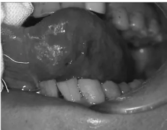
きるからである。しかしながら,この focal defect は病変 での位置や臓器によってもすべてが壊死組織と限定できず, 何かしらの原因で画像上に現れた所見と認識していなけれ ばならない。 そこで,われわれは偶然にも造影 MRI の撮像時に周辺 性に強く造影され,この focal defect に類似する所見を呈 した病変 2 症例に遭遇し,最終的に病理組織学的に摘出 物と比較し focal defect 様所見の生じた原因を推察するこ とができた 2 症例の概要について報告する。 症 例 症例 1 患者は 72 歳の女性である。6 ヶ月前に舌尖に口内炎が でき近医にて治療を受けたが,3 ~ 4 ヶ月前から徐々に硬 結を触れる様になり,違和感があったため本学口腔外科を 紹介され受診した。初診時の視診では,舌下面の舌尖付近 に 10
×
15mm大の境界明瞭で上皮と癒着する球形の腫 瘤がみられ,腫瘤の周囲が 7 mm ほど隆起し表面平滑で 一部潰瘍形成がみられた(Figure 1)。患者はときどき舌 尖にピリピリ感を併発していた。超音波画像では比較的境 界明瞭でほぼ均一な中等度の内部エコ-を示し,ドプラ像 では周囲に強い血流信号を有する低エコ-像を示した。 MRIでは左側の舌尖付近に T1 強調画像で低信号(FigureFigure 2 MRIs in case 1
a. T1-weighted axial MRI(TR/TE : 500/14) showed low signal intensity in the right tip of the tougue.
b. Fat-suppression T2-weighted axial MRI(TR/ TE : 4263/102)showed moderately high intensity. c. Fat-suppression post-Gd-enhanced T1- weight-ed axial MRI(TR/TE : 856/12)showweight-ed thin en-hanced high intensity with marked enen-hanced margin.
a
b
c
Figure 1 Tumor mass shows ulcer on the surface of
2a),T2 強調脂肪抑制画像で内部がわずかに高信号を示 す円形の辺縁が高信号に富み(Figure 2b),Gd(gadrini-um)-DTPA(diethylenetriamine-pentaacetic acid) 造 影 T1 強調脂肪抑制画像(造影 T1 強調脂肪抑制画像とす る)では内部も弱い造影効果を示し周囲を増強する focal defect様 所 見 を 呈 し て い た(Figure 2c)。 超 音 波,CT, MRI画像診断からは顎下部や頸部には有意に腫大したリ ンパ節は存在しなかった。画像検査の終了した時点で臨床 的には上皮性悪性腫瘍が考えられたため生検を実施した。 病理組織所見では,腫瘍細胞は多くの小嚢胞様腫瘍胞巣が 形成され,腫瘍内部には毛細血管がわずかに散在しており, いわゆる線維性の被膜と言われるものは存在せず腫瘍周辺 には小動脈の存在が多数みられた。病理組織診断は粘表皮 癌と診断された(Figure 3)。初診から 4 週間後に本学口 腔外科にて舌の部分切除術が施行された。現在術後 2 年 6ヶ月を経過するが再発はみられていない。 症例 2 患者は 26 歳の女性である。近医を受診し下顎の智歯の 抜歯を勧められて本学口腔外科に来院した。口腔内診査時 に左側の顎下部の腫脹がみられたため,担当医はさらに精 査することを勧めた。現病歴として 4 年前から腫脹に気 づいていたが放置していたが,最近,増大傾向があること を自覚したとこのことである。超音波画像では比較的境界 明瞭でほぼ均一な低エコ-像を示し,さらに造影 CT では 左側顎下腺の前方部に focal defect 所見を呈した。MRI で は左側顎下腺の前下方部に T1 強調画像で低信号(Figure 4a),T2 強調脂肪抑制画像で内部に不均一な中等度の信 号を伴う低信号(Figure 4b),造影 T1 強調脂肪抑制画像 (Figure 4c, d)では周囲を増強する内部壊死も考えられる focal defect所見を呈していた。臨床的に顎下腺の腫瘍性 病変が疑われたため,初診から 2 ヶ月後に左側顎下腺良 性腫瘍の臨床診断名で腫瘍摘出術が施行された。摘出時に 腫瘍は周囲軟組織からの剥離は容易であり,顎下腺との連 続性がみられ一塊として摘出を行った。病理組織所見では, 腫瘍は境界明瞭で線維性の被膜を有し,腫瘍中心部に多量 の軟骨様組織が占拠し辺縁部に腫瘍細胞が変位してみられ, 腫瘍の内部には毛細血管がわずかに散在するのみで,腫瘍 周辺に小動脈が存在していた。病理組織診断は多形性腺腫 と診断された(Figure 5)。現在再発はみられず患者の予 後は良好である。 考 察
造影 CT や造影 MRI における focal defect の所見は病 変の内部変化を示す特異性の高い画像所見であり,頸部リ ンパ節転移の診断にも応用され,リンパ節内に置いては低 吸収(MRI では低信号)領域を示す。この focal defect の 所見は中心壊死(central necrosis)として診断されてき た経緯があるが,今日 focal defect の原因は壊死のみでな く,線維化,類骨,嚢胞,浮腫,ケラチンの蓄積,腫瘍細 胞などさまざまな原因により発生することが知られてい る2,6,7。また,場所によっても中心部でなくて症例 2 で見 られたように偏在するものもある。現在,とくに顎下部な どの転移巣の有無に対する初期検査では,超音波,CT, MRIによる画像診断が主体に行われ,たとえば転移リン Figure 3 Case 1 was diagnosed as mucoepidermoid carcinoma.
a. The tumor has numerous cystic nests and lack fibrous capsule(HE stain ×3.1). b. Small arteries are found nearby the tumor(arrows)(HE stain ×10).
a
パ節の診断には focal defect 所見,リンパ節長短径の変化 と形態,リンパ節の集簇性,節外進展などが診断基準と なっている3が,決定的なものはなく総合的に考慮してい るものが多い8。また,頸部のリンパ節の腫大を示す疾患 は多くみられ,扁平上皮癌のリンパ節転移の他に菊池病や 結核でもリンパ節の壊死を伴うことがあり,MRI で focal defectが画像上の重要な所見となることも述べられてい る9。さらに Bartles ら10は MRI が軟部組織解像度に優れ 病理組織診断名との一致率では CT よりも勝れたことから, 穿刺吸引細胞診と同等の成績であったとの報告をしている。 また,耳下腺腫瘍では MRI は有用性が高い画像診断法と 考えられ11–13,扁平上皮癌のリンパ節転移では CT の感度 と正診率が高く MRI と比較して優れていたという報告14 と,MRI も CT と同等であったという報告もある15。 近年 MRI に新たな因子を加えて診断に応用し始めた拡 散 MRI の利用が増加してきた。拡散強調画像(DWI)と は分子の拡散運動の程度を画像化する方法で通常のパルス 系列に 2 個の逆向きの傾斜磁場を印加して拡散の程度を 定量化したもので,いわゆる見かけの拡散係数(Appar-ent Diffusion Coeffici定量化したもので,いわゆる見かけの拡散係数(Appar-ent, ADC)と呼ばれている16,17。近 年 MRI 拡散強調画像から ADC の測定により鑑別を試み たものがある15,18–24が,完全に明確な結論は出されてはい ないのが現状である。 MRIの特徴に,信号強度から組織像をある程度推測す ることも可能となってきている。骨腫瘍では壊死組織と腫 瘍組織部の信号強度の違いにおいて,T1 強調画像で低信 号と T2 強調画像で高信号を呈することは類似する。造影 後に T1 強調画像で高信号となるのが腫瘍組織部,低信号 となるのが壊死組織でありさらに壊死組織の辺縁は高信号 を呈する。このことについては,腫瘍組織は T1 強調画像 a c b d
Figure 4 MRIs in case 2 a. T1-weighted axial MRI(TR/TE: 415/14)showed low signal intensity.
b. T2-weighted axial MRI(TR/TE: 3826/90)showed low intensity with heterogeneous moderate signal intensity. c. Fat-suppression post-Gd-enhanced T1 weighted axial MRI(TR/TE: 4.6/2.4)showed a focal defect with mark-edly enhanced margin at the edge of the left submandibular gland.
d. Fat-suppression post-Gd-enhanced T1 weighted coronal MRI(TR/TE : 4.6/2.4)showed a focal defect with markedly enhanced margin at the edge of anterior and inferior to the left submandibular gland.
では血流に富む腫瘍組織が存在するためであり,造影効果 の程度は構成される組織により多少異なるためである。一 方,腫瘍の中心部に壊死組織がある場合,ほとんど造影さ れないためこのような特徴的な focal defect 所見となる。 今回の唾液腺疾患では,悪性腫瘍では悪性度の高い腫瘍は 低いものに比べて T2 強調画像で低信号を呈する傾向が強 いとの報告もある25,26。また,多形性腺腫は T1 強調画像 で低信号,T2 強調画像で高信号を呈するものが多く27, 内部に形成される粘液基質,軟骨成分などの組織により T2強調画像で不均一な内部性状を表す。さらに,欠畑 ら28は耳下腺腫瘍の良悪性の質的診断に heavily T2 強調 画像と dynamic MRI を組み合わせることも有用であると 指摘している。杉尾ら29は,頸部の病変で focal defect 様 所見を有する 3 症例を報告し,その中で MRI は CT や超 音波画像検査の補足的な手段とも述べ,質的診断を MRI のみで行うことは難しいとの報告もある。 今回,MRI の周辺性に強く造影された症例の病理組織 像から,症例 1,2 共に腫瘍の内部には毛細血管がわずか に散在するのみで,腫瘍周辺に小動脈が存在していたこと が,造影 MRI で腫瘍周辺に増強した所見の生じた原因と 考えられた。さらに,症例 1 では小嚢胞状の腫瘍胞巣が 多数形成されていたこと(Figure 2a)が T2 強調画像で 高信号を呈し,造影 T1 強調脂肪抑制画像造影で内部にも 造影効果がみられたのは,悪性腫瘍であるであるために周 囲の小血管が栄養血管として病変内部に入り込んでいたた めではないかと推察できた。症例 2 では腫瘍中心部に多 量の硝子軟骨組織が形成され,血管組織が少なかった (Figure 5a)ため focal defect に近い所見を呈したものと
考えられた。
さらに壊死組織と腫瘍組織を鑑別するためには,dy-namic造影 MRI による腫瘍血流状態の時間差で区別され るといわれている6。また,唾液腺腫瘍で多形性腺腫など
Figure 5 Case 2 was diagnosed as pleo-morphic adenoma.
a. The tumor is well-circumscribed by fibrous capsule, and composed of central cartilagi-nous element and peripheral epithelial ele-ment(HE stain Rupe).
b. There are few capillaries in cartilaginous element(HE stain×50).
c. Small arteries are found nearby the tumor (arrow heads)(HE stain ×10).
a
c
の病変内に嚢胞成分を多く含む場合においては dynamic 造影 MRI の時間 – 信号強度曲線から成分の違いなどの鑑 別 を 試 み た も の も あ る9。 し か し な が ら dynamic 造 影 MRIにおいては,漸増型が多形腺腫,急増漸減型が悪性 腫瘍と報告されている9,28が,例外も多くみられる。 今回の 2 症例は,舌尖付近と顎下腺に造影 MRI 所見で 周辺性に強く造影され focal defect にも類似する所見を有 したもので,症例 1 は悪性腫瘍,症例 2 は良性腫瘍の一 部に存在したものであった。症例 1 では,舌に関する MRIの診断は,ほとんどが外舌筋と内舌筋からなる筋肉 が主体となって構成されるため,CT より組織分解能の高 い MRI のほうが有用であることが示された。症例 2 では 造影 CT も撮像し focal defect に近い所見が左側顎下腺下 方に偏在していた。病変における focal defect のできる位 置によっては,内部壊死組織の存在が強く疑われ,良悪性 病変の診断にも大きく影響が出てくる。そのため,MRI の DWI や dynamic 造影 MRI など適切なる方法を使用す ることも,今後 focal defect に対する診断に大きく役立つ ものと考える。
文 献
1. 渡辺嘉之,御供政紀.MRA, dynamic MRI, 野村恭也,小松 崎篤,本庄巌総編集,飯沼壽孝担当編集.画像診断.第 1 版. 東京.中山書店.2001:60–67.
2. Nakayama E, Yonetsu K, Yoshiura K, Araki K, Kanda S, Yoshida K. Diagnostic value of magnetic resonance imag-ing for malignant tumors in the oral and maxillofacial re-gion. Oral Surg Oral Med Oral pathol Oral Radiol Endod. 1996;82:691–697. 3. 尾尻博也著.頭頸部の臨床画像診断学.第 1 版.東京.南江 堂.2005:253–256. 4. 田中 修,大澤忠.骨・関節・軟部組織.高橋睦正編著.全 身の MRI.第 1 版.東京.南江堂.1993:582–636. 5. 尾尻博也.頸部リンパ節転移.多田信平,黒崎喜久編集.頭 頸部の CT・MRI.第 1 版.東京.メディカルサイエンス・ インタ-ナショナル.2002:506–542.
6. Erlemann R, Reiser MF, Peters PE, Vasallo P, Nommensen B, Kusnierz-Glaz CR, Ritter J, Roessner A. Musculoskele-tal neoplasms : static and dynamic Gd-DTPA-enhanced MR imaging. Radiology. 1989;171:767–773.
7. 田中宏子,河野 敦,遠藤寛子,五味直哉,松枝 清,藤原 良将,山田恵子,川端一嘉.転移の画像診断 頭頸部.臨床 画像.2007;23:734–741.
8. Som PM. Detection of metastasis in cervical lymph nodes: CT and MRI criteria and differential diagnosis. Am J Roentgenol. 1992;158:961–969.
9. 阪本真弥.唾液腺腫瘍の MRI.歯科放射線.2003;43: 206–210.
10.Bartles S, Talbot JM, DiTomasso J, et al. The relative val-ue of fine-needle aspiration and imaging in the preopera-tive evaludation of parotid masses. Head Neck. 2000;22: 781–786.
11.岩井 大.CT,MRI の有用性と限界,山下敏夫編集.耳下 腺 腫 瘍 臨 床 の 最 前 線 Q & A. 第 1 版. 東 京. 金 原 出 版. 2004:44–48.
12.Sakamoto M, Sasano T, Higano S, Takahashi S, Iikubo M, Kakehata S. Usefulness of heavily T(2)weighted
mag-netic resonance images for the differential diagnosis of pa-rotid tumors. Dentomaxillofac Radiol. 2003;32:259–259. 13.Ross MR, Schomer DF, Chappell P, Enzmann DR. MR im-aging of head and neck tumors: comparison of T1-weighted contrast-enhanced fat-suppressed images with convention-al T2-weighted and fast spin-echo T2-weighted images. Am J Roentgenol. 1994;163:173–178.
14.Yousem DM, Som PM, Hackney DB, Schwaibold F, Hendix RA. Central nodal necrosis and extracapsular neoplastic spread in cervical lymph nodes : Mr imaging versus CT. Radiology. 1992;182:753–759.
15.King AD, Ahuja AT, Yeung DKW, Fong DKY, Lee YYP, Lei KIK, Tse GMK. Malignant cervical lymphadenopathy : Di-agnostic accuracy of diffusion-weighted MR imaging. Radi-ology. 2007;245:806–813. 16.荒木 力著.拡散 MRI ブラウン運動拡散 テンソルから q 空 間へ.第 1 版.東京.秀潤社.2006:138–140,200–203. 17.多田信平監修.田中 宏,石井千佳子,佐藤希光共著.MRI テクニックマニュアル.第 1 版.東京.南江堂.2002:14– 15,235.
18.Srinivasan A, Dvorak R, Perni K, Rohrer S, Mukherji SK. Differentiation of benign and malignant pathology in the head and neck using 3T apparent diffusion coefficient val-ues : Early experience. AM J Neuroradiol. 2008;29:40– 44.
19.Matsushima N, Maeda M, Takamura M, Takeda K. Appar-ent diffusion coefficiAppar-ents of benign and malignant salivary gland tumor. Comparison to histopathological findings. J Neuroradiol. 2007;34:183–189.
20.Eida S, Sumi M, Sakihara N, Takahashi H, Nakamura T. Apparent diffusion coefficients mapping of salivary gland tumors: Prediction of the benignancy and malignancy. AM J Neuroradiol. 2007;28:116–121.
21.Maeda M, Kato H, Sakuma H, Maier SE, Takeda K. Use-fulness of the apparent diffusion coefficient in line scan diffusion-weighted imaging for distinguishing between squamous cell carcinoma and malignant lymphomas of the head and neck. AM J Neuroradiol. 2005;26:1186–1192. 22.Kawai Y, Sumi M, Kitamori H, Takagi Y, Nakamura T.
Diffusion-weighted MR microimaging of the lacrimal glands in patients with sjögren's syndrome. Am J Roent-genol. 2005;184:1320–1325.
23.Sumi M, Sakihama N, Sumi T, Morikawa M, Uetani M, Kabasawa H, Shigeno K, Hayashi K, Takahashi H, Naka-mura T. Discrimination of metastatic cervical lymph nodes with diffusion-weighted MR imaging in patients with head and neck cancer. AM J Neuroradiol. 2003;24:1627– 1634.
24.Sumi M, Takagi Y, Uetani M, Morikawa M, Hayashi K, Kabasawa H, Aikawa K, Nakamura T. Diffusion-weighted echoplanar MR imaging of the salivary gland. Am J Roent-genol. 2002;178:959–965.
25.池田耕士.唾液腺,小玉隆男編集,眼窩・耳鼻咽頭・口腔領 域の MRI.第 1 版.東京.メジカルビュ-社.2004:184– 203.
26.Som PM, Biller HF. High-grade malignancies of the parot-id gland: Identication with MR imaging. Radiology. 1989; 173:823–826. 27.石島 健,佐藤宏昭,中里龍彦.副(旁)咽頭間隙腫瘍. Johns.2005;21:459–464. 28.欠畑誠治,阪本真弥.耳下腺悪性腫瘍.Johns.2005;21: 452–458. 29.杉尾雄一郎,吉田厚子,稲垣幹矢,州崎春海.頸部の炎症性 疾患.Johns.2005;21:465–470.