Title
Heparan sulfate on intestinal epithelial cells plays a critical role
in intestinal crypt homeostasis via Wnt/β-catenin signaling(
Dissertation_全文 )
Author(s)
Yamamoto, Shuji
Citation
Kyoto University (京都大学)
Issue Date
2014-03-24
URL
http://dx.doi.org/10.14989/doctor.k18141
Right
Type
Thesis or Dissertation
Textversion
ETD
Heparan sulfate on intestinal epithelial cells plays a critical role in intestinal
crypt homeostasis via Wnt/
-catenin signaling
Shuji Yamamoto,1,2Hiroshi Nakase,1Minoru Matsuura,1Yusuke Honzawa,1Kayoko Matsumura,1 Norimitsu Uza,1Yu Yamaguchi,3Emiko Mizoguchi,4and Tsutomu Chiba1
1Department of Gastroenterology and Hepatology, Graduate School of Medicine, Kyoto University, Kyoto;2Japan Society for
the Promotion of Science, Tokyo, Japan;3Sanford Children’s Health Research Center, Sanford-Burnham Medical Research
Institute, La Jolla, California;4Gastrointestinal Unit, Department of Medicine, Massachusetts General Hospital and Harvard
Medical School, Boston, Massachusetts
Submitted 18 December 2012; accepted in final form 24 May 2013
Yamamoto S, Nakase H, Matsuura M, Honzawa Y, Matsumura K, Uza N, Yamaguchi Y, Mizoguchi E, Chiba T. Heparan sulfate on
intestinal epithelial cells plays a critical role in intestinal crypt ho-meostasis via Wnt/-catenin signaling. Am J Physiol Gastrointest Liver Physiol 305: G241–G249, 2013. First published June 6, 2013; doi:10.1152/ajpgi.00480.2012.—Heparan sulfate (HS), a constituent of HS proteoglycans (HSPGs), is a linear polysaccharide present on the cell surface. HSPGs modulate functions of several growth factors and signaling molecules. We examined whether small intestinal epi-thelial HS plays some roles in crypt homeostasis using intestinal epithelium cell (IEC)-specific HS-deficient C57Bl/6 mice. Survival rate after total body irradiation was significantly reduced in HS-deficient mice due to profound intestinal injury. HS-HS-deficient IECs exhibited Wnt/-catenin pathway disruption, decreased levels of -catenin nuclear localization, and reduced expression of Wnt target genes, including Lgr5 during crypt regeneration. Moreover, epithelial HS increased Wnt binding affinity of IECs, promoted phosphorylation of Wnt coreceptor LRP6, and enhanced Wnt/-catenin signaling following ex vivo stimulation with Wnt3a, whereas activation of canonical Wnt signaling following direct inhibition of glycogen syn-thase kinase-3 by lithium chloride was similar between HS-deficient and wild-type mice. Thus HS influences the binding affinity of IECs to Wnt, thereby promoting activation of canonical Wnt signaling and facilitating regeneration of small intestinal crypts after epithelial injury.
heparan sulfate; intestinal regeneration; Wnt signaling
THE SMALL INTESTINAL EPITHELIUM comprises a single layer of
columnar cells that are organized into villi and crypts. Villi extend into the intestinal lumen and contain three types of terminally differentiated cells: enterocytes, goblet cells, and enteroendocrine cells. Enterocytes absorb nutrients, goblet cells secrete mucin, and enteroendocrine cells release gastro-intestinal hormones. Crypts are formed by epithelial invagina-tions into the connective tissue of the intestine and contain stem cells, their transit-amplifying daughter cells, and Paneth cells, which secrete antibacterial peptides into the crypt lumen (99 –13). An intricate cell-replacement process maintains the integrity of the mucosal epithelium of the intestine. Vigorous proliferation occurs in crypt compartments. When the commit-ted transit-amplifying cells reach the crypt-villus junction, they rapidly and irreversibly differentiate. The differentiated cells
migrate up to the villus tip, where they are shed. In mouse, the small intestinal epithelium is renewed every 5 days (2).
Several intracellular signaling pathways, such as the Wnt/ -catenin, bone morphogenic protein, phosphoinositide 3-ki-nase (PI3K)/Akt, and Notch pathways, have critical roles in crypt-villous homeostasis (2). In addition, several extracellular components have also been shown to regulate intestinal crypt homeostasis (36, 39), but their precise roles remain obscure.
Heparan sulfate (HS) is a linear polysaccharide constituting repeating disaccharide units of glucuronic acid and N-acetylg-lucosamine. HS is found on the surface of most cells as a constituent of HS proteoglycans (HSPGs), which comprise a core protein with covalently attached HS chains. In the small intestinal epithelial cells (IECs) of humans (42) and mice (5), HSPGs are predominantly located on the basolateral surface of the cells. HSPGs bind to various growth factors, including Wnt, Hedgehog, transforming growth factor-, and FGF and modulate the biologic activities of these molecules (4, 58).
The importance of HSPG in embryological development is established. In Drosophila, HS is required for distribution and signaling of morphogens such as Wingless, Hedgehog, and the Drosophila homologue of bone morphogenetic protein, Deca-pentaplegic, in the embryonic epidermis and the wing disc (8, 20, 22). HS is also involved in FGF signaling during the migration of mesodermal and tracheal cells (32). In vertebrate, HS influences FGF function in zebrafish limb development (40) and is essential for Wnt11 during zebrafish and Xenopus gastrulation (41). In mice, HS is required for proper Indian Hedgehog distribution during endochondral bone development (30) and Fgf8 function in brain development (26). Despite many evidences demonstrating that HSPGs play pivotal roles in embryogenesis, it remains unclear how HSPGs are involved in intestinal epithelial homeostasis.
In the present study, we used the intestine-specific HS-deficient mouse model (5) to elucidate the role of HS in the small intestine.
MATERIALS AND METHODS
Mice. We used C57Bl/6 mice. Conditional Ext1 allele (Ext1flox)
was created as described previously (26). Villin-Cre mice (16) were obtained from the Jackson Laboratory. Mice were maintained on a 12-h:12-h light/dark cycle and fed standard laboratory mouse chow ad libitum in specific pathogen-free conditions. All experiments were performed with 2- to 4-mo-old mice. All animal experiments in this study were approved by the Review Board of Kyoto University and adhered to their institutional ethical guidelines. For survival data, mice were euthanized if moribund or seriously injured (e.g.,
vocaliz-Address for reprint requests and other correspondence: H. Nakase, Dept. of Gastroenterology and Hepatology, Graduate School of Medicine, Kyoto Univ., 54 Shogoin Kawahara-cho, Sakyo-ku, Kyoto, 606-8507, Japan (e-mail: hiropy_n @kuhp.kyoto-u.ac.jp)
ing, lack of grooming, or more than 20% weight loss) to prevent suffering.
Histopathology and immunohistochemical staining. Mice were killed, and the small intestine was removed, fixed in 10% formalde-hyde, and embedded in paraffin. Sections were stained with hematox-ylin and eosin reagent. Immunohistochemical staining with anti-HS mAb (10E4; Seikagaku, Tokyo, Japan), bromodeoxyuridine (BrdU) antibody (Sigma-Aldrich, Tokyo, Japan), anti-FGF receptor (FGFR) 1 (phospho Tyr154) antibody (Novus Biologicals, Littleton, CO), or anti-pFGFR3 (Tyr 724) antibody (Santa Cruz Biotechnology, Santa Cruz, CA) was performed on paraffin-embedded slices using the avidin-biotin immunoperoxidase method. Sections were deparaf-finized and rehydrated through a graded series of xylene-ethanol washes. Endogenous peroxidase was blocked with 0.3% H2O2. After
biotin was conjugated with secondary antibodies, sections were incu-bated with the avidin-biotin peroxidase complex (ABC Elite Kit; Vector Laboratories, Burlingame, CA) before reaction with 3,3= -diaminobenzidine/H2O2. Nuclei were counterstained with
hematox-ylin. For immunofluorescence staining, specimens were incubated with anti--catenin antibody (Cell Signaling Technology, Tokyo, Japan) and followed by staining with Alexa Fluor 488 anti-rabbit-IgG (H⫹L) (Invitrogen, Tokyo, Japan). Nuclei were visualized by DAPI staining.
Total body␥-irradiation and bone marrow transplantation. Total body irradiation (TBI) was performed using a137Cs source emitting at
a fixed-dose rate of 1.11 Gy/min (Gammacell 40 Exactor; MDS Nordion International, Ontario, Canada). For bone marrow transplan-tation, Ext1flox/flox(Ext1F/F) donor mice were killed, and marrow cells
were harvested from the medullary cavities of the femur by flushing with Hanks’ balanced salt solution (GIBCO, Tokyo, Japan). Cells were counted, diluted to 5⫻ 106cells/200l, and injected into the tail
vein of recipient mice 16 h after TBI. Mice were killed, and the proximal jejunum was rapidly dissected, fixed in 10% neutral buffered formalin, and embedded in paraffin. Paraffin sections (4m) were cut perpendicular to the long axis of the intestine before immunohisto-chemical analysis. Immunohistological staining was performed as described above.
In vivo microcolony assay of crypts. Crypt stem cell survival was determined 3.5 days after TBI based on BrdU incorporation into proliferating crypt cells, as previously reported (59). Each mouse received 120 mg/kg BrdU intraperitoneally 2 h before harvesting the tissue to permit identification of replicating S phase cells by immu-nohistochemistry using the avidin-biotin immunoperoxidase method. The number of surviving crypts per cross-section was scored. A surviving crypt was defined as a crypt with five or more BrdU-labeled epithelial cells.
Isolation of IECs. IECs were isolated as described previously (27). Mice were killed, and the small intestine was removed and placed in ice-cold PBS flushed through the lumen of the intestines using a syringe. The intestines were then cut longitudinally with scissors and rinsed with cold PBS. The small intestines were placed in PBS with 3 mM EDTA and 50 mM DTT and stored on ice for 1 h in 50-ml conical tubes. The supernatant was filtered and centrifuged for 5 min at 400 g, and the cell pellet was resuspended in cold PBS. Finally, primary IECs were collected by centrifugation through a 20/40% discontinuous Percoll gradient at 600 g for 30 min. Purity of IECs was assessed by flow cytometric analysis using rat anti-mouse E-Cadherin mAb (R & D Systems, Tokyo, Japan) and was confirmed more than 90% pure. Primary mouse IECs were collected in sample buffer for subsequent RNA isolation as well as immunoblotting or in PBS for ex vivo stimulation.
Immunoblotting. Primary mouse IECs were lysed with Tris·HCl buffer containing 1% Triton X-100 (lysis buffer). Cell lysate proteins (15g) were subjected to SDS-PAGE separation followed by immu-noblotting using primary antibody. The primary antibodies used for immunoblot analysis were as follows: anti--catenin, anti-cyclin D1, anti-c-Myc, anti-Survivin, anti-phospho--catenin (Ser552), anti-Akt,
phospho-Akt (Thr308), Erk1/2, phospho-Erk1/2, anti-SAPK/JNK, anti-phospho-anti-SAPK/JNK, anti-p38 MAPK, anti-phos-pho-p38 MAPK, anti-phospho-FGF receptor substrate (FRS) 2␣, anti-active-catenin, anti-LRP6, anti-phospho-LRP6 (Cell Signaling Technology), and anti-Lgr5 antibody (Abcam, Tokyo, Japan). The bound antibody was detected by horseradish peroxidase-conjugated secondary antibody (GE Healthcare, Piscataway, NJ) followed by chemiluminescence using an ECL Plus kit (GE Healthcare). Blotting with anti--actin antibody (Sigma-Aldrich) was run as a loading control.
Quantitation of gene expression using real-time PCR. The total RNA isolated with TRIzol reagent (Invitrogen) was reverse-tran-scribed using Superscript First-Strand Synthesis System for RT-PCR (Invitrogen) according to the manufacturer’s instructions. PCR am-plification of cDNA (1g/20 l of PCR reaction) was carried out in the LightCycler (Roche Diagnostics, Tokyo, Japan) using Fast-Start Universal SYBR Green Master (Rox; Roche Diagnostics). Results are expressed as the ratio of each molecule to Gapdh. Primer pairs were as follows: Gapdh: AGCCTTCTCCATGGTG-GTGAAGAC (forward), CGGAGTCAACGGATTTGGTCGTAT (reverse); Lgr5: CTACTTGACTTTGAGGAAGACC (forward), AG-GAAAGCGCCAGTACTGC (reverse); cyclin D1: TCCGCAAGCA TGCACAGA (forward), GGTGGGTTGGAAATGAACTTCA (re-verse); c-Myc: GCTCGCCCAAATCCTGTA (forward), AGGAC-TCGGAGGACAGCA (reverse); Survivin: GCGGAGGCTGGC TTCA (forward), AAAAAACACTGGGCCAAATCA (reverse); Ker-atin8: TCATCCTATGGGGGACTCAC (forward), TCTTCACAAC-CACAGCCTTG (reverse); Frizzled4: GACAACTTTCACGCCGC-TCATC (forward), CCAGGCAAACCCAAATTCTCTCAG (reverse); Frizzled6: TGTTGGTATCTCTGCGGTCTTCTG (forward), CTCG-GCGGCTCTCACTGATG (reverse); Frizzled7: ATATCGCCTA-CAACCAGACCATCC (forward), AAGGAACGGCACGGAG-GAATG (reverse); and Axin2: TCACAGCCCTTGTGGTTCAAG (for-ward), GGTAGATTCCTGATGGCCGTAGT (reverse).
Wnt3a binding assay and ex vivo stimulation of primary mouse IECs. Recombinant mouse Wnt3a (R&D Systems, Minneapolis, MN) was labeled with Alexa Fluor 488 dye using the Alexa Fluor 488 Microscale Protein Labeling Kit (Invitrogen) according to the manu-facturer’s instructions. Primary mouse IECs (1⫻ 105) were incubated
with 30 ng/ml Alexa Fluor 488-conjugated Wnt3a for 60 min at 37°C. The cells were filtered through nylon filters (Becton Dickinson, Tokyo, Japan) and washed three times with PBS, and mean fluores-cent intensity was measured using the FACS Canto II (Becton Dick-inson). For ex vivo stimulation, primary mouse IECs were incubated with 30 ng/ml or 100 ng/ml recombinant mouse Wnt3a, 200 ng/ml recombinant mouse Wnt5a (R&D Systems), or 60 mM lithium chlo-ride (LiCl) at 37°C and harvested for immunoblotting or PCR anal-ysis.
Statistical analysis. The results are represented as means⫾ SE. A Student’s t-test was performed for statistical evaluation. Actuarial survival was calculated by the Kaplan-Meier method, and P values were evaluated by log-rank test. A P value of⬍0.05 was considered statistically significant.
RESULTS
Generation of intestine-specific HS-deficient mice. To
exam-ine whether HS on IEC of the small intestexam-ine affects crypt homeostasis, we generated mice with intestine-specific condi-tional Ext1 knockout by crossing Ext1F/Fmice with Villin-Cre
mice as reported previously (5). Ext1 encodes glycosyltrans-ferase, which polymerizes alternating glucuronic acid and
N-acetylglucosamine sugar residues in the HS biosynthetic
process (35), and is indispensable for HS synthesis because cells lacking a functional Ext1 allele do not synthesize HS (34, G242 HEPARAN SULFATE IN INTESTINAL CRYPT REGENERATION
37). Intestine-specific Ext1 knockout mice are referred to as
Ext1⌬/⌬mice, and Ext1F/Fmice were used as controls.
PCR and immunoblot analyses of IEC separated from the small intestine of Ext1⌬/⌬mice revealed minimal levels of Ext1 mRNA (Fig. 1A) and Ext1 protein (Fig. 1B), respectively. Immunohistochemical study with a mAb to HS (10E4) re-vealed that HS was expressed on the basolateral surface of IEC in Ext1F/F mice (Fig. 1C, left), whereas HS expression was
undetectable in the IEC of Ext1⌬/⌬mice (Fig. 1C, right). Both macroscopic and microscopic findings were normal in Ext1⌬/⌬ mice as previously reported (5), suggesting that HS on IECs
are not critically involved in crypt homeostasis in the steady state.
Epithelial HS is required for intestinal regeneration after TBI. We first examined the effects of intestine-specific deletion of Ext1 on radiation damage to the small intestine in vivo. Ext1F/F
and Ext1⌬/⌬mice received TBI followed by bone marrow trans-plantation to rescue from bone marrow death induced by lethal irradiation. Of interest, all Ext1⌬/⌬ mice with 12 Gy TBI died within 6 days despite bone marrow transplantation, whereas 71% of Ext1F/Fmice were alive more than 30 days after TBI (log-rank
test; P⬍ 0.01; Fig. 2A). Histological evaluation 4 days after 12 Gy TBI revealed the small intestinal mucosa with regenerative crypts covering most of the inner intestinal surface in Ext1F/Fmice
(Fig. 2B, top), whereas severe intestinal damage with almost no villi or crypts in the small intestinal mucosa of Ext1⌬/⌬mice was observed (Fig. 2B, bottom). This finding suggests that
Ext1⌬/⌬mice after TBI died of intestinal damage induced by irradiation. Therefore, we further evaluated roles of epithelial surface HS in crypt regeneration in the small intestine follow-ing radiation injury usfollow-ing a crypt microcolony assay (59). Because most of Ext1⌬/⌬mice died within 4 days after 12 Gy TBI (Fig. 2A), dose of irradiation was reduced to 10 Gy in this experiment. Intestine-specific deletion of Ext1 significantly decreased the number of surviving crypts, which were labeled with BrdU 3.5 days after 10 Gy TBI (Fig. 2, C and D). Taken together, these results suggest that HS on IECs has protective effects against irradiation and plays critical roles in crypt regeneration after TBI-induced epithelial injury.
HS is involved in Wnt/-catenin signaling in intestinal epi-thelium. Given that IEC-specific Ext1 deletion results in decreased
proliferation of IECs, we investigated the Wnt/-catenin and the MAPK pathways that are important for regulating IEC prolifera-tion (17, 50). Immunoblot analysis demonstrated no significant difference in the phosphorylation of the MAPKs, including p38 MAPK, Erk1/2, and JNK1/2, in IECs 4 days after TBI between
Fig. 1. Heparan sulfate (HS) biosynthesis in intestinal epithelial cells (IECs) was defective in intestine-specific Ext1-deficient mice. A and B: Analysis of PCR products of Ext1 (A) and immunoblotting of Ext1 (B) in IECs isolated from Ext1F/Fand Ext1⌬/⌬mice. Both mRNA and protein expression of Ext1
were disrupted in Ext1⌬/⌬mice. C: immunohistochemistry for HS in the small intestine of Ext1F/F(left) and Ext1⌬/⌬(right) mice. Scale bar⫽ 200 m.
Fig. 2. Intestine-specific HS-deficient mice were sensitive to total body irradiation (TBI)-induced gastrointestinal toxicity. A: Kaplan-Meier sur-vival analysis of Ext1F/Fand Ext1⌬/⌬mice after
12 Gy TBI (n⫽ 7). By log-rank test, P ⫽ 0.0004.
B: upper jejunal sections of Ext1F/F (top) and Ext1⌬/⌬(bottom) mice 4 days after 12 Gy TBI stained with hematoxylin and eosin. C: represen-tative bromodeoxyuridine (BrdU) labeling of sur-viving crypts. Scale bar⫽ 100 m. D: surviving crypts per cross-section. A surviving crypt was defined as a crypt with 5 or more BrdU-labeled epithelial cells. 6 cross-sections per mouse were scored and compared between Ext1F/Fand Ext1⌬/⌬
Ext1⌬/⌬ and Ext1F/F mice (Fig. 3A). In contrast, the protein
levels of total-catenin were lower in IECs isolated from the small intestine of Ext1⌬/⌬ mice 4 days after 10 Gy TBI than those of Ext1F/Fmice (Fig. 3B). In addition, nonphosphorylated
-catenin (active -catenin) was also lower in Ext1⌬/⌬ mice
(Fig. 3C). Moreover, immunofluorescence staining revealed lower levels of-catenin nuclear localization in crypt cells of
Ext1⌬/⌬mice 4 days after TBI, compared with those of Ext1F/F
mice (Fig. 3, D and E). These data suggest the decreased
protein levels of cytoplasmic-catenin with attenuated canon-ical Wnt signaling in IECs of Ext1⌬/⌬ mice after TBI.
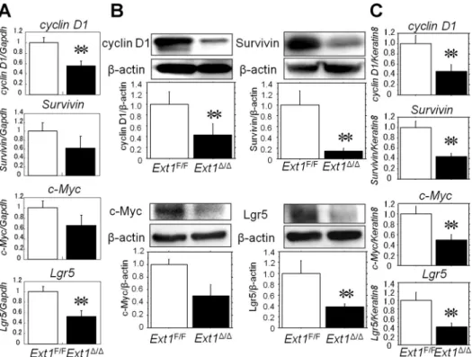
To address whether decreased levels of-catenin had func-tional consequences, we examined the expression of Wnt target genes including, cyclin D1, c-Myc, Survivin, and Lgr5, in IECs isolated from Ext1F/Fand Ext1⌬/⌬mice. Both the mRNA and
protein levels of these genes in IECs with disrupted HS biosynthesis were lower than those in HS-competent IECs after TBI (Fig. 4, A and B). To exclude the possibility that reduced
Fig. 3. The protein levels of total and active -catenin were decreased in intestine-specific HS-deficient mice after TBI. A: immunoblotting anal-ysis of phosphorylated and total MAPKs. A rep-resentative blot from 5 independent experiments is shown. B and C: immunoblotting analysis of total-catenin (B) and activated nonphosphory-lated -catenin (C). A representative blot is shown, and graph bars are expressed as the ratio of each molecule to-actin. (n ⫽ 5) **P ⬍ 0.05.
D: representative images of immunofluorescence
staining for -catenin in the small intestine of
Ext1F/Fand Ext1⌬/⌬mice.-catenin was stained
with Alexa Fluor 488, and nuclei were visualized by DAPI staining. Scale bar⫽ 100 m. E: the ratio of crypt cells with -catenin-positive nu-cleus per crypt. More than 50 crypts per mouse were scored and compared between Ext1F/Fand Ext1⌬/⌬
mice (n⫽ 3). *P ⬍ 0.01.
Fig. 4. The mRNA and protein levels of Wnt-target genes were lower in IECs from intestine-specific Ext1 knockout mice than those from
Ext1F/Fmice 4 days after 10 Gy TBI. A: The
mRNA levels of Wnt-target genes cyclinD1,
c-Myc, Survivin, and Lgr5. Results are
ex-pressed as the ratio of each molecule to Gapdh. (n ⫽ 4–7) **P ⬍ 0.05. B: immunoblotting analysis of Wnt-target genes in IECs from
Ext1F/Fand Ext1⌬/⌬mice. A representative blot
is shown, and graph bars are expressed as the ratio of each molecule to-actin. (n ⫽ 5) **P ⬍ 0.05. C: mRNA levels of Wnt-target genes normal-ized by Keratin8. (n⫽ 4–7) **P ⬍ 0.05.
G244 HEPARAN SULFATE IN INTESTINAL CRYPT REGENERATION
numbers of crypt cells reflected lower levels of expression of Wnt target genes in Ext1⌬/⌬mice, we used Keratin8, the major intermediate filament proteins in the intestinal epithelia (57), to normalize the data in addition to a housekeeping gene. As expected, the mRNA levels of Wnt target genes were signifi-cantly lower in IECs harvested from Ext1⌬/⌬mice than those from Ext1F/F mice even when normalized by Keratin8 (Fig.
4C). These data indicate that disruption of Ext1 in IECs impaired Wnt/-catenin signaling and that HS plays an impor-tant role in regeneration of intestinal crypts following TBI-induced intestinal injury.
In addition, we evaluated the mRNA expression of Frizzled receptors, acting as receptors for Wnt proteins (2, 44, 50), which resulted in similar levels between Ext1F/Fand Ext1⌬/⌬
mice (Fig. 5, A–C).
Deletion of HS in IECs does not affect the FGF-FGFR axis or the PI3K-Akt pathway. Next, we investigated the
mecha-nism by which intestinal epithelial HS mediates Wnt/-catenin signaling. It was recently reported that FGFR3 signaling reg-ulates crypt epithelial stem cell expansion and crypt morpho-genesis partly through -catenin/T cell factor-4-dependent pathways (56). In addition, HS binds to both FGFs and FGFRs (43, 49) and is required for FGF signal transduction (32). Therefore, we first evaluated the FGF-FGFR axis in radiation
enteritis. Immunohistochemical analysis revealed no signifi-cant difference in the phosphorylation of FGFR3 and FGFR1 in crypts 4 days after 10 Gy TBI between Ext1⌬/⌬and Ext1F/F
mice (Fig. 6, A and B). Moreover, levels of phosphorylation of the docking-protein FRS2␣, which plays a critical role in FGFR-mediated signal transduction pathways (19, 21), were similar in IECs between Ext1F/Fand Ext1⌬/⌬mice (Fig. 6C).
Given that the PI3K-Akt pathway activates Wnt/-catenin signaling by phosphorylating-catenin at Ser552 (24, 31), we next examined whether PI3K-Akt pathway-mediated-catenin activation was involved in the disruption of HS in IECs. Phosphorylation of Akt at Ser 308 was a similar level in IECs of Ext1⌬/⌬mice and Ext1F/Fmice. In addition, protein levels of
phosphorylated-catenin at Ser552 in IECs from Ext1⌬/⌬mice were comparable with those from Ext1F/F mice (Fig. 6D).
These data suggest that neither the FGF-FGFR axis nor the PI3K-Akt pathway is involved in HS-dependent Wnt/-catenin signaling in IECs from HS-deficient mice after irradiation.
HS affects binding affinity of IECs to Wnt3a and enhanced Wnt/-catenin signaling. Because HS chains are capable of
binding Wnt proteins (1, 7, 46), we hypothesized that HS on IEC binds Wnt proteins and increases the dose of the ligands on the cell surface, which may result in enhanced Wnt/ -catenin signaling. Therefore, we assessed Wnt3a binding in
Fig. 5. The gene expressions of Frizzled4, 6, and 7 were similar between Ext1F/Fand Ext1⌬/⌬mice 4
days after 10 Gy TBI. Quantitative PCR analysis of
Frizzled4 (A), 6 (B), and 7 (C). Results are expressed as
the ratio of each molecule to Gapdh. (n⫽ 4–7).
Fig. 6. FGF-FGFR axis and phosphoinositide 3-kinase (PI3K)-Akt pathway were similar between Ext1F/Fand Ext1⌬/⌬mice 4 days after 10 Gy TBI. A and B: im-munohistochemistry for pFGFR3 (A) and pFGFR1 (B) in the small intestine of Ext1F/Fand Ext1⌬/⌬ mice. A
representative image from 6 mice is shown. Scale bar⫽ 100m. C: immunoblotting analysis of phospho-FGF receptor substrate (pFRS)2␣ (Tyr196 and Tyr436) in IECs isolated from Ext1F/Fand Ext1⌬/⌬mice. A
represen-tative blot is shown from 5 independent experiments.
D: immunoblotting analysis of pAkt, p-catenin (Ser552),
and total-catenin in IECs isolated from Ext1F/Fand Ext1⌬/⌬mice. A representative blot is shown, and graph bars are expressed as the ratio of each molecule to -actin. (n ⫽ 5).
IECs harvested from Ext1F/Fand Ext1⌬/⌬mice. Flow
cytomet-ric analysis indicated that the binding of fluorescent-labeled Wnt3a to IECs of Ext1⌬/⌬ mice was significantly lower than that of Ext1F/Fmice (Fig. 7, A and B). Moreover, immunoblot
analysis revealed that stabilization of -catenin by ex vivo stimulation with Wnt3a was attenuated in IECs from Ext1⌬/⌬ mice compared with Ext1F/Fmice, whereas-catenin stability
was not affected by deletion of HS sugar chains when IECs were incubated with LiCl (Fig. 7C), which directly inhibits the activity of glycogen synthase kinase-3 in a Wnt-independent manner (25, 28). In addition, phosphorylation of Wnt corecep-tor LRP6 at Ser 1,490 (2, 44, 50) after ex vivo stimulation with Wnt3a was attenuated in IECs harvested from Ext1⌬/⌬ mice (Fig. 7D). The mRNA levels of Wnt target gene Axin2 were lower in IECs of Ext1⌬/⌬mice than those of Ext1F/Fmice after
incubation with Wnt3a, and the difference of Axin2 expression between Ext1F/Fand Ext1⌬/⌬ mice after stimulation with 100
ng/ml of Wnt3a was augmented, whereas similar levels of
Axin2 gene expression were observed between two groups after
ex vivo stimulation with LiCl (Fig. 7E). These results suggest that HS on IECs increases cell surface binding affinity of IECs to Wnt ligands, enhances Wnt/-catenin signaling, and facili-tates crypt regeneration after intestinal epithelial injury.
DISCUSSION
In this study, we showed that intestine-specific HS-deficient mice were more sensitive to TBI-induced intestinal injury than
Ext1F/Fmice and that HS influenced Wnt binding affinity of
IECs and subsequent Wnt/-catenin signaling. Our data sug-gested that HS plays an important role in Wnt/-catenin signaling during regeneration of the small intestinal crypts in mice.
First, we assessed the role of epithelial HS in regeneration of small intestine after TBI. The gross intestinal phenotype and histology of intestine-specific HS-deficient mice without TBI appeared normal in our study, being in agreement with the findings by Bode et al. (5). However, our study demonstrated that the survival rate of these mice after TBI was significantly reduced compared with that of Ext1F/Fmice due to severe small
intestinal injury. Furthermore, in in vivo crypt colony assay, which reflects the capacity of regeneration of intestinal crypts after irradiation (59), the number of surviving crypts in intes-tine-specific Ext1-deficient mice was significantly lower than that in Ext1F/Fmice. These findings suggest that HS is essential
for crypt regeneration after intestinal epithelial injury. Next, we investigated whether HS plays some roles in MAPK pathways or Wnt/-catenin signaling in IECs. Several
Fig. 7. The binding affinity with Wnt3a was decreased, and Wnt/-catenin signaling was disturbed in IECs defective in HS biosynthesis in ex vivo stimulation with Wnt3a but not with LiCl. A and B: Wnt3a binding assay of IECs harvested from Ext1F/Fand Ext1⌬/⌬mice. IECs from Ext1F/For Ext1⌬/⌬mice were incubated
with 30 ng/ml Alexa Fluor 488-conjugated Wnt3a for 60 min at 37°C. A: representative flow cytometry histogram. Fluorescent intensities of HS-competent IECs incubated with (Ext1F/FWnt; solid line, open histogram) or without (Ext1F/FCtl; solid line, solid histogram) Wnt3a and those of HS-deficient IECs with (Ext1⌬/⌬
Wnt; dashed line, open histogram) or without (Ext1⌬/⌬Ctl; dashed line, solid histogram) Wnt3a are shown. B: mean fluorescence intensity of IECs incubated with fluorescent-conjugated Wnt3a after subtracting that of the untreated IECs (n⫽ 3). **P ⬍ 0.05. C: immunoblotting analysis of total and active -catenin in IECs after ex vivo stimulation with Wnt3a, LiCl, or Wnt5a. Primary mouse IECs were incubated with 30 ng/ml or 100 ng/ml Wnt3a, 60 mM LiCl, or 200 ng/ml Wnt5a for 240 min at 37°C and harvested for immunoblotting. Untreated IECs were used as controls. A representative blot from 3 independent experiments is shown. D: immunoblotting analysis of phosphorylated and total LRP6 in IECs after ex vivo stimulation with Wnt3a. IECs from Ext1F/For Ext1⌬/⌬mice were
incubated with 100 ng/ml Wnt3a at 37°C and harvested after indicated time for immunoblotting. A representative blot from 3 independent experiments is shown.
E: quantitative PCR analysis of Axin2 in IECs incubated with Wnt3a or LiCl for 240 min. Untreated IECs were used as controls. The gene expressions were
normalized by Gapdh. (n⫽ 6) **P ⬍ 0.05 compared with Ext1F/Fcontrol; ††P⬍ 0.05 compared with Ext1F/FWnt3a 100 ng/ml; ‡‡P⬍ 0.05 compared with Ext1⌬/⌬control.
G246 HEPARAN SULFATE IN INTESTINAL CRYPT REGENERATION
lines of evidence indicate that MAPK pathways are involved in, not only intestinal cell proliferation, but also intestinal tumorigenesis (17). Our study demonstrated that activation of MAPK pathways in IECs was similar between Ext1F/F and Ext1⌬/⌬ mice after TBI, suggesting that HS does not play a significant role in MAPK signaling during the regeneration of intestinal crypts after lethal irradiation. In contrast, protein levels of total as well as active form of-catenin were lower, and -catenin nuclear localization was reduced in IECs iso-lated from the small intestine of Ext1⌬/⌬ mice than Ext1F/F
mice. To confirm whether decreased levels of -catenin had functional consequences, we examined the expression of Wnt target genes in IECs isolated from Ext1F/Fand Ext1⌬/⌬mice.
Expressions of Wnt target genes in IECs in Ext1⌬/⌬mice were lower than those in Ext1F/F mice. Thus the Wnt/-catenin
pathway was disturbed, and the expression of Wnt target genes was reduced in HS-deficient IECs. Wnt/-catenin signaling plays a central role in the regeneration of intestinal epithelium (29, 44). In addition, the role of HSPGs in the regulation of Wnt pathway has been extensively studied in cell signaling during development. In Drosophila, the abrogation of HSPG activity by mutation of the EXT family genes leads to reduced extracellular Wingless levels and loss of Wnt target gene expression (6, 23, 52). In Xenopus, HSPGs have been shown to interact with Wnt11 during gastrulation (41) and axis forma-tion (53). Taken together, our data and previous findings indicate that HS on IECs has an important function in canon-ical Wnt signaling and is essential for the proliferation of small intestinal epithelium. Notably, our data demonstrated that both mRNA expression and protein levels of c-Myc and Lgr5 were reduced when intestinal epithelial HS was disrupted. c-Myc is essential for accelerating the cell cycle of crypt progenitor cells (38), and Lgr5 is one of the intestinal stem cell markers (3). These findings may suggest that HS on IEC is required for the expansion of intestinal stem cells, as well as for the prolifera-tion of transit-amplifying cells after crypt injury.
Finally, we examined the effect of HS on cell-binding affinity of Wnt proteins, which may result in enhanced Wnt/ -catenin signaling. Our results revealed that Wnt binding affinity of IECs and Wnt/-catenin signaling in ex vivo stim-ulation with Wnt3a were clearly reduced by HS deficiency. On the other hand, activation of Wnt/-catenin signaling following direct inhibition of glycogen synthase kinase-3 by LiCl was not affected by HS deficiency although the possibility cannot be denied that other effects of LiCl than inhibition of glycogen synthase kinase-3, such as inducing autophagy by inhibiting inositol monophosphatase (48), are influenced. Furthermore, phosphorylation of LRP6 by stimulation with Wnt3a was reduced in IECs from Ext1⌬/⌬ mice. These findings strongly suggest that HS on the cell surface enhanced binding of Wnt ligands to IECs and thereby promoted the canonical Wnt pathway in the regeneration of intestinal epithelium.
Modification of HS structures has been reported to be important for binding to Wnt ligands or receptors. The Dro-sophila mutants of sulfateless, HS N-deacetylase/N-sulfotrans-ferase, are completely deficient in HS sulfation and have disrupted Wingless signaling (33, 54). Reducing 6-O-sulfation of the HS chains results in the reduction of Wnt binding to HS, facilitating the interaction between Wnt ligand and receptor and promotes canonical Wnt signal transduction (1, 18). Be-cause we disrupted the Ext1 gene, which is indispensable for
HS synthesis, HS was almost completely eliminated on the surface of IECs in our study. Therefore, specific conformations of HS for binding to Wnt and facilitating ligand-receptor signal transduction in crypt regeneration remain to be elucidated.
Furthermore, in our study, HS synthesis was disturbed irre-spective of the family of HSPGs. Cell surface HSPGs are classified into two major families based on their core protein structure, glypicans and syndecans. Glypicans are linked to the plasma membrane by a glycosylphosphatidylinositol linkage and syndecans by a transmembrane domain. Several studies demonstrated that glypicans play an important role in the interaction between HS and Wnt signaling (33, 55), whereas syndecans are important for wound repair (15, 51) and IEC proliferation (14). Further studies are needed to determine which HSPGs are most involved in intestinal crypt regenera-tion.
Recently, several populations of intestinal stem cells, includ-ing Lgr5⫹cells (3), Bmi1⫹cells (47), and Lrig1⫹ cells (45), have been identified. Each of these populations has its own distinctive adjacent niches and plays separate but cooperative functions in homeostasis of intestinal crypt (3, 45, 47). In this study, we focused Lgr5 expression in IECs after TBI. At the minimum, our data suggested that gene expression and protein levels of Lgr5 were attenuated in IECs with disrupted HS biosynthesis although there was one limitation that specificity of anti-Lgr5 antibody used in this study was not validated by immunohistochemistry. Thus further investigations on the in-teraction between HS on IECs and intestinal stem cells would be required.
In conclusion, we elucidated an important role of HS in extracellular regulation of Wnt signaling in crypt regeneration of the small intestine. Further studies on the interactions of HSPGs with Wnt ligands and their receptors will provide new insights into the homeostatic mechanisms of the intestine.
GRANTS
This work was supported by a Grant-in-Aid for the Japan Society for the Promotion of Science Fellows (to S. Yamamoto); a Grant-in-Aid for Scientific Research (C) from the Ministry of Culture and Science of Japan (grant 18590677), the Kato Memorial Trust for Nambyo Research, the Japan Foundation for Applied Enzymology, the Shimizu Foundation for the Promotion of Immunology Research (to H. Nakase); a research grant (ROI DK 80070) from the National Institutes of Health in the U.S. (to E. Mizoguchi); Grants-in-Aid for Scientific Research (16017240, 16017249, 17013051, 17659212, and 18012029) from the Ministry of Education, Culture, Sports, Science, and Technology of Japan, Grants-in-Aid for Scientific Research (15209024 and 18209027) from JSPS, and a Grant-in-Aid for Research on Measures for Intractable Disease, and Research on Advanced Medical Technology (nano005) from the Ministry of Health, Labor, and Welfare, Japan (to T. Chiba).
DISCLOSURES
No conflicts of interest, financial or otherwise, are declared by the authors.
AUTHOR CONTRIBUTIONS
Author contributions: S.Y., H.N., and T.C. conception and design of research; S.Y., M.M., Y.H., K.M., N.U., and Y.Y. performed experiments; S.Y., M.M., Y.H., K.M., and N.U. analyzed data; S.Y., H.N., M.M., Y.H., K.M., N.U., Y.Y., E.M., and T.C. interpreted results of experiments; S.Y. prepared figures; S.Y. drafted manuscript; S.Y., H.N., Y.Y., E.M., and T.C. edited and revised manuscript; S.Y., H.N., M.M., Y.H., K.M., N.U., Y.Y., E.M., and T.C. approved final version of manuscript.
REFERENCES
1. Ai X, Do AT, Lozynska O, Kusche-Gullberg M, Lindahl U, Emerson
sulfate proteoglycans to promote Wnt signaling. J Cell Biol 162: 341–351, 2003.
2. Barker N, van de Wetering M, Clevers H. The intestinal stem cell.
Genes Dev 22: 1856 –1864, 2008.
3. Barker N, van Es JH, Kuipers J, Kujala P, van den Born M, Cozijnsen
M, Haegebarth A, Korving J, Begthel H, Peters PJ, Clevers H.
Identification of stem cells in small intestine and colon by marker gene Lgr5. Nature 449: 1003–1007, 2007.
4. Bernfield M, Götte M, Park PW, Reizes O, Fitzgerald ML, Lincecum
J, Zako M. Functions of cell surface heparan sulfate proteoglycans. Annu Rev Biochem 68: 729 –777, 1999.
5. Bode L, Salvestrini C, Park PW, Li JP, Esko JD, Yamaguchi Y,
Murch S, Freeze HH. Heparan sulfate and syndecan-1 are essential in
maintaining murine and human intestinal epithelial barrier function. J Clin
Invest 118: 229 –238, 2008.
6. Bornemann DJ, Duncan JE, Staatz W, Selleck S, Warrior R. Abro-gation of heparan sulfate synthesis in Drosophila disrupts the Wingless, Hedgehog and Decapentaplegic signaling pathways. Development 131: 1927–1938, 2004.
7. Bradley RS, Brown AM. The proto-oncogene int-1 encodes a secreted protein associated with the extracellular matrix. EMBO J 9: 1569 –1575, 1990.
8. Bulow HE, Hobert O. The molecular diversity of glycosaminoglycans shapes animal development. Annu Rev Cell Dev Biol 22: 375–407, 2006. 9. Cheng H, Leblond CP. Origin, differentiation and renewal of the four main epithelial cell types in the mouse small intestine. III. Entero-endocrine cells. Am J Anat 141: 503–519, 1974.
10. Cheng H, Leblond CP. Origin, differentiation and renewal of the four main epithelial cell types in the mouse small intestine. II. Mucous cells.
Am J Anat 141: 481–501, 1974.
11. Cheng H, Leblond CP. Origin, differentiation and renewal of the four main epithelial cell types in the mouse small intestine. V. Unitarian Theory of the origin of the four epithelial cell types. Am J Anat 141: 537–561, 1974.
12. Cheng H, Leblond CP. Origin, differentiation and renewal of the four main epithelial cell types in the mouse small intestine. I. Columnar cell.
Am J Anat 141: 461–479, 1974.
13. Cheng H, Leblond CP. Origin, differentiation and renewal of the four main epithelial cell types in the mouse small intestine. IV. Paneth cells.
Am J Anat 141: 521–535, 1974.
14. Day R, Ilyas M, Daszak P, Talbot I, Forbes A. Expression of synde-can-1 in inflammatory bowel disease and a possible mechanism of heparin therapy. Dig Dis Sci 44: 2508 –2515, 1999.
15. Elenius K, Vainio S, Laato M, Salmivirta M, Thesleff I, Jalkanen M. Induced expression of syndecan in healing wounds. J Cell Biol 114: 585–595, 1991.
16. el Marjou F, Janssen KP, Chang BH, Li M, Hindie V, Chan L,
Louvard D, Chambon P, Metzger D, Robine S. Tissue-specific and
inducible Cre-mediated recombination in the gut epithelium. Genesis 39: 186 –193, 2004.
17. Fang JY, Richardson BC. The MAPK signaling pathways and colorectal cancer. Lancet Oncol 6: 322–327, 2005.
18. Freeman SD, Moore WM, Guiral EC, Holme AD, Turnbull JE,
Pownall ME. Extracellular regulation of developmental cell signaling by
XtSulf1. Dev Biol 320: 436 –445, 2008.
19. Gotoh N, Manova K, Tanaka S, Murohashi M, Hadari Y, Lee A,
Hamada Y, Hiroe T, Ito M, Kurihara T, Nakazato H, Shibuya M, Lax I, Lacy E, Schlessinger J. The docking protein FRS2␣ is an essential
component of multiple fibroblast growth factor responses during early mouse development. Mol Cell Biol 25: 4105–4116, 2005.
20. Hacker U, Nybakken K, Perrimon N. Heparan sulfate proteoglycans: the sweet side of development. Nat Rev Mol Cell Biol 6: 530 –541, 2005. 21. Hadari YR, Gotoh N, Kouhara H, Lax I, Schlessinger J. Critical role for the docking-protein FRS2␣ in FGF receptor-mediated signal transduc-tion pathways. Proc Natl Acad Sci USA 98: 8578 –8583, 2001. 22. Haltiwanger RS, Lowe JB. Role of glycosylation in development. Annu
Rev Biochem 73: 491–537, 2004.
23. Han C, Belenkaya TY, Khodoun M, Tauchi M, Lin X, Lin X. Distinct and collaborative roles of Drosophila EXT family proteins in morphogen signalling and gradient formation. Development 131: 1563–1575, 2004. 24. He XC, Yin T, Grindley JC, Tian Q, Sato T, Tao WA, Dirisina R,
Porter-Westpfahl KS, Hembree M, Johnson T, Wiedemann LM, Barrett TA, Hood L, Wu H, Li L. PTEN-deficient intestinal stem cells
initiate intestinal polyposis. Nat Genet 39: 189 –198, 2007.
25. Hedgepeth CM, Conrad LJ, Zhang J, Huang HC, Lee VM, Klein PS. Activation of the Wnt signaling pathway: a molecular mechanism for lithium action. Dev Biol 185: 82–91, 1997.
26. Inatani M, Irie F, Plump AS, Tessier-Lavigne M, Yamaguchi Y. Mammalian brain morphogenesis and midline axon guidance require heparan sulfate. Science 302: 1044 –1046, 2003.
27. Kirsch DG, Santiago PM, di Tomaso E, Sullivan JM, Hou WS, Dayton
T, Jeffords LB, Sodha P, Mercer KL, Cohen R, Takeuchi O, Kors-meyer SJ, Bronson RT, Kim CF, Haigis KM, Jain RK, Jacks T. p53
controls radiation-induced gastrointestinal syndrome in mice independent of apoptosis. Science 327: 593–596, 2010.
28. Klein PS, Melton DA. A molecular mechanism for the effect of lithium on development. Proc Natl Acad Sci USA 93: 8455–8459, 1996. 29. Korinek V, Barker N, Moerer P, van Donselaar E, Huls G, Peters PJ,
Clevers H. Depletion of epithelial stem-cell compartments in the small
intestine of mice lacking Tcf-4. Nat Genet 19: 379 –383, 1998. 30. Koziel L, Kunath M, Kelly OG, Vortkamp A. Ext1-dependent heparan
sulfate regulates the range of Ihh signaling during endochondral ossifica-tion. Dev Cell 6: 801–813, 2004.
31. Lee G, Goretsky T, Managlia E, Dirisina R, Singh AP, Brown JB, May
R, Yang GY, Ragheb JW, Evers BM, Weber CR, Turner JR, He XC, Katzman RB, Li L, Barrett TA. Phosphoinositide 3-Kinase Signaling
Mediates-Catenin Activation in Intestinal Epithelial Stem and Progen-itor Cells in Colitis. Gastroenterology 139: 869 –881, 2010.
32. Lin X, Buff EM, Perrimon N, Michelson AM. Heparan sulfate pro-teoglycans are essential for FGF receptor signaling during Drosophila embryonic development. Development 126: 3715–3723, 1999.
33. Lin X, Perrimon N. Dally cooperates with Drosophila Frizzled to trans-duce Wingless signaling. Nature 400: 281–284, 1999.
34. Lin X, Wei G, Shi Z, Dryer L, Esko JD, Wells DE, Matzuk MM. Disruption of gastrulation and heparan sulfate biosynthesis in EXT1-deficient mice. Dev Biol 224: 299 –311, 2000.
35. Lind T, Tufaro F, McCormick C, Lindahl U, Lidholt K. The putative tumor suppressors EXT1 and EXT2 are glycosyltransferases required for the biosynthesis of heparan sulfate. J Biol Chem 273: 26265–26268, 1998. 36. Lorentz O, Duluc I, Arcangelis AD, Simon-Assmann P, Kedinger M,
Freund JN. Key role of the Cdx2 homeobox gene in extracellular
matrix-mediated intestinal cell differentiation. J Cell Biol 139, 1553–1565, 1997.
37. McCormick C, Leduc Y, Martindale D, Mattison K, Esford LE, Dyer
AP, Tufaro F. The putative tumour suppressor EXT1 alters the expression
of cell-surface heparan sulfate. Nat Genet 19: 158 –161, 1998.
38. Muncan V, Sansom OJ, Tertoolen L, Phesse TJ, Begthel H, Sancho E,
Cole AM, Gregorieff A, de Alboran IM, Clevers H, Clarke AR. Rapid
loss of intestinal crypts upon conditional deletion of the Wnt/Tcf-4 target gene c-Myc. Mol Cell Biol 26: 8418 –8426, 2006.
39. Nguyen HT, Dalmasso G, Yan Y, Laroui H, Dahan S, Mayer L,
Sitaraman SV, Merlin D. MicroRNA-7 modulates CD98 expression
during intestinal epithelial cell differentiation. J Biol Chem 285: 1479 – 1489, 2010.
40. Norton WH, Ledin J, Grandel H, Neumann CJ. HSPG synthesis by zebrafish Ext2 and Extl3 is required for Fgf10 signalling during limb development. Development 132: 4963–4973, 2005.
41. Ohkawara B, Yamamoto TS, Tada M, Ueno N. Role of glypican 4 in the regulation of convergent extension movements during gastrulation in Xenopus laevis. Development 130: 2129 –2138, 2003.
42. Oshiro M, Ono K, Suzuki Y, Ota H, Katsuyama T, Mori N. Immuno-histochemical localization of heparan sulfate proteoglycan in human gastrointestinal tract. Histochem Cell Biol 115: 373–380, 2001. 43. Pellegrini L, Burke DF, von Delft F, Mulloy B, Blundell TL. Crystal
structure of fibroblast growth factor receptor ectodomain bound to ligand and heparin. Nature 407: 1029 –1034, 2000.
44. Pinto D, Gregorieff A, Begthel H, Clevers H. Canonical Wnt signals are essential for homeostasis of the intestinal epithelium. Genes Dev 17: 1709 –1713, 2003.
45. Powell AE, Wang Y, Li Y, Poulin EJ, Means AL, Washington MK,
Higginbotham JN, Juchheim A, Prasad N, Levy SE, Guo Y, Shyr Y, Aronow BJ, Haigis KM, Franklin JL, Coffey RJ. The pan-ErbB
negative regulator Lrig1 is an intestinal stem cell marker that functions as a tumor suppressor. Cell 149: 146 –158, 2012.
46. Reichsman F, Smith L, Cumberledge S. Glycosaminoglycans can mod-ulate extracellular localization of the wingless protein and promote signal transduction. J Cell Biol 135: 819 –827, 1996.
G248 HEPARAN SULFATE IN INTESTINAL CRYPT REGENERATION
47. Sangiorgi E, Capecchi MR. Bmi1 is expressed in vivo in intestinal stem cells. Nat Genet 40: 915–920, 2008.
48. Sarkar S, Floto RA, Berger Z, Imarisio S, Cordenier A, Pasco M,
Cook LJ, Rubinsztein DC. Lithium induces autophagy by inhibiting
inositol monophosphatase. J Cell Biol 170: 1101–1111, 2005.
49. Schlessinger J, Plotnikov AN, Ibrahimi OA, Eliseenkova AV, Yeh BK,
Yayon A, Linhardt RJ, Mohammadi M. Crystal structure of a ternary
FGF-FGFR-heparin complex reveals a dual role for heparin in FGFR binding and dimerization. Mol Cell 6: 743–750, 2000.
50. Scoville DH, Sato T, He XC, Li L. Current view: intestinal stem cells and signaling. Gastroenterology 134: 849 –864, 2008.
51. Stepp MA, Gibson HE, Gala PH, Iglesia DD, Pajoohesh-Ganji A,
Pal-Ghosh S, Brown M, Aquino C, Schwartz AM, Goldberger O, Hinkes MT, Bernfield M. Defects in keratinocyte activation during wound healing in
the syndecan-1-deficient mouse. J Cell Sci 115: 4517–4531, 2002. 52. Takei Y, Ozawa Y, Sato M, Watanabe A, Tabata T. Three Drosophila
EXT genes shape morphogen gradients through synthesis of heparan sulfate proteoglycans. Development 131: 73–82, 2004.
53. Tao Q, Yokota C, Puck H, Kofron M, Birsoy B, Yan D, Asashima M,
Wylie CC, Lin X, Heasman J. Maternal wnt11 activates the canonical
wnt signaling pathway required for axis formation in Xenopus embryos.
Cell 120: 857–871, 2005.
54. Toyoda H, Kinoshita-Toyoda A, Fox B, Selleck SB. Structural analysis of glycosaminoglycans in animals bearing mutations in sugarless, sulfate-less, and tout-velu. J Biol Chem 275: 21856 –21861, 2000.
55. Tsuda M, Kamimura K, Nakato H, Archer M, Staatz W, Fox B,
Humphrey M, Olson S, Futch T, Kaluza V, Siegfried E, Stam L, Selleck SB. The cell-surface proteoglycan Dally regulates Wingless
sig-nalling in Drosophila. Nature 400: 276 –280, 1999.
56. Vidrich A, Buzan JM, Brodrick B, Ilo C, Bradley L, Fendig KS,
Sturgill T, Cohn SM. Fibroblast growth factor receptor-3 regulates
Paneth cell lineage allocation and accrual of epithelial stem cells during murine intestinal development. Am J Physiol Gastrointest Liver Physiol 297: G168 –G178, 2009.
57. Wang L, Srinivasan S, Theiss AL, Merlin D, Sitaraman SV. Interleu-kin-6 induces keratin expression in intestinal epithelial cells: potential role of keratin-8 in interleukin-6-induced barrier function alterations. J Biol
Chem 282: 8219 –8227, 2007.
58. Whitelock JM, Iozzo RV. Heparan sulfate: a complex polymer charged with biological activity. Chem Rev 105: 2745–2764, 2005.
59. Withers HR, Elkind MM. Microcolony survival assay for cells of mouse intestinal mucosa exposed to radiation. Int J Radiat Biol Relat Stud Phys