FDG-PET/CT が施行された抗酸菌症に関する検討
1, 4
宇留賀公紀
2石原眞木子
1花田 豪郎
1高谷 久史
1
宮本 篤
1諸川 納早
3藤井 丈士
5黒崎 敦子
1, 4岸 一馬
緒 言
18F-fluorodeoxyglucose-positron emission tomography (FDG-PET/CT)は,ブトウ糖を放射性同位元素 18F で標 識した FDG を用いることにより,体内でのグルコース代 謝を可視化する検査で,肺結節の良悪性鑑別,リンパ節 転移診断,遠隔転移診断,再発診断などに有用である1) ∼ 3)。 一方,結核や真菌感染症,サルコイドーシス等のさまざ まな良性疾患で偽陽性を示すことがある4) ∼ 7)。良悪性疾 患の鑑別方法として,悪性疾患では Standardized uptake value(SUV)max 2.5 以上を示す傾向があること8) 9)や, 前期相から後期相に集積の増加が見られることが報告さ れている10) 11)。しかし,抗酸菌症の FDG-PET/CT 所見に 関する詳細な報告は少なく,今回 FDG-PET/CT が施行さ れた抗酸菌症について検討した。 方 法 2008 年 4 月から 2010 年 7 月までに当院で FDG-PET/ CT が施行され,活動性の悪性腫瘍の合併がなく,治療 の行われた抗酸菌症 10 例のうち,検査前の空腹時血糖 が 297 mg/dl と高値であった 1 例を除く 9 例を対象とし た。FDG-PET/CT の施行目的は,胸部異常影の精査が 4 例で,残り 5 例は大腸癌や乳癌などの術後再発の検索で あった。抗酸菌症の診断は,病変部位の抗酸菌培養陽 性,または喀痰検査が 2 回以上抗酸菌培養陽性とした。こ れらについて,患者背景,FDG-PET/CT の SUVmax,前 期相から後期相への取り込みの上昇の有無,高分解能 CT(HRCT)所見,診断方法などについて,後ろ向きに 検討を行った。 FDG-PET/CT は検査 5 時間前から絶食したうえで,検 査前に血糖値を測定した。その後,FDG 185MBq の注射 を行い,東芝 Aquiduo 16 を用いて前期相(約 1 時間後) と後期相(約 2 時間後)の 2 点で撮影した。なお,当院 ではデリバリー FDG を用いている。SUV は,全身に均 一に FDG が分布した放射線濃度を 1 とした時の,評価を 行いたい病変での濃度であり,数式で表すと〔組織での 取り込み(MBq ⁄組織量[ml] )/注射したFDGの量(MBq/ 体重 [g] )〕となる。 対象の背景については,性別が男性 3 例,女性 6 例, 年齢中央値は 72 歳(46∼81),検査前の血糖値の中央値 国家公務員共済組合連合会虎の門病院1呼吸器センター内科, 2放射線科,3病理部,4冲中記念成人病研究所,5結核予防会複 十字病院臨床放射線科 連絡先 : 宇留賀公紀,虎の門病院呼吸器センター内科,〒 105 _ 8470 東京都港区虎ノ門 2 _ 2 _ 2 (E-mail : [email protected])
(Received 20 Aug. 2013 / Accepted 14 Oct. 2013)
要旨:2008 年 4 月から 2010 年 7 月までに当院で18F-fl uorodeoxyglucose-positron emission tomography
(FDG-PET/CT)を施行し,細菌学的に抗酸菌症と診断された 9 例を検討した。診断は,肺非結核性抗 酸菌症 4 例,肺結核 2 例,リンパ節結核 2 例,胸膜結核腫が 1 例であった。全例が Standardized uptake value max 2.5 以上であり,かつ前期相から後期相への上昇を認めた。結核の 1 例は,結核治療後の胸 膜結核腫であったが,他の症例と同様に FDG の高い集積を認めた。今回の検討からは,FDG-PET/CT のみで抗酸菌症と悪性疾患を鑑別することは困難で,確定診断をつけることが大切であると考えられ た。
キーワーズ:18F-fl uorodeoxyglucose-positron emission tomography : FDG-PET/CT,結核症,非結核性抗酸
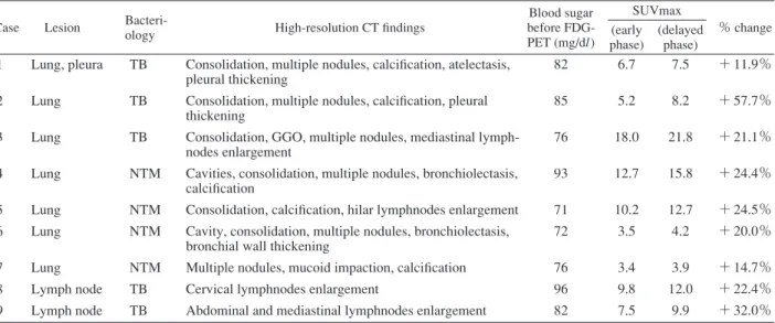
Table High-resolution CT and FDG-PET/CT fi ndings
Case Lesion
Bacteri-ology High-resolution CT fi ndings
Blood sugar before FDG-PET (mg/dl) SUVmax % change (early phase) (delayed phase) 1 2 3 4 5 6 7 8 9 Lung, pleura Lung Lung Lung Lung Lung Lung Lymph node Lymph node TB TB TB NTM NTM NTM NTM TB TB
Consolidation, multiple nodules, calcifi cation, atelectasis, pleural thickening
Consolidation, multiple nodules, calcifi cation, pleural thickening
Consolidation, GGO, multiple nodules, mediastinal lymph-nodes enlargement
Cavities, consolidation, multiple nodules, bronchiolectasis, calcifi cation
Consolidation, calcifi cation, hilar lymphnodes enlargement Cavity, consolidation, multiple nodules, bronchiolectasis, bronchial wall thickening
Multiple nodules, mucoid impaction, calcifi cation Cervical lymphnodes enlargement
Abdominal and mediastinal lymphnodes enlargement
82 85 76 93 71 72 76 96 82 6.7 5.2 18.0 12.7 10.2 3.5 3.4 9.8 7.5 7.5 8.2 21.8 15.8 12.7 4.2 3.9 12.0 9.9 + 11.9% + 57.7% + 21.1% + 24.4% + 24.5% + 20.0% + 14.7% + 22.4% + 32.0% TB : Mycobacterium tuberculosis, NTM : non-tuberculous mycobacteria, GGO : ground glass opacity
40 結核 第 89 巻 第 2 号 2014 年 2 月 は 82 mg/dl(71∼96)であった。抗酸菌症の診断は,肺 非結核性抗酸菌症(Mycobacterium avium)4 例,肺結核 2 例,リンパ節結核 2 例,胸膜結核腫 1 例であった。診 断方法は,気管支鏡が 4 例,手術が 3 例(胸腔鏡下肺ま たはリンパ節生検が 2 例,頸部リンパ節生検が 1 例),CT ガイド下肺生検が 1 例,喀痰が 1 例であった。なお,症 例 3 については,既に症例報告を行っている12)。 結 果 HRCT では,結節や浸潤影,気管支拡張,空洞など多 彩な所見が混在していた(Table)。FDG-PET/CT では,9 例いずれも SUVmax は 2.5 以上であり(中央値 9.9),また 前期相から後期相への SUVmax の上昇を認めた。 結核治療後の胸膜結核腫(症例 1 )および非結核性抗 酸菌症(症例 4 )の 2 例を提示する。 症例 1 は 70 歳女性で喘鳴と倦怠感を主訴に当院を受 診し,喀痰検査でガフキー 1 号,培養検査で M. tubercu-losisが同定され,肺結核・結核性胸膜炎(病型 rⅢ1 Pl) と診断した。薬剤感受性検査はすべて感受性であり,イ ソニアジド,リファンピシン,エタンブトール,ピラジ ナミドの 4 剤による標準治療を終了した。治療終了後の 胸部 CT で胸膜直下に新たな結節を認めたため,FDG-PET/CT が 行 わ れ,SUVmax 6.7( 前 期 相)→ 7.5( 後 期 相)と高い FDG の集積を認めた(Fig. 1)。同部位に対し て CT ガイド下生検を行い,肥厚した胸膜組織中に乾酪 壊死を伴う類上皮細胞肉芽腫が認められたが,抗酸菌の 培養・PCR 検査はいずれも陰性であり,結核治療後の胸 膜結核腫による病変と考えられた。その後,結節は自然 に縮小した。 症例 4 は 76 歳女性で,胃癌と右乳癌術後の経過観察 中に,HRCT で空洞や気管支拡張を伴う浸潤影,胸膜に は一部石灰化を認め,再発の検査のため FDG-PET/CT が 施行された。FDG-PET/CT ではSUVmax 12.7(前期相)→ 15.8(後期相)と高い取り込みを認めた(Fig. 2)。喀痰検 査でM. aviumが検出され,非結核性抗酸菌症と診断した。 考 察 われわれは,FDG-PET/CT が施行された抗酸菌症 9 例 の FDG-PET/CT 所見を検討し,全例で病変部の SUVmax は 2.5 以上であり,かつ前期相から後期相への SUVmax の上昇を認め,FDG の集積からの悪性疾患との鑑別は困 難であった。このうち肺結核の 1 例は,初期悪化による 病変であったが,他の症例と同様に FDG の高い集積が 観察された。 FDG-PET/CT は,グルコース代謝の高い種々の良性疾 患でも偽陽性となることが報告されている4)∼7) 13)。偽陽 性を解決する目的で,さまざまな評価方法が検討されて いる。最も汎用されているのが,SUVmax 2.5 以上であれ ば悪性腫瘍である可能性が高いと判断する方法である9)。 しかし,良性病変でもSUVmax 2.5 以上を示したとする報 告も多い14) 15)。また,悪性病変では FDG 集積のピークが 2 時間以降になるのを利用し,約 1 時間後の前期相より 2 ∼ 3 時間後の後期相に FDG 集積のピークが認められた 場合には悪性を疑う方法が頭頸部の病変で考案され16), その後,肺病変にも応用された10) 11)。集積のピークの時 間が良悪性疾患で異なる理由について明確な機序は分か っていないが,細胞内の FDG をリン酸化または脱リン 酸化するグルコース 6 リン酸化酵素の分布が,悪性病変 では低濃度であるのに対して良性病変では高濃度である ことが影響していると推測されている6)。
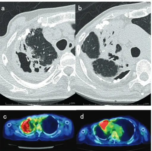
Fig. 1 Case 1: 70-year-old woman
with pulmonary tuberculosis and tuberculous pleurisy.
(a) High-resolution CT before anti-tuberculosis therapy.
(b) After anti-tuberculosis therapy, high-resolution CT showed a new nodule (arrow head).
(c) FDG-PET/CT scan showed remarkable uptake of FDG in it [SUVmax 6.7 (early phase) → 7.5 (delayed phase)]. It was diagnosed as a lesion occurred by pleural tuberculoma after treatment.
Fig. 2 Case 4: 76-year-old woman with non-tuberculosis mycobacterial infection.
(a, b) High-resolution CT showed cavitary consolidation with bronchiectasis. (c, d) FDG-PET/CT scan showed avid FDG uptake [SUVmax 12.7 (early phase) →15.8 (delayed phase)].
42 結核 第 89 巻 第 2 号 2014 年 2 月 抗酸菌症の病変でも,FDG が集積することが報告され ている。Demura ら17)は,肺結核 25 例と非結核性抗酸菌 症 22 例の FDG-PET/CT 所見を解析し,SUVmax 中央値は 5.05(2.5 ∼ 7.6)であった。このうち 14 例については治 療後に再度 FDG-PET/CT を行い,FDG の集積が治療前よ り低下していて,活動性の評価にも有用であると述べて いる。また Goo ら18)は,FDG-PET/CT を行った 10 例の結 核腫について検討し,SUVmax 中央値は 4.2(1.9 ∼ 3.7) であった。さらに,リンパ節結核についても,肺結核や 非結核性抗酸菌と同様に FDG の集積を認めることが報 告されている7)。 一方,抗酸菌症を対象とした前期相と後期相の比較に ついての報告は少ない。Abdul ら19)は結核性脊椎炎の症 例で,前期相から後期相に FDG の集積の低下が認めら れたと報告している。しかし,Sathekge ら20)は結核の 30 例について FDG-PET/CT 所見を検討し,前期相から後期 相への FDG の集積の変化率は悪性疾患と有意差を認め ず,われわれと同じ結果であった。 本検討の限界として,まず単施設での後ろ向きの少数 例の検討であることが挙げられる。また,本検討での FDG-PET/CT の解析は SUV 値を中心に行ったが,実際 の臨床における放射線科医による読影は CT 所見を含め て行っていることが多いと考えられ,本検討と実際の読 影とは異なる可能性がある。さらに,HRCT 所見のみで 抗酸菌の可能性が高いという診断が可能であったかにつ いては,検証を行えていない。 今回の検討から,FDG-PET/CT での FDG の集積度によ り抗酸菌症と悪性疾患を鑑別することは難しく,HRCT 所見も参考にして確定診断をつけることが大切であると 考えられた。 この論文の要旨は,第 86 回日本結核病学会総会(2011 年 6 月 3 日,東京)にて発表した。
著者の COI(confl icts of interest)開示:本論文発表内 容に関して特になし。
文 献
1 ) 小川洋二:肺癌におけるFDG-PET/CT. 肺癌. 2010 ; 50 : 853 859.
2 ) Fischer B, Lassen U, Mortensen J, et al.: Preoperative stag-ing of lung cancer with combined PET-CT. N Engl J Med. 2009 ; 361: 32 39.
3 ) Ung YC, Maziak DE, Vanderveen JA, et al.:18
Fluorodeoxy-glucose positron emission tomography in the diagnosis and staging of lung cancer: a systematic review. J Natl Cancer Inst. 2007 ; 99 : 1753 1767.
4 ) Hara T, Kosaka N, Suzuki T, et al.: Uptake rates of 18
F-fl uorodeoxyglucose and 11C-choline in lung cancer and
pulmonary tuberculosis: a positron emission tomography study. Chest. 2003 ; 124 : 893 901.
5 ) Kubota K, Matsuzawa T, Fujiwara T, et al.: Differential diagnosis of lung tumor with positron emission tomography: a prospective study. J Nucl Med. 1990 ; 31 : 1927 1932. 6 ) Alavi A, Gupta N, Alberini JL, et al.: Positron emission
tomography imaging in nonmalignant thoracic disorders. Semin Nucl Med. 2002 ; 32 : 293 321.
7 ) Chang JM, Lee HJ, Goo JM, et al.: False positive and false negative FDG-PET scans in various thoracic diseases. Korean J Radiol. 2006 ; 7 : 57 69.
8 ) Patz EF Jr, Lowe VJ, Hoffman JM, et al.: Focal pulmonary abnormalities: evaluation with F-18 fl uorodeoxyglucose PET scanning. Radiology. 1993; 188: 487 490.
9 ) Al-Sugair A, Coleman RE: Applications of PET in lung cancer. Semin Nucl Med. 1998 ; 28 : 303 319.
10) Matthies A, Hickeson M, Cuchiara A, et al.: Dual time point
18F-FDG PET for the evaluation of pulmonary nodules. J
Nucl Med. 2002 ; 43 : 871 875.
11) Demura Y, Tsuchida T, Ishizaki T, et al.: 18F-FDG accumulation with PET for differentiation between benign and malignant lesions in the thorax. J Nucl Med. 2003 ; 44 : 540 548.
12) 宇留賀公紀, 村瀬享子, 黒崎敦子, 他:FDG-PETにて高 い集積を認めた乾酪性肺炎の 1 例. 日呼吸会誌. 2010 ; 48 : 247 252.
13) Shim SS, Lee KS, Kim BT, et al.: Focal parenchymal lung lesions showing a potential of positive and false-negative interpretations on integrated PET/CT. AJR Am J Roentgenol. 2006 ; 186 : 639 648.
14) Knight SB, Delbeke D, Stewart JR, et al.: Evaluation of pulmonary lesions with FDG-PET. Comparison of fi ndings in patients with and without a history of prior malignancy. Chest. 1996 ; 109 : 982 988.
15) Kapucu LO, Meltzer CC, Townsend DW, et al.: Fluorine-18-fl uorodeoxyglucose uptake in pneumonia. J Nucl Med. 1998 ; 39 : 1267 1269.
16) Hustinx R, Smith RJ, Benard F, et al.: Dual time point fl uorine-18 fl uorodeoxyglucose positron emission tomogra-phy: a potential method to differentiate malignancy from infl ammation and normal tissue in the head and neck. Eur J Nucl Med. 1999 ; 26 : 1345 1348.
17) Demura Y, Tsuchida T, Uesaka D, et al.: Usefulness of
18F-fl uorodeoxyglucose positron emission tomography for
diagnosing disease activity and monitoring therapeutic response in patients with pulmonary mycobacteriosis. Eur J Nucl Med Mol Imaging. 2009 ; 36 : 632 639.
18) Goo JM, Im JG, Do KH, et al.: Pulmonary tuberculoma evaluated by means of FDG PET: fi ndings in 10 cases. Radiology. 2000 ; 216 : 117 121.
19) Abdul H, Abdul N, Nordin A.: Dual time point imaging of FDG PET/CT in a tuberculous spondylodiscitis. Biomed Imaging Interv J. 2010 ; 6 : e18.
20) Sathekge MM, Maes A, Pottel H, et al.: Dual time-point FDG PET-CT for differentiating benign from malignant
solitary pulmonary nodules in a TB endemic area. S Afr Med J. 2010 ; 100 : 598 601.
Abstract 18F-fl uorodeoxyglucose-positron emission
tomog-raphy (FDG-PET/CT) is a useful technique for distinguishing malignant and benign lesions, although the occurrence of false-positive results in cases involving benign lesions is possible. We evaluated nine patients with mycobacterial infections who underwent PET/CT from April 2008 to July 2010. FDG-PET/CT was performed 1_2h (during the early and late phases) after administration of FDG at a dose of 185 MBq/individual after fasting for at least 5h. Out of the nine patients, four were diagnosed with pulmonary nonmycobacterium tuberculosis, two with pulmonary tuberculosis, two with tuberculous lymph-adenopathy, and one with pleural tuberculoma. All patients had a maximum standardized uptake value (SUVmax) of >2.5, and the SUVmax increased from the early to the late phase. One lesion that occurred due to tuberculous pleurisy after treatment demonstrated high FDG uptake, similar to the other cases. It is diffi cult to distinguish mycobacterial infections from malignant diseases using FGD-PET alone ; hence, the use
of high-resolution CT and bacteriological tests is required for diagnosis and distinction.
Key words:18F-fl uorodeoxyglucose-positron emission
tomog-raphy: FDG-PET/CT, Tuberculosis, Non-tuberculous myco-bacterial infection, Mycobacteriosis, Pleural tuberculoma 1Department of Respiratory Medicine, Respiratory Center, Toranomon Hospital, 2Department of Radiology, and 3 Pathol-ogy, Toranomon Hospital, 4Okinaka Memorial Institute for Medical Research, 5Department of Clinical Radiology, Fukujuji Hospital, Japan Anti-Tuberculosis Association Correspondence to: Hironori Uruga, Department of Respi-ratory Medicine, RespiRespi-ratory Center, Toranomon Hospital, 2_ 2_ 2, Toranomon, Minato-ku, Tokyo 105_ 8470 Japan. (E-mail: [email protected])
−−−−−−−−Original Article−−−−−−−−
EVALUATION OF MYCOBACTERIAL INFECTIONS USING
18
F-FLUORODEOXYGLUCOSE-POSITRON EMISSION TOMOGRAPHY:
RESULTS OF NINE CASES
1, 4Hironori URUGA, 2Makiko ISHIHARA, 1Shigeo HANADA, 1Hisashi TAKAYA, 1Atsushi MIYAMOTO, 1Nasa MOROKAWA, 3Takeshi FUJII, 5Atsuko KUROSAKI,