Human Skeletal Remains from the Teauma Site,
Marakei Island, Gilbert Islands, Republic of
Kiribati
著者
OGATA Takahiko, KAWAJI Noritomo, MINE
Kazuharu, YAMAMOTO Miyoko
journal or
publication title
南海研紀要
volume
6
number
1
page range
72-92
URL
http://hdl.handle.net/10232/15662
72 Mem. Kagoshima Univ. Res. Center S. Pac, Vol.6, No. I, 1985
Human Skeletal Remains from the Teauma Site,
Marakei Island, Gilbert Islands, Republic
of Kiribati*
Takahiko OGATA**, Noritomo Kawaji**, Kazuharu MINE**
and Miyoko YAMAMOTO**
Abstract
Several human skeletal remains excavated from Marakei Island, Republic of
Kiribati in 1984 were examined. It was shown that these were recently burried and at least five individuals were recognizable. As to the cranial indices, one is meso-, hypsi- and acrocranic, and another is dolicho-, ortho- and acrocranic. All of the mandibles (three males and two females) have an ante-gonial notch, not exhibiting the so-called "rocker jaw". The postcranial skeletons were large for the males so that the estimated heights, calculated by Pearson's formula, were very tall (169.0 cm and 173.3 cm). Whereas the postcranial skeletons of the females were small and slender and the statures relatively short (149.7 cm and 155.5 cm). On several bones
osteoarthritic changes and healed fracture were observed.
Introduction
The Gilbert Islands, including Marakei Island, are located at the easternmost end of Micronesia, bounded on the southeast by Polynesia and on the southwest
by Melanesia (Fig. 1). The location of the Gilbert Islands is important to elucidate
the origin of Polynesians as well as Micronesians. However, there have been few reports concerned with skeletal remains of the Gilbert Islanders, e. g. Krause (1881).
In the present paper, we examine some skeletal materials obtained from Marakei
Island, and discuss the relationships between them and those of other Oceanic
populations.
Materials and Methods
The skeletal remains were excavated from the Teauma site, Marakei Island, the *This study was reported to Mr. B. Eritaia, Cultural Officer, Ministry of Home Affairs and Decentraliza
tion, Republic of Kiribati in March, 1985.
Mem. Kagoshima Univ. Res. Center S. Pac, Vol.6, No. 1, 1985 JAPAN140
V
NORTHERN-MARIANA ISLANDS ;Saipan ' Guam BELAU FEDERATED STATES OF ~T~ 160' 170' iao" no-MARSHALL ISLANDS \ Midway MICRONESIA GILBERT ISLANDS ., , . Marake Makin, i SOLOMON 1~ Qs, ISLANDS 73 160' Oahu °S> O'Fig. 1. Map of Micronesia showing location of Teauma site, Marakei Island
Gilbert Islands in 1984. This site is located at the southeast part of the Marakei Atoll. It is a "Te Baro", a grave for the members of a family or a clan, about 80-100 years ago. The human skeletal materials were found in a jumbled state, so that they were reassembled into individuals according to the morphological
characters and dimensions of bones. The measurements were carried out after Martin
& Sailer (1957).
Results
At least five individuals are recognized. The observations and measurements
for those skeletons are as follows.
1. Cranium
Three calvariae (Nos. 1-3) and five mandibles (Nos. 1-5) can be measured.
Although many fragments of crania are preserved, there were no complete ones to be measured. The upper facial skeletons were almost lost except for fragments of
maxilla and zygomatic bones.
No. 1 calvaria and No. 1 mandible are supposed to belong to the same individual,
whereas others are obscure. The measurements and indices are shown in Tables 1
74 OGATA et al. : Human Skeletal Remains from Marakei Island
No. 1 calvaria (Plate 1)
Sex : Male, from well-developed brow-ridges, mastoid processes and external occipital protuberance.
Age: Adult, from complete ossification of spheno-occipital synchondrosis and no
occurrence of sutural obliteration.
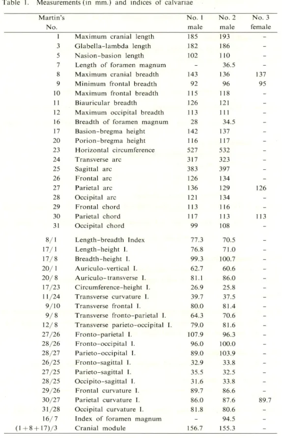
Comment: Only cerebral cranium is preserved. Cranial dimensions are large in
general. As measurements, maximum length is normal, while maximum
breadth and basion-bregma height are large. The indices show meso- (77.3),
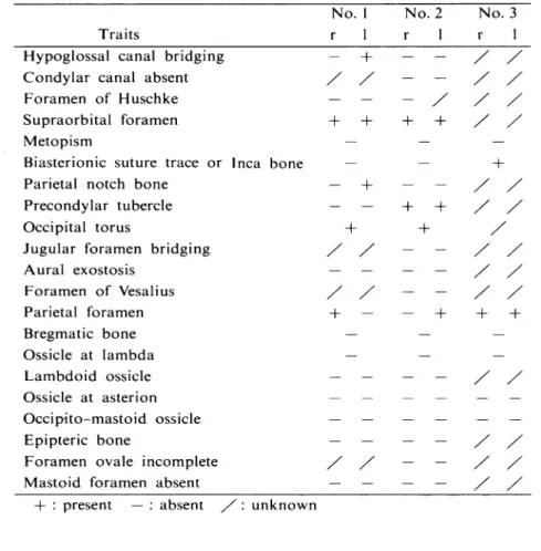
hypsi- (76.8) and acrocranic (99.3). The breadth of the frontal region is narrow and the transverse fronto-parietal index is rather small (64.3). As non-metric variations, hypoglossal canal bridging (1), supraorbital foramina (r, 1), parietal notch bone (1), occipital torus and parietal foramen (r) are
found.
No. 2 calvaria (Plate 1)
Sex : Male, from well-developed brow-ridges and external occipital protuberance. Age: Mature, from complete obliteration of the main cranial sutures.
Comment: Only cerebral cranium is preserved. As measurements, maximum length is fairly large, while maximum breadth is small and basion-bregma height
is normal. The indices show dolicho- (70.5), ortho- (71.0) and acrocranic
(100.7); considerably different from No. 1 calvaria. The frontal arc is longer than the parietal arc. Namely the fronto-parietal index is small (96.3). As non-metric variations, supraorbital foramina (r, 1), precondylar tubercles (r, 1), occipital torus and parietal foramen (1) are found. Supramastoid crests are remarkably developed.
No. 3 calvaria
Sex : Female, from small size in general.
Age: Adult, from slight occurrence of the main sutural obliteration.
Comment: Only cranial vault is preserved, lacking cranial base and face. A few
items can be measured. Maximum cranial breadth is somewhat broad. It
appears mesocranic on observation and resembles No. 1 calvaria in norma
lateralis. Inca bone and parietal foramina (r, 1) are found.
No. 1 mandible (Plate 2)
Sex : Male, from large dimensions and rugged appearance.
Age : Adult, from slight attrition of teeth and condition of 8 8.
Dentition:
8 7 6 5 4 0 0 0 | 0 0 0 4 5 6 7 ®
( O post-mortem loss; ® almost impacted tooth)
Comment: This mandible is preserved nearly intact except post-mortem loss of six anterior teeth. It is robust and mental protuberance is developed. Attrition
Mem. Kagoshima Univ. Res. Center S. Pac, Vol.6, No. I, 1985 75
of remaining teeth is limited to enamel. 8 has already erupted, whereas
I 8 tilts mesially and is almost impacted.
Dental caries is not found.
No. 2 mandible (Plate 2)
Sex : Male, from large dimensions and eversion of mandibular angle. Age : Mature, from its dentition.
Dentition :
/ /
V O O / 0 | 0 0 0 0 5 XXO
(X ante-mortem loss; / both tooth and socket missing)
Comment: Only left half is preserved. 6 7 are lost ante-mortem and 4 3 1 1 2 3 4 8 post-mortem. Remaining 5 tilts distally and attrition is slight.
No. 3 mandible (Plate 2)
Sex : Female, from smooth contour and small size in general. Age : Mature, from its dentition.
Dentition : A X X X X O / / / O O x O X X A
(A probably congenital absence)
Comment: This lacks ascending rami bilaterally. All premolars and molars except
5 are lost ante-mortem and alveolar bone is absorbed. The alveoli of
2 3 are enlarged probably due to periodontal lesion.
No. 4 mandible (Plate 2)
Sex : Female, from its dimension and slenderness.
Age : Old, from indications of what appear to have been edenturous.
Dentition : / / / / / / X X | X X X X X X X A
Comment: The alveolar ridge of the right posterior teeth is broken. Absorption of
the alveolar part reaches to the level of the mental foramen. Ascending rami
tilt backward and the gonial angle is obtuse (143°).
No. 5 mandible (Plate 2)
Sex : Male, from its robustness and roughness of muscular attachment area.
Age : Adult, from eruption of 8
Dentition :
/ / / / / / / /
\ / / / / / O Z O
Comment : Only left ascending ramus is preserved. Minimum ramal breadth is
76 Ogata et al. : Human Skeletal Remains from Marakei Island
Table 1. Measurements (in mm.) and indices of calvariae
Martin's No. 1 No. 2 No. 3
No. male male female
1 Maximum cranial length 185 193
-3 Glabella-lambda length 182 186
-5 Nasion-basion length 102 110
-7 Length of foramen magnum - 36.5
-8 Maximum cranial breadth 143 136 137 9 Minimum frontal breadth 92 96 95 10 Maximum frontal breadth 115 118
-11 Biauricular breadth 126 121
-12 Maximum occipital breadth 113 111
-16 Breadth of foramen magnum 28 34.5
-17 Basion-bregma height 142 137 -20 Porion-bregma height 116 117 -23 Horizontal circumference 527 532 -24 Transverse arc 317 323 -25 Sagittal arc 383 397 -26 Frontal arc 126 134 -27 Parietal arc 136 129 126 28 Occipital arc 121 134 -29 Frontal chord 113 116 -30 Parietal chord 117 113 113 31 Occipital chord 99 108 -8/1 Length-breadth Index 77.3 70.5 -17/1 Length-height I. 76.8 71.0 -17/8 Breadth-height I. 99.3 100.7 -20/1 Auriculo-vertical I. 62.7 60.6 -20/8 Auriculo-transverse I. 81.1 86.0 -17/23 Circumference-height I. 26.9 25.8 -11/24 Transverse curvature I. 39.7 37.5 -9/10 Transverse frontal I. 80.0 81.4 -9/8 Transverse fronto-parietal I. 64.3 70.6 -12/8 Transverse parieto-occipital I. 79.0 81.6 -27/26 Fronto-parietal I. 107.9 96.3 -28/26 Fronto-occipital I. 96.0 100.0 -28/27 Parieto-occipital I. 89.0 103.9 -26/25 Fronto-sagittal I. 32.9 33.8 -27/25 Parieto-sagittal I. 35.5 32.5 -28/25 Occipito-sagittal I. 31.6 33.8 -29/26 Frontal curvature I. 89.7 86.6 -30/27 Parietal curvature I. 86.0 87.6 89.7 31/28 Occipital curvature I. 81.8 80.6
-16/7 Index of foramen magnum - 94.5
-Mem. Kagoshima Univ. Res. Center S. Pac, Vol.6, No. 1, 1985 77 Table 2. Measurements and indices of mandibles
Martin's No.
No. 1 No. 2 No. 3 No. 4 No. 5 male male female female male
65 Bicondylar breadth 125 - - -
-66 Bigonial breadth 105 - 87 -
-67 Anterior mandibular breadth 56 - 44 -
-68 Mandibular length 80 (84) 74 -
-68(1) Mandibular length 112 - - 98
-69 Symphyseal height 32 (30) - -
-69(1) Mandibular body height 34 32 25 -
-69(3) Mandibular body thickness 13 1 1 10 9
-70 Ramal height 74 - - (49)
-70(1) Anterior ramal height - 69 52 - 62
70(2) Minimum ramal height 56 54 - (33) (56)
70a Condylar height 69 - - -
-71 Ramal breadth 35 - - -
-71a Minimum ramal breadth 33.5 35.5 - - 36
79 Gonial angle 115° - - 143° -68/65 Breadth-length I. 64.0 - - - -71/70 Ramal I. 47.3 - - - -71a/70(2) Ramal I. 59.8 65.7 - - (64.3 66/65 Mandibular breadth I. 84.0 - - - -69(3)/69(l) Body height-thickness I. 38.2 34.4 40.0 - -Note : The estimated values are in parentheses.
2. Postcranial Skeleton (Plates 3 and 4)
Many shafts of long bones and fragments are preserved (Table 4). At least
three males (A, B and C) and two females (D and E) are recognized. Measurements
and indices are shown in Tables 5-10. Identification of each individual according
to the morphological characters of bones are as follows:
Individual A
Material: A left humerus (H-l), a left radius (R-l), a left ulna(U-l), shafts of
the femora (F-l, 2) and a shaft of the left tibia (T-l). Sex : Male, from bone dimensions.
Age: Mature (?)
Comment: Materials show large dimensions in general. Osteoarthritic bone lipping and pitting occur in the articular facets of the left elbow joint. Capitulum
of humerus and head of radius are eburnated and show the ivory polish
(Plate 5). The femora exhibit well-developed pilastering (Fig. 2) and tend to
be platymeric (platymeric indices : r 87.5, 1 84.4). The tibia is not platycnemic
78 Ogata et al.: Human Skeletal Remains from Marakei Island
Table 3-1. Non-metric variations (calvariae)
No. 1 No. 2 No. 3
Traits r 1 r 1 r 1
Hypoglossal canal bridging - + - - / /
Condylar canal absent / / - - / /
Foramen of Huschke — - - / / /
Supraorbital foramen + + + + / /
Metopism - -
-Biasterionic suture trace or Inca bone — — +
Parietal notch bone - + — — / /
Precondylar tubercle - — + + / /
Occipital torus + + /
Jugular foramen bridging / / - - / /
Aural exostosis — — — — / / Foramen of Vesalius / / — — / / Parietal foramen + - - + + + Bregmatic bone - — — Ossicle at lambda — — — Lambdoid ossicle — — — — / / Ossicle at asterion Occipito-mastoid ossicle Epipteric bone - — - - / /
Foramen ovale incomplete / / - — / /
Mastoid foramen absent - - — — / / + : present — : absent / : unknown
Table 3-2. Non-metric variations (mandibles)
No. 1 No. 2 No. 3 No. 4 No. 5 Traits r 1 r 1 r 1 r . 1 r 1
Mylohyoid bridging Accessory mental foramen
Mandibular torus
estimated to be 173.3 cm (Table 11). It seems that No. 2 calvaria belongs
to this individual.
Individual B
Material: Shafts of the humeri (H-2, 3), portions of the radii (R-2, 3) and ulnae (U-2, 3), portion of the left femur (F-3), an almost complete left tibia (T-2)
Mem. Kagoshima Univ. Res. Center S. Pac, Vol. 6, No. 1, 1985
Table 4. Postcranial skeletal remains
Bone Remains
Vertebra many fragments Sternum a fragment Rib many fragments Scapula some fragments
Clavicle some fragments
Humerus five shafts and fragments
H-l : left (A, male): head is broken
H-2 : right (B. male) : shaft only
H-3 : left (B, male): shaft only H-4: right (E, female) : shaft only H-5 : left (E, female) : shaft only Radius seven shafts and fragments
R-I : left (A, male) R-2: right (B, male) R-3: left (B, male) R-4: right (C, male) R-5: left (D, female) R-6 : right (E, female)
R-7: left (E, female)
Ulna six shafts and fragments
U-l : left (A, male) U-2: right (B, male)
U-3 : left (B, male)
U-4 : right (D, female) U-5 : right (C. male)
U-6: left (C, male)
Pelvis many fragments
Femur six shafts and fragments F-l : right (A, male)
F-2 : left (A, male)
F-3 : left (B, male)
F-4: left (D. female): almost complete F-5 : right (E, female): shaft only F-6 : left (E, female) : almost complete
Patella a right one
Tibia four shafts and fragments
T-l : left (A, male) : shaft only T-2 : left (B. male) : almost complete
T-3 : left (C, male) : shaft only
T-4: left (E, female): shaft only
Fibula a shaft and fragments
Fb-1 : left (B, male): shaft only
Others some carpal bones, metacarpal bones, tarsal bones,
metatarsal bones and so on
complete
portion of the shaft portion of the shaft almost complete shaft only shaft only shaft only complete shaft only
portion of the shaft portion of the shaft almost complete almost complete
shaft only
shaft only
lower portion only
80 Ogata et al. : Human Skeletal Remains from Marakei Island
F - l ( r ) F-2(l) F-3(l)
F-4(l) F-5(r) F-6(l)
Fig. 2. Cross-sectional shape of the midshaft of femur
Sex : Male, from bone dimensions.
Age : Adult ( ? )
Comment: Since the long bones have large dimensions and strong muscle marks, this
individual should be a big and strong man. The femur shows considerably
developed pilastering (Fig. 2). The pilasteric index (125.9) is large. The tibia is not so platycnemic (cnemic index : 66.7) and development of the soleal line
is moderate. The fibula is robust. The height is estimated to be 169.0 cm
(Table 11). It seems that No. 1 calvaria belongs to this individual.
Individual C
Material: A right radius (R-4). ulnae (U-5, 6) and a shaft of the left tibia (T-3).
Sex : Male, from bone dimensions.
Age : Adult ( ? )
Comment : The ulnae are robust. The tibia exhibits strong muscle marks and medial
bowing at the midshaft and is not so platycnemic (cnemic index : 64.9).
Individual D
Material : A shaft of the left radius (R-5), a portion of the right ulna (U-4) and an almost complete left femur (F-4).
Sex : Female, from bone dimensions, Age : Mature ( ? )
Comment: The radius is slender but extension of interosseous crest is well marked.
The ulna has an old healed fracture on the shaft and osteoarthritic lipping
Mem. Kagoshima Univ. Res. Center S. Pac, Vol. 6, No. 1985
Table 5. Measurements and indices of humerus
Martin's No.
5 Max. dia. midshaft 6 Min. dia. midshaft
7 Least girth of shaft
7a Girth in middle 6/5 Cross section I. H-I(l) male H-2(r) male H-3(l) male H-4(r) female H-5(l) female 24 26 27 21 20 19 20 20 15 15 65 70 73 56 54 70 75 76 59 57 79.2 76.9 74.1 71.4 75.0
Table 6. Measurements and indices of radius
Martin's No.
R-I(l) R-2(r) R-3(l) R-4(r) R-5( 1) R-6(r) R-7(l)
male male male male female female female
1 Maximum length 267 2 Physiological length 252 3 Least girth of shaft 43
4 Trans, dia. of shaft 17 4a Trans, dia. in middle 17
5 Sag. dia. of shaft 14 5a Sag. dia. in middle 14
5(5) Girth in middle 46
3/2 Length-thickness I. 17.1 5/4 Cross section I. 82.4
17
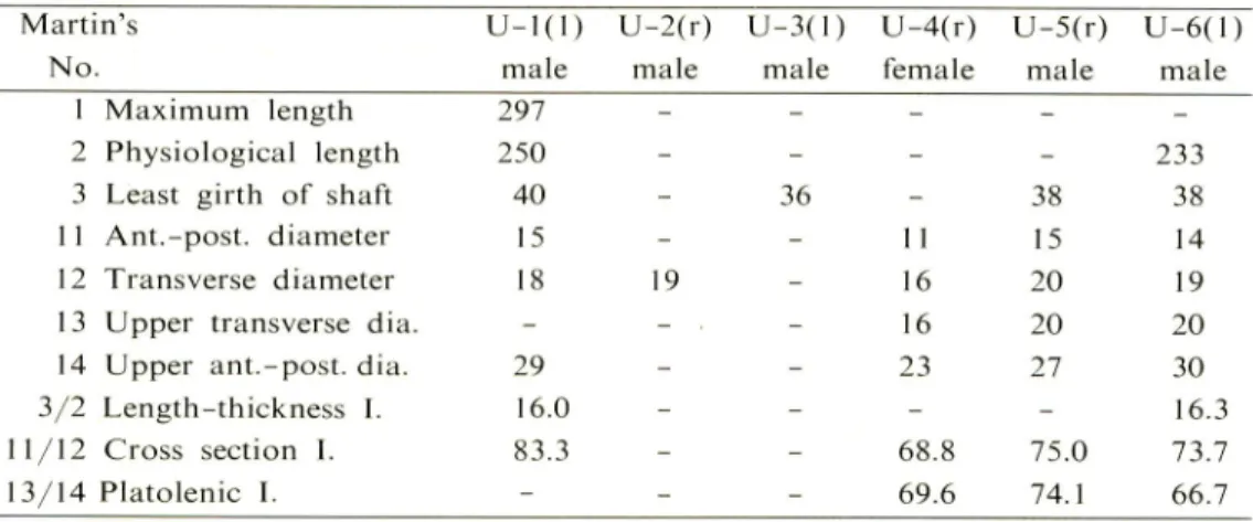
Table 7. Measurements and indices of ulna
13 68.4 240 - - -44 37 - 32 17 18 13 14 16 13 - -14 10 10 10 14 11 - -46 37 - -18.3 - - -82.4 55.6 76.9 71. Martin's No.
U-I(l) U-2(r) U-3(l) U-4(r) U-5(r) U-6(l)
male male male female male male
1 Maximum length 2 Physiological length 3 Least girth of shaft 1 1 Ant.-post. diameter 12 Transverse diameter 13 Upper transverse dia. 14 Upper ant.-post. dia. 3/2 Length-thickness I. 11/12 Cross section I. 13/14 Platolenic I. 297 250 40 15 29 16.0 83.3 36 19 - - 233 - 38 38 11 15 14 16 20 19 16 20 20 23 27 30 - - 16.3 68.8 75.0 73.7 69.6 74.1 66.7
Ogata et al. : Human Skeletal Remains from Marakei Island
Table Measurements and indices of femur
Marti r i's F-I(r) F-2(l) F-3( 1) F-4( 1) F-5(r) F-6(l)
No. male male male female female female
1 Maximum length - - - 425 - (395)
2 Physiological length - - - 421 - (388)
6 Sag. dia. midshaft 29 30 34 26 24 24
7 Trans, dia. midshaft 26 26 27 25 22 23 8 Girth in middle 84 86 96 81 72 72
9 Upper trans, dia. shaft 32 32 - 28 30 28
10 Upper sag. dia. shaft 28 27 - 24 24 21
13 Upper breadth - - - 87 -
-14 Head length - - - 58 -
-15 Vertical dia. neck - - - 28 - 25
16 Sagittal dia. neck - - - 25 -
-17 Girth of neck - - - 92 -
-23 Max. length lat. cond. - - - 61 -
-25 Post, height lat. cond. - - - 36 -
-29 Neck-shaft angle - - - 135° -
-8/2 Length-thickness 1. - - - 19.2 - (18.6)
6/7 Pilasteric I. 111.5 115.4 125.9 104.0 109.1 104.3
10/9 Platymeric 1. 87.5 84.4 - 85.7 80.0 75.0
16/15 Cross section I. neck - - - 89.3 -
-Table 9. Measurements and indices of tibia
Martin's No. 1 Total length la Maximum length 3 Upper breadth 6 Lower breadth
7 Sag. dia. of lower epiph. 8 Max. dia. in middle 8a Max. dia. in nut. foramen 9 Trans, dia. in middle 9a Trans, dia. in nut. foramen 10 Girth of shaft
10a Girth in nut. foramen 10b Least girth of shaft 9a/8a Cnemic I. 9/8 Cross section I. 10/la Length-thickness 1. T-I(l) male T-2(l) male T-3( 1) male T-4( I ) female - 373 - -- 380 - -- 70 - -- 50 - -- 38 - -32 34 34 26 38 39 37 30 23 23 23 20 30 26 24 20 86 92 87 72 - 102 93 79 78 80 78 63 78.9 66.7 64.9 66.7 71.9 67.6 67.6 76.9 - 24.2 _ _
Mem. Kagoshima Univ. Res. Center S. Pac, Vol.6, No. I, 1985 83
Table 10. Measurements and indices of fibula
Martin's Fb-l(l)
No. male
2 Max. dia. in middle 19
3 Min. dia. in middle 13 4 Girth in middle 54
3/2 Cross section I. 68.4
Table 11. Estimated stature according to Pearson's formula
Individual Sex Bone Estimated
examined stature (cm)
A male 1. radius 173.3
B male 1. tibia 169.0
D female 1. femur 155.5
E female 1. femur 149.7
to be 155.5 cm (Table 11). It seems that No. 3 mandible belongs to this
individual.
Individual E
Material: Shafts of the humeri (H-4, 5) and radii (R-6, 7), almost complete femora
(F-5, 6) and a shaft of the left tibia (T-4). Sex : Female, from bone dimensions.
Age : Old (?)
Comment : As the long bones have small dimensions and weak muscle marks, this
individual seems to be a small and gracile woman.
The femora are not very
pilasteric (Fig. 2). The tibia is not very platycnemic (cnemic index : 66.7).
The height is estimated to be 149.7 cm (Table 11). It seems that No. 4
mandible belongs to this individual.
Discussion
Although the Teauma crania lack an upper face, the main measurements and indices of Nos. 1 and 2 calvariae (both male) could be compared with the available
data of other Oceanic populations (Table 12).
Table 12. Comparison of main cranial measurements and indices (Male) Micronesia Melanesia Polynesia Teauma Teauma Gilbert Saipan Guam New Britain Fiji Auckland Mokapu Martin's No. 1 No. 2 Is. Maori No. n=16 n=5 n=27 n=229 n=13 n=90 n=139 1 Maximum cranial length 185 193 182.8 185.0 180.5 184.3 192.6 185.9 184.3 c 0 8 Maximum cranial breadth 143 136 136.8 143.8 140.5 132.4 134.2 135.7 145.0
£
9 Minimum frontal breadth 92 96 -97.2 96.8 93.3 96.5 94.0 95.6 m 10 Maximum frontal breadth 115 118 -118.0 -112.4 -110.5 117.6 — 17 Basion-bregma height 142 137 141.9 144.3 143.6 -138.1 137.9 143.0 X i Pi E3 23 Horizontal circumference 527 532 -529.2 511.8 518.7 526.9 525.0 523.2 24 Transverse arc 317 323 -323.6 316.7 304.1 314.0 313.0 334.4 C/3 25 Sagittal arc 383 397 -381.8 378.9 374.5 370.4 377.0 380.2 5" 26 Frontal arc 126 134 -136.6 -124.5 -130.1 135.9 70 27 Parietal arc 136 129 -131.4 -132.5 -126.6 126.1 | 28 Occipital arc 121 134 -113.2 -117.5 -119.9 118.2 a 8/1 Length-breadth 1. 77.3 70.5 74.8 77.8 78.5 71.9 69.6 73.0 79.2 o 3 <: 17/1 Length-height I. 76.8 71.0 77.6 77.9 79.6 -71.7 74.2 77.7 17/8 Breadth-height I. 99.3 100.7 103.7 100.3 102.2 -97.2 101.8 98.3 9/10 Transverse frontal I. 80.0 81.4 -82.0 -83.0 -85.2 81.8 ST 9/8 Transverse fronto-parietal I. 64.3 70.6 -66.4 68.9 70.5 71.9 69.5 66.3 27/26 Fronto-parietal I. 107.9 96.3 -96.1 -106.4 -97.9 92.8 28/26 Fronto-occipital I. 96.0 100.0 -83.3 -94.4 -92.4 87.0 Krause Sarai Marshall Bonin Marshall Shima Snow Author (1881) (1951) & Snow (1956) (1936) & Snow (1956) & Suzuki (1967) (1974)Mem. Kagoshima Univ. Res. Center S. Pac, Vol. 6. No. I, 1985 85 whereas No. 2 calvaria is dolicho-. ortho- and acrocranic, and the parietal arc is
short.
The characteristics of No. I resemble those of Saipan and Mokapu.
On the
other hand, those of No. 2 resemble Fiji and Auckland Maori. The narrowness
of the frontal region is characteristic in Polynesians (Shima & Suzuki, 1967).
Whether it is also characteristic in the Teauma population is obscure since only
three remains have been examined.
All of the mandibles have an ante-gonial notch,
not the so-called "rocker jaw" which is the most frequent form of Polynesian
mandible (Marshall & Snow, 1956). Thus the characteristics of the Teauma crania
are not uniform from one to another.
The postcranial skeletons have large dimensions and strong muscle marks in
the male.
The femur tends to be pilasteric in both sexes.
Those of Mokapu also
possess the primitive characteristics in limb bones, for example, the pilaster formation
of femora (Snow, 1974).
From the pathological point of view, osteoarthritic changes on several bones and healed fracture of the ulna can be seen.
Acknowledgement
We wish to thank Prof. J. Takayama, Tezukayama University, for offering
these skeletal materials.
References
Bonin, G. von, 1936: On the craniology of Oceania. Crania from New Britain. Biometrika 2: 123-148.
Krause, R., 1881 : Ein Beitrag zur Kunde der Sudsee-Volker.
Die ethn-anthrop.
Abteilung des Museum Godeffory. Hamburg.
Marshall, D. S. & C. E. Snow, 1956: An evaluation of Polynesian craniology.
Am.
J. Phys. Anthrop. 14:405-426.
Martin, R. & K. Sailer, 1957 : Lehrbuch der Anthropologic
Gustav Fischer,
Stuttgart.
Sarai.C, 1951 : Mikuroneshia-jin Togaikotsuno Jinruigaku-tekiKenkyu{Anthropologi
cal study on the Micronesian skulls). Tokyo Jikeikai Ikadaigaku
Kaibogaku-kyoshitsu Gyosekishu (Rep. Dep. Anat., Jikei Univ. Sch. Med.) 4: 1-57 (in
Japanese).
Shima, G., 1966: The physical anthropology of the Polynesians. Acta Anatomica
Nipponica 41 : 139-141 (in Japanese).
Shima, G., 1968: Das Verhaltnis der Polynesier zu den Mikronesiern in Betracht der Kephaldimensionen und Kephalindizes. Osaka City Medical Journal 14:
86 OGATA et al. : Human Skeletal Remains from Marakei Island
Shima, G. & M.Suzuki, 1967: Problems of race formation of the Maori and Moriori in terms of skulls. Osaka City Medical Journal 13: 9-54.
Snow, C. E., 1974: Early Hawaiians. University Press of Kentucky, Lexington,
Kentucky.
Mem. Kagoshima Univ. Res. Center S. Pac, Vol. 6, No. 1, 1985 87
Explanations of Plates
Plate 1 No. 2 calvaria (A~D) and No. 1 calvaria (E~H).
Plate 2 Mandibles. No. 1 (A), No. 2 (B), No. 3 (C), No. 4 (D) and No. 5 (E).
Plate 3 Skeletons of the upper limb. Humeri (H-l-H-5), ulnae (U-l-U-6) and
radii (R-NR-7).
Plate 4 Skeletons of the lower limb. Femora (F-NF-6), tibiae (T-NT-4) and
fibula (Fb-1).
Plate 5 Osteoarthritic changes at the left elbow joint (A), the cervical vertebra (B-a),
88 OGATA et al. : Human Skeletal Remains from Marakei Island
Mem. Kagoshima Univ. Res. Center S. Pac, Vol. 6, No. 1, 1985 89
90 OGATA et al. : Human Skeletal Remains from Marakei Island
9m^§§§S^"§l!U^^^
O
41
Mem. Kagoshima Univ. Res. Center S. Pac, Vol.6, No. 1, 1985 91
:*$?;# #f
llll
*Sil!p;flpM?iRil!jMpiJ|ii smI *V. :&• W -W;M- •&*•-'-:.#?4;'-*#v# #-•
IliillilSI
iiBllltllliliBgiil ISSIMSSk iSwiiillif^NffijIi^iS*"^'-•: >• •«&'• .& is*
EPS
OS Sgfifsn pil|i!!^l|l!^ii|iil!li|^illl= ilea! llil;illls&l§ &^&P&M&£$£;*••*•#'**- #.*:
92 OGATA et al. : Human Skeletal Remains from Marakei Island
? '^m-0:
«