緒 言
げっ歯類を用いる小核試験(以下小核試験)は,幼若
赤血球中の小核の有無を観察することにより,化学物質
の染色体異常誘発作用を間接的に評価する in vivo 遺伝
毒性試験の一つとして広く用いられている(Hayashi et
al., 2000; Krishna and Hayashi, 2000).小核試験には骨髄
細胞と末梢血を用いる 2 種類の方法があり,骨髄細胞で
は,ギムザあるいはアクリジンオレンジ(AO)染色法
(Hayashi et al., 1983)を用い,幼若赤血球を観察して小
核出現頻度を求めている.末梢血では,AO 超生体染色
法(Hayashi et al., 1990)を用い,幼若赤血球の中で観
Environ. Mutagen Res., 27: 171-175(2005)
原 著
ニューメチレンブルー/アクリジンオレンジ染色法を
用いたラット骨髄におけるフェナセチンの
小核誘発作用について
杉原 忠一
*,澤田 繁樹,羽倉 昌志,内田 加奈子,
堀 雄二,野々垣 泰,青木 豊彦
エーザイ
(株)
安全性研究所川島研究室 〒 501-6195 岐阜県各務原市川島竹早町 1
Micronucleus formation in the bone marrow of rats treated with phenacetin
using new methylene blue/acridine orange staining method
Tadakazu Sugihara
*, Shigeki Sawada, Atsushi Hakura, Kanako Uchida, Yuji Hori,
Yasushi Nonogaki and Toyohiko Aoki
Kawashima Research, Drug Safety Research Laboratories, Eisai Co., Ltd. 1 Kawashimatakehaya-machi, Kakamigahara-shi, Gifu 501-6195, Japan
Summary
In the rat micronucleus test, it was reported that phenacetin induced micronuclei in peripheral blood, but not in bone marrow. In this study, we used the new methylene blue/acridine orange (N/AO) staining method and the acridine orange (AO) staining method to examine whether phenacetin induces micronuclei in rat bone marrow. Utilizing the method of N/AO staining, reticulocytes were classified into Type I to IV, and then micronucleated reticulocytes (MNRETs) of each type were counted. Micronucleated polychromat-ic erythrocytes (MNPCEs) were detected in AO-stained specimens. After a single oral administration of phenacetin, MNRETs were mainly increased in Type II and Type III reticulocytes at 24 and 48 hours, respec-tively, but were not increased at 72 h after administration at a dose of 2000 mg/kg. In addition, the frequen-cy of MNRETs in reticulofrequen-cytes of Type I and Type II was increased at doses of 500, 1000 and 2000 mg/kg, and the MNRETs frequency at 2000 mg/kg was 3-fold higher than that of MNPCEs at the same dose. After two or four repeated doses of phenacetin, MNRETs were mainly increased in both Type II and Type III retic-ulocytes at 24 h after final administration. Similar magnitude increases in the frequencies of MNRETs in Type I and Type II reticulocytes, and in Type III reticulocytes and MNPCEs were found for both two and four repeated doses. These results indicate that phenacetin can induce micronuclei in rat bone marrow and that the N/AO staining method in combination with micronucleus scoring in the restricted type(s) of reticu-locytes is useful in the rat micronucleus test.
Keywords: micronucleus test, bone marrow, new methylene blue/acridine orange staining, reticulocyte,
phenacetin
*E-mail: [email protected]
受付: 2005 年 8 月 31 日 改訂稿受領: 2005 年 10 月 12 日 受理: 2005 年 10 月 12 日
察細胞を限定して小核出現頻度を求めている.すなわち,
マウスの末梢血では,観察細胞を網赤血球 I 型,II 型お
よび III 型のみに限定することによって,骨髄細胞と同
様に末梢血においても化合物の小核誘発性を検出できる
と報告されている(CSGMT, 1992)
.ラットの末梢血で
は,観察細胞を網赤血球 I 型および II 型のみに限定して
小核出現頻度を求めているが,脾臓で小核を有する幼若
赤血球が破壊されることもあり,骨髄細胞の方が末梢血
に比べて検出力が高いと報告されている(Wakata et al.,
1998).
解 熱 鎮 痛 薬 で あ る フ ェ ナ セ チ ン は , A m e s 試 験
(Nohmi et al.,
1983),染色体異常試験(Granberg-Ohman et al., 1980)およびマウス小核試験(骨髄細胞お
よび末梢血)
(CSGMT, 1990; 1992)では陽性と報告され
ている.しかし,ラット小核試験の骨髄細胞(2 回反復
投与の 500 mg/kg 群)では陰性,末梢血(単回投与の
2000 mg/kg 群)では陽性と報告されている(Wakata et
al., 1998).本論文では,ラットにおける骨髄細胞と末梢
血との小核誘発性の相違が観察対象細胞,投与量あるい
は投与回数の相違に基づくものであるかを確認するた
め,著者らが開発したニューメチレンブルー/アクリジ
ンオレンジ(N/AO)染色法(Sugihara et al., 2000)を
用い,骨髄細胞においても観察細胞を限定することによ
って,フェナセチンの小核誘発性を検出できるかを調べ
るとともに,投与回数の影響も併せて検討した.
実験材料および方法
1.動物
日本チャールス・リバー
(株)より Crj:CD
(SD)
IGS 雄
ラットを購入し,2 週間馴化した後,ランダムに各群 5
例に分け,8 週齢で使用した.飼料は 1 日約 21 g の制限
給餌で与え,水は自由に摂取させた.
2.フェナセチンの投与
フェナセチン(phenacetin, CAS No. 62-44-2,和光純
薬工業)をオリーブ油で懸濁した後,単回経口投与,2
回および 4 回反復経口投与した.単回では,250,500,
1000,2000 mg/kg を投与し,投与 24,48 および 72 時間
後に動物を頸椎脱臼で安楽死させ,大腿骨から骨髄細胞
を採取し(Yamamoto and Kikuchi, 1981)
,牛胎仔血清
(Invitrogen Corp.)で骨髄細胞浮遊液を調製した.2 回
および 4 回反復では,単回投与の 2000 mg/kg 群で死亡
動物(5/15 例)が認められたため,250,500,1000
mg/kg を 24 時間間隔で 2 回あるいは 4 回投与し,最終
投与 24 時間後に単回と同様に骨髄細胞浮遊液を調製し
た.対照群にはオリーブ油を用いた.投与容量は,単回
投与群では 2 mL/100 g,2 回および 4 回反復投与群では
1 mL/100 g とした.
3.小核標本の作製と観察
N/AO 染色法では,骨髄細胞浮遊液に同量(20
µ
L)
の 0.5%ニューメチレンブルー溶液を混合し,15 分間染
色した後,スライドグラスに塗抹し,メタノールで 10
分間固定した.観察直前に 0.007%アクリジンオレンジ
溶液で 3 分間染色し,ゼーレンセン緩衝液で洗浄した後,
カバーグラスをかけ,カバーグラスの周囲をシールした.
観察は,BP-495 および O-515 フィルターを備えた蛍光顕
微鏡(倍率: 1000 倍)で,1 個体当たり 2000 個の網赤
血球を成熟度別の I ∼ IV 型に分類し(CSGMT, 1992),
小核を有する型別網赤血球数を調べた.また,1 個体当
たり網赤血球 I 型と II 型を合わせて 2000 個あるいは網赤
血球 III 型を 2000 個観察し,小核を有する細胞数を調べ
た.
AO 染色法では,骨髄細胞浮遊液をスライドグラスに
塗抹した後,N/AO 染色法と同様に以後の操作を行った.
観察は,BP-495 および O-515 フィルターを備えた蛍光顕
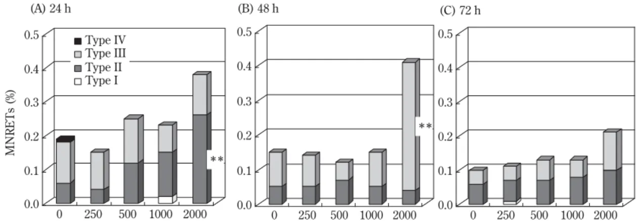
0.0 0.1 0.2 0.3 0.4 0.5 MNRETs (%) 0 250 500 1000 2000 (A) 24 h ** 0.0 0.1 0.2 0.3 0.4 0.5 0 250 500 1000 2000 (B) 48 h 0.0 0.1 0.2 0.3 0.4 0.5 0 250 500 1000 2000 (C) 72 h ** Type IV Type III Type II Type IFig. 1 Distribution of micronucleated reticulocytes (MNRETs) after a single dose of phenacetin. Rats were killed 24 (A), 48 (B) and 72 (C) h after a single administration of phenacetin at doses from 250 to 2000 mg/kg, and N/AO stained spec-imens were prepared from the bone marrow cells. Significant difference compared to respective control, **
p<0.01 (Fisher’s exact test).
微鏡(倍率: 1000 倍)で,1 個体当たり幼若赤血球を
2000 個観察し,小核を有する細胞数を調べた.
4.統計処理
網赤血球 I 型と II 型を合わせた小核出現頻度,網赤血
球 III 型の小核出現頻度および幼若赤血球の小核出現頻
度は Fisher 直接確率検定を行った.また,全網赤血球
中の網赤血球 II 型および III 型の型別小核出現頻度につ
いても Fisher 直接確率検定を行った.有意水準は両側
検定で 5%および 1%で行った.
結 果
1.単回投与による小核出現頻度
フェナセチンを単回投与し,投与 24,48 および 72 時
間後に N/AO 染色標本を作製した後,網赤血球 I ∼ IV 型
の型別小核出現頻度について観察した結果を Fig. 1 に示
した.小核出現頻度は,投与量とともに増加し,投与
24 時間後では網赤血球 II 型に,48 時間後では網赤血球
III 型に有意な増加が認められ,72 時間後では対照群と
同程度になった.
N/AO 染色標本では網赤血球 I 型と II 型を合わせた小
核出現頻度および網赤血球 III 型の小核出現頻度,AO 染
色標本では幼若赤血球の小核出現頻度を観察し,フェナ
セチンを単回投与した際の N/AO 染色法および AO 染色
法による小核出現頻度の違いについて比較した結果を
Table 1 に示した.網赤血球 I 型と II 型を合わせた小核出
現頻度は,投与 24 時間後では投与量とともに増加し,
500 mg/kg 以上の群で有意差が認められた.網赤血球
III 型の小核出現頻度は,投与 48 時間後の死亡動物の認
められた 2000 mg/kg 群でのみ,有意な増加が認められ
た.幼若赤血球の小核出現頻度は,投与 24 および 48 時
間後の 2000 mg/kg 群でのみ,わずかな増加ではあるが
有意差が認められた.小核出現頻度は網赤血球 I 型と II
型を合わせた N/AO 染色法の方が幼若赤血球の AO 染色
法より約 3 倍高い値を示した.
2.反復投与による小核出現頻度
フェナセチンを 2 回あるいは 4 回反復投与し,最終投
与 24 時間後に N/AO 染色標本を作製した後,網赤血球 I
∼ IV 型の型別小核出現頻度について観察した結果を
Fig. 2 に示した.小核出現頻度は,2 回および 4 回反復投
与とも投与量とともに増加し,網赤血球 II 型および III
型に有意な増加が認められた.
N/AO 染色標本では網赤血球 I 型と II 型を合わせた小
核出現頻度および網赤血球 III 型の小核出現頻度,AO 染
色標本では幼若赤血球の小核出現頻度を観察し,フェナ
Table 1 Comparison of N/AO and AO stainings after a single doseSampling
Dose No. of
N/AO staining AO staining time
(mg/kg) rats Type I and II Type III
(hours) observed MNRETs (%) MNRETs (%) MNPCEs (%) 24 0 5 0.14 0.18 0.15 250 5 0.11 0.24 0.16 500 5 0.27* 0.22 0.18 1000 5 0.65** 0.11 0.26 2000a 4 0.86** 0.21 0.29* 48 0 5 0.10 0.18 0.15 250 5 0.13 0.13 0.18 500 5 0.13 0.08 0.10 1000 5 0.14 0.22 0.19 2000a 4 0.23* 0.46** 0.31* 72 0 5 0.06 0.11 0.19 250 5 0.10 0.09 0.09 500 5 0.10 0.13 0.08 1000 5 0.12 0.12 0.13 2000a 2 0.19* 0.20 0.20
Rats were killed 24, 48 and 72 h after a single administration of phenacetin at doses from 250 to 2000 mg/kg, and N/AO and AO stained specimens were prepared from the bone marrow cells.
Values are means from 2∼5 rats.
Type I and II MNRETs=micronucleated Type I and II reticulocytes/Type I and II retic-ulocytes.
Type III MNRETs=micronucleated Type III reticulocytes/Type III reticulocytes. MNPCEs=micronucleated polychromatic erythrocytes/polychromatic erythrocytes.
a
Death=each one rat at 24 and 48 h, and 3 rats at 72 h. Significant difference compared to respective control, *
p<0.05,**
p<0.01 (Fisher’s exact test).
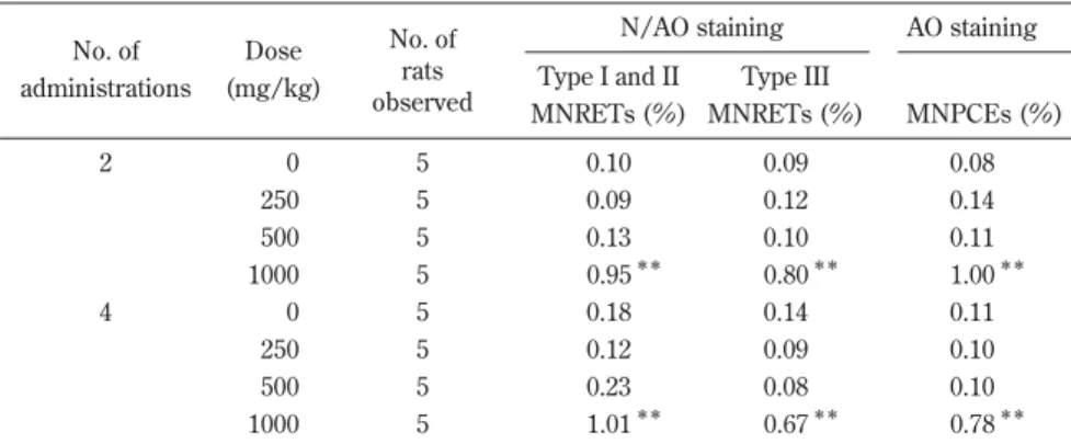
セチンを 2 回あるいは 4 回反復投与した際の N/AO 染色
法および AO 染色法による小核出現頻度の違いについて
比較した結果を Table 2 に示した.網赤血球 I 型と II 型
を合わせた小核出現頻度,網赤血球 III 型の小核出現頻
度および幼若赤血球の小核出現頻度は,それぞれ 2 回お
よび 4 回反復投与の 1000 mg/kg 群で有意な増加が認め
られ,染色法による違いはなかった.
考 察
フ ェ ナ セ チ ン は , マ ウ ス 小 核 試 験 の 骨 髄 細 胞
(CSGMT, 1990; Hayashi et al., 1989; Sutou et al., 1990)
および末梢血(CSGMT, 1992; Higashikuni et al., 1992)
では陽性と報告されている.しかし,ラット小核試験の
骨髄細胞では陰性,末梢血では陽性と報告されている
(Wakata et al., 1998)
.本研究では,ラットの骨髄細胞に
おける phenacetin の小核誘発性の有無について著者ら
が開発した N/AO 染色法(Sugihara et al., 2000)を用い
て検討した.
Wakata et al.(1998)の報告では,500 mg/kg の 2 回
反復投与による骨髄細胞および 2000 mg/kg の単回投与
による末梢血を観察していたことから,AO 染色法を用
いて幼若赤血球を観察した結果,単回投与の小核出現頻
度は,死亡動物の認められた 2000 mg/kg 群でのみ,対
照群の 2 倍程度のわずかな増加が認められたにすぎなか
っ た ( Table 1). 反 復 投 与 の 小 核 出 現 頻 度 は , 500
mg/kg 群では 2 回および 4 回反復投与ともに Wakata et
al.(1998)の報告と同様に増加は認められなかった.し
かし,1000 mg/kg 群では 2 回および 4 回反復投与ともに
対照群の 7 倍以上の小核出現頻度の増加が認められた
(Table 2).このことから,フェナセチンの骨髄細胞と
0.0 0.2 0.4 0.6 0.8 1.0 MNRETs (%) 0 250 500 1000 (A) Two repeated doses** ** 0.0 0.2 0.4 0.6 0.8 1.0 0 250 500 1000 (B) Four repeated doses
** ** Type IV Type III Type II Type I
Fig. 2 Distribution of micronucleated reticulocytes (MNRETs) after two or four repeated doses of phenacetin. Rats were killed 24 h after two (A) or four (B) days of administration of phenacetin at doses from 250 to 1000 mg/kg, and N/AO stained specimens were prepared from the bone marrow cells. Significant difference compared to respective control, **
p<0.01 (Fisher’s exact test).
Table 2 Comparison of N/AO and AO stainings after 2 or 4 repeated doses No. of Dose No. of N/AO staining AO staining administrations (mg/kg) observedrats Type I and II Type III
MNRETs (%) MNRETs (%) MNPCEs (%) 2 0 5 0.10 0.09 0.08 250 5 0.09 0.12 0.14 500 5 0.13 0.10 0.11 1000 5 0.95** 0.80** 1.00** 4 0 5 0.18 0.14 0.11 250 5 0.12 0.09 0.10 500 5 0.23 0.08 0.10 1000 5 1.01** 0.67** 0.78**
Rats were killed 24 h after 2 or 4 days of administration of phenacetin at doses from 250 to 1000 mg/kg, and N/AO and AO stained specimens were prepared from the bone marrow cells. Values are means from 5 rats.
Type I and II MNRETs=micronucleated Type I and II reticulocytes/Type I and II reticulocytes. Type III MNRETs=micronucleated Type III reticulocytes/Type III reticulocytes.
MNPCEs=micronucleated polychromatic erythrocytes/polychromatic erythrocytes. Significant difference compared to respective control, **
末梢血での小核誘発性の相違は,投与量の相違によるも
のと考えられた.
単回投与において,フェナセチンの小核誘発性を AO
染色法より高感度に検出できるか否かを調べるため,
N/AO 染色法(Sugihara et al., 2000)を用いて観察細胞
を網赤血球 I 型および II 型のみに限定した結果,小核出
現頻度は 500 mg/kg 以上の群で有意な増加が認められ,
2000 mg/kg 群では対照群の 6 倍高い値を示した.網赤
血球 I 型と II 型を合わせた小核出現頻度は,幼若赤血球
の小核出現頻度よりも約 3 倍高く,Wakata et al.(1998)
の報告の末梢血と同程度(単回投与の 2000 mg/kg 群で
最大 0.80 %)の小核出現頻度を確認できた(Table 1).
このことから,骨髄細胞においても N/AO 染色を用いて
観察細胞を限定することによって,AO 染色より高感度
に化合物の小核誘発性を検出できる可能性が示唆され
た.
赤血球の成熟過程は,骨髄中で前赤芽球が数回分裂し
た後,核を失って網赤血球となり,その後 1 日から 2 日
で成熟した後,骨髄中から末梢血に流出し,成熟赤血球
になるといわれている(其田および一条,1979)
.Fig. 1
から,フェナセチンの小核出現頻度は,投与 24 時間後
では網赤血球 II 型が,48 時間後では網赤血球 III 型が増
加し,72 時間後では対照群と同程度になった.このこ
とから,フェナセチンによって誘発された小核を有する
細胞は,投与 24 時間後に網赤血球 II 型となり,48 時間
後に網赤血球 III 型まで成熟し,72 時間後には末梢血に
流出すると考えられた.本研究から,小核を有する細胞
は経時的に成熟するため,投与 24 時間後では網赤血球 I
型および II 型を,投与 48 時間後では網赤血球 III 型を観
察すれば,AO 染色法より高感度に小核誘発性を検出で
きる可能性が示唆された(Table 1)
.
N/AO 染色法は,幼若赤血球を成熟度別に分類できる
ように,ニューメチレンブルー超生体染色で RNA を網
状に染め,メタノールで RNA の網状構造を固定すると
ともにニューメチレンブルー色素を脱色し,骨髄細胞で
一般的に用いられている AO 染色(Hayashi et al., 1983)
を施す染色法である(Sugihara et al., 2000)
.本研究か
ら,N/AO 染色法を骨髄細胞に応用し,観察細胞を限定
することによって,AO 染色より高感度に小核を検出で
きる可能性が示されたことから,本染色法は,骨髄細胞
を用いる小核試験において,有用な染色法の一つである
と考えられた.
結 語
ラット骨髄小核試験において,N/AO 染色法を用いて
観察細胞を限定することによって,フェナセチンの小核
誘発性を AO 染色法より高感度に検出できることを確認
した.
参 考 文 献
CSGMT (1990) Single versus multiple dosing in the micronucleus test: the summary of the fourth collaborative study by CSGMT/JEMS・MMS, Mutat. Res., 234, 205-222.
CSGMT (1992) Micronucleus test with mouse peripheral blood ery-throcytes by acridine orange supravital staining: The summary report of the 5th collaborative study by CSGMT/JEMS・MMS, Mutat. Res., 278, 83-98.
Granberg-Ohman, I., S. Johansson and A. Hjerpe (1980) Sister-chro-matid exchanges and chromosomal aberrations in rats treated with phenacetin, phenazone and caffeine, Mutat. Res., 79, 13-18. Hayashi, M., T. Sofuni and M. Ishidate Jr. (1983) An application of
acridine orange fluorescent staining to the micronucleus test, Mutat. Res., 120, 241-247.
Hayashi, M., S. Sutou, H. Shimada, S. Sato, Y.F. Sasaki and A. Wakata (1989) Difference between intraperitoneal and oral gavage application in the micronucleus test: The 3rd collaborative study by CSGMT/JEMS・MMS, Mutat. Res., 223, 329-344.
Hayashi, M., T. Morita, Y. Kodama, T. Sofuni and M. Ishidate Jr. (1990) The micronucleus assay with mouse peripheral blood retic-ulocytes using acridine orange-coated slides, Mutat. Res., 245, 245-249.
Hayashi, M., J.T. MacGregor, D.G. Gatehouse, I.-D. Adler, D.H. Blakey, S.D. Dertinger, G. Krishna, T. Morita, A. Russo and S. Sutou (2000) In vivo rodent erythrocyte micronucleus assay. II. Some aspects of protocol design including repeated treatments, integration with toxicity testing, and automated scoring, Environ. Mol. Mutagen., 35, 234-252.
Higashikuni, N., T. Baba, T. Nakamura and S. Sutou (1992) The micronucleus test with peripheral reticulocytes from phenacetin-treated mice, Mutat. Res., 278, 159-164.
Krishna, G. and M. Hayashi (2000) In vivo rodent micronucleus assay: protocol, conduct and data interpretation, Mutat. Res., 455, 155-166.
Nohmi, T., K. Yoshikawa, M. Nakadate and M. Ishidate Jr. (1983) Species difference in the metabolic activation of phenacetin by rat and hamster liver microsomes, Biochem. Biophys. Res. Commun., 110, 746-752.
Sugihara, T., S. Sawada, A. Hakura, Y. Hori, K. Uchida and F. Sagami (2000) A staining procedure for micronucleus test using new methylene blue and acridine orange: specimens that are supravi-tally stained with possible long-term storage, Mutat. Res., 470, 103-108.
Sutou, S., M. Kondo and Y. Mitsui (1990) Effects of multiple dosing of phenacetin in the micronucleus test, Mutat. Res., 234, 183-186. 其田三夫,一条 茂(1979)獣医血液学 II,医歯薬出版,東京,
pp. 403-456.
Wakata, A., Y. Miyamae, S. Sato, T. Suzuki, T. Morita, N. Asano, T. Awogi, K. Kondo and M. Hayashi (1998) Evaluation of the rat micronucleus test with bone marrow and peripheral blood: Summary of the 9th collaborative study by CSGMT/JEMS・MMS, Environ. Mol. Mutagen., 32, 84-100.
Yamamoto, K.I. and Y. Kikuchi (1981) Studies on micronuclei time response and on the effects of multiple treatments of mutagens on induction of micronuclei, Mutat. Res., 90, 163-173.