INTRODUCTION
Retrocaval ureter is a rare congenital anomaly that requires surgical correction in the sympto-matic patient.1,2) Recent technological develop-ments in the field of laparoscopic surgery have had a significant impact on modern urology. Developed surgical devices, has enabled laparo-scopic procedures to be applied to plastic sur-gery on the upper urinary tract, including pyeloplasty, which requires watertight anasto-moses. Many physicians acknowledge the
deceased invasiveness and the cosmetic advan-tages of laparoscopic surgery, however laparo-scopic plastic surgery for the urinary tract still has limitations because of the difficulty in the intracorporeal suturing.3-5)For patients with small stone burdens in kidneys, shockwave lithotripsy (SWL), ureteroscopy, or both may be reasonable treatment options, but minimally invasive treatment for patient with larger stone burdens presents a special challenge. The com-bination of laparoscopic and percutaneous approaches allows the establishment of the per-cutaneous tract into the retrocaval ureter under direct vision, thus avoiding repetitive therapy for renal stone and retrocaval ureter.
Herein, we report our experience of success-ful retroperitoneoscopic ureteroureterostomy for retrocaval ureter with simultaneous flexible
Simultaneous Management for Retrocaval Ureter and Ipsilateral Renal
Stone Using Retroperitoneoscopic Approach: Report of a Case
Shouji SUZUKI*, Norifumi SAWADA, Yaburu HANEDAHidenori ZAKOJI, Takayuki TSUCHIDA, Masami AIKAWA, Isao ARAKIand Masayuki TAKEDA Department of Urology, Interdisciplinary Graduate School of Medicine and Engineering,
University of Yamanashi, Chuo, Yamanashi, Japan
Abstract: A 32-year-old male presented with a month history of right back and flank pain and a possibility of gross hematuria. As part of the initial evaluation abdominal ultra-sound revealed marked right hydronephrosis. An excretion urogram showed delayed excretion of contrast material extending to the L4 level distally. Computed tomography revealed right renal pelvic stone and right retrocaval ureter. At surgery, a right-sided dou-ble-J ureteric stent was placed under fluoroscopic guidance.
Retroperitoneal approach was used and flexible ureteroscopic removal of the right renal stone via a trocar port followed by retroperitoneoscopic right ureteroureterostomy using an intracorporeal suture technique. So far, none has reported simultaneous treat-ment of retroperitoneoscopic repair with retrocaval ureter and removal for the renal pelvic stone via a trocar port. We showed that this simultaneous management via a trocar port can be the better therapeutic choice for the renal stone with retrocaval ureter.
Key words: retrocaval ureter, retroperitoneoscopic management, renal stone
*Correspondence to: S. Suzuki, Department of
Urolo-gy, Interdisciplinary Graduate School of Medicine and Engineering, University of Yamanashi, Shimokato 1110, Chuo, Yamanashi 409-3898, Japan, E-mail: nsawada@yamanashi.ac.jp
Received January 29, 2009 Accepted April 9, 2009
ureteroscopic management for renal pelvic stone via a trocar port.
CASE REPORT
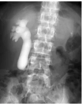
A 32-year-old male presented with a month history of right back and flank pain and a possi-bility of gross hematuria. As part of the initial evaluation abdominal ultrasound revealed marked right hydronephrosis. An intravenous pyelography (IVP) showed delayed excretion of contrast material extending to the L4 level dis-tally (Fig.1). Computed tomography revealed a right renal stone with 9 mm long and right retrocaval ureter (Fig.2). Blood creatinine con-centration was 0.97 mg/dL. Cystoscopy and right retrograde pyelography demonstrated par-tial ureteral obstruction.
General anesthesia was induced and the patient was placed in the lithostomy position and a 7 Fr D-J ureteral stent was inserted as far as the renal pelvis using a cystourethroscope under
fluoro-scopic guidance. The stent was first inserted to clarify the positions of inferior vena cava (IVC) and retrocaval ureter. The patient was then placed in the right lateral flank position and a 2 cm skin incision was made at the middle border of the right iliac crest in the posterior axillary line. The muscles were split to expose the retroperitoneum, the peritoneum was reflected medially. Working space was made by a balloon dissector. A balloon trocar was inserted through the skin incision and the retroperitoneal space was pressurized to 10 mmHg with CO2. Total of 4 trocars were inserted into the retroperitoneal spaces. When the operation was started, the CO2 pressure was reduced to 8 mmHg. The ureter was found to override the vena cava, then run behind the vessel. The lower part of the ureter distal to the retrocaval portion was freed from the surrounding tissue. The ureter was then brought up anteriorly following removal of the ureteral catheter. The dilated upper part of the ureter was separated from IVC using sharp
Fig. 1. Preoperative excretory urography.
Fig. 2. Computed tomography during excretory urog-raphyshows existence of a retrocaval ureter.
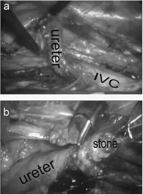
and blunt dissection. A stay suture was put in the proximal ureter and the stent was with-drawn from the renal pelvis for insertion of the flexible ureteroscope. A 6.9 Fr flexible uretero-scope was inserted in the retroperitoneal space via a trocar, and put into the renal pelvis via proximal ureteral end. A renal stone was caught using the basket catheter, and tried to put out of the ureter (Fig.3). The ureteral stump was spatulated for 1 cm, then, the stone was retrieved. The retrocaval portion was transected just above the retrocaval segment, which was then moved anterior to the vena cava. After double-J stent was inserted as far as the renal pelvis, end-to-end anastomosis of the ureter was performed endoscopically. The anastomosis was created using 8 interrupted 4–0 polyglatin sutures. A penrose drain tube was placed near the anastomosis. The operative time for this procedure is 288 minutes and estimated blood loss was 20 mL. The patient was ambulant and on oral intake from two days after surgery. There was no urine leakage and the drain was removed 7 weeks after operation and 8 months after operation, ultrasound revealed no hydronephrosis in the right kidney (Fig.4). Ten
months after operation, blood creatinine con-centration showed a little improvement and was 0.86 mg/dL.
Fig. 3. (a) The retrocaval ureter found to override the vena cava. (b) The upper portions of the right ureter was identified and renal pelvic stone was held with basket catheter.
DISCUSSION
Retrocaval ureter is a rare congenital anom-aly, usually associated with upper urinary tract stasis and an ‘S’ deformity of the ureter. Its ex-act prevalence is unknown. Retrocaval ureter results from the posterior cardinal vein persist-ing as a segment of the infrarenal vena cava dur-ing development. This anomaly entraps a seg-ment of the proximal ureter as it wraps around the vena cava, often resulting in obstruction and hydronephrosis. Individuals who become symptomatic typically present in the third or fourth decade of life. When symptoms develop or renal function progressively decreases, surgi-cal correction should be performed.
The efficacy of laparoscopy for ureteral sur-gery has been established. Moore et al reported on 30 patients who underwent laparoscopic pyeloplasty for ureteropelvic junction obstruc-tion.6)At a mean of 16 months after laparoscop-ic pyeloplasty 97% of their 30 patients had a patent anastomosis on radiographic evaluation. Preoperative assessment of the distal ureter is essential to ensure a normal caliber distal seg-ment, allowing primary ureteroureterostomy.
Simforoosh et al reported in their series that a case of right renal pelvic stone with retrocaval ureter and the stone was removed while tran-secting the renal pelvis using a transperitoneal laparoscopy.7)Few reports document other att-empted laparoscopic repairs of a retrocaval ureter with renal pelvic stone. Retrocaval ureter is a rare congenital anomaly, which sometimes causes symptomatic hydronephrosis. The laparoscopic treatment is appealing, because it has advantages of reduced analgesic require-ment, shorter hospital stay, early convalescence, and better cosmesis, in addition to excellent safety and efficacy profiles. Ureteroscopic treat-ment of stones in a renal pelvis is another
mini-mally invasive procedure. However, this requires a flexible ureteroscope and the skills to use it in addition to the long time needed to fragment larger stones.
We showed that retroperitoneoscopic repair with simultaneous flexible ureteroscopic man-agement via a trocar port can be the therapeu-tic choice for the renal stone with retrocaval ureter. Although retroperitoneal space is limit-ed and difficult to perform suturing, retroperi-toneoscopic surgery seems to be superior to conventional transperitoneal laparoscopic sur-gery in reducing the potential risk of abdominal complications.8)Furthermore, retroperitoneo-scopic approach provides more direct and less invasive access to the diseased ureter, we recom-mend that in case with congenital upper ureter-al or pelvic-ureterureter-al obstruction with renureter-al pelvic stone simultaneous flexible ureteroscopic management via a trocar port and retroperito-neoscopic ureteroplastic surgery.
REFERENCES
1) Matsuda T, Yasumoto R, Tsujino T: Laparoscopic treatment of a retrocaval ureter. Eur Urol, 29: 115–118, 1996.
2) Ameda K, Kakizaki H, Harabayashi T, Watarai Y, Nonomura K, Koyanagi T: Laparsocopic ureter-oureterostomy for retrocaval ureter. Int J Urol, 8: 71–74, 2001.
3) Ishitoya S, Okubo K, Arai Y: Laparoscopic ureter-olysis for retrocaval ureter. Br J Urol, 77: 162–163, 1996.
4) Miyazato M, Kimura T, Ohyama C, Hatano T, Miyazato T, Ogawa Y: Retroperitoneoscopic ureteroureterostomy for retrocaval ureter. Hinyokika Kiyo, 48: 25–28, 2002.
5) Mugiya S, Suzuki K, Ohhira T, Un-No T, Takaya-ma T, Fujita K: Retroperitoneoscopic treatment of a retrocaval ureter. Int J Urol, 6: 419–422, 1999.
6) Moore RG, Averch TD, Schulam PG, Adams JB, 2nd, Chen RN, Kavoussi LR: Laparoscopic pyelo-plasty: experience with the initial 30 cases. J Urol, 157: 459–462, 1997.
Laparoscopic pyelopyelostomy for retrocaval ureter without excision of the retrocaval seg-ment: first report of 6 cases. J Urol, 175: 2166–2169; discussion 2169, 2006.
8) Polascik TJ, Chen RN: Laparoscopic ureter-oureterostomy for retrocaval ureter. J Urol, 160: 121–122, 1998.