Case Report
Hepatopulmonary Syndrome Improved by Living‑related Liver Transplantation:A Case Report
Takeshi GOCHO,Yuichi ISHIDA,Yoshiaki KITA,Tomonori IIDA,Taro SAKAMOTO, Ryusuke ITO,Hiroaki SHIBA,Shigeki WAKIYAMA,Tadashi UW AGAW A,Shoichi HIROHARA,
Takeyuki MISAW A,Mikio ZENIYA,and Katsuhiko YANAGA Department of Surgery, The Jikei University School of Medicine
Division of Gastroenterology, Department of Internal Medicine, The Jikei University Graduate School of Medicine
ABSTRACT
Progressive dyspnea devel oped in a 61‑year‑old man with cryptogenic liver cirrhosis,and hepatopulmonary syndrome was diagnosed. Phys ical examination revealed jaundice(total bilir- ubin,4.5 mg/dl),spider nevi,and clubbed fingers. Arterial blood gas analysis showed an arterial oxygen pressure of 57.5 mm Hg and an alveolar‑ar terial oxygen difference of 42 mm Hg. A Tc‑
macroaggregated albumin pulmonary perfusion scan demonstrated uptake in the brain and kidneys suggestive of an intrapulmonary shunt. The right ‑to‑left shunt ratio was calculated to be 30.6%.
The patient underwent living‑related liver transplantation using an extended right lobe graft donated by his younger brother. Both the arterial oxygen pressure(102 mm Hg)and the right‑to‑
left shunt ratio(7.4%)had normalized by 8 months after liver transplantation,and the patient has since been weaned off home oxygen therapy. (Jikeikai Med J 2010;57:39‑41)
Key words:hepatopulmonary syndrome,liver transplantation,liver cirrhosis
INTRODUCTION
Hepatopulmonary syndrome(HPS)was first re- ported by Kennedy and Knudson in 1977 and is defined as the triad of li ver disease,arterial deox-
ygenation,and intrapulmonary vascular dilatation (IPVD). The prevalence of HPS in patients with chronic liver disease ranges f rom 15% to 32% . Spontaneous resolution of HPS is rare,and the only effective surgical treatment is liver transplantation.
We report herein a case of HPS which showed marked improvement after living‑related liver trans- plantation(LRLT).
CASE REPORT
A 61‑year‑old man with a 5‑year history of liver dysfunction had received a di agnosis of cryptogenic liver cirrhosis at the age of 58 years. Serum markers for viral hepatitis,anti‑nucl ear antibody,and anti‑ mitochondrial antibody were negative,and the patient had no history of alcohol abus e. He was treated with ursodeoxycholic acid until the age of 60 years,when he complained of progressi ve dyspnea,for which home oxygen therapy was started. When he was referred to our hospital for further eval uation,HPS was diagnosed and LRLT was s cheduled.
Jikeikai Med J 2010;57:39‑41
Received for publication,December 21,2009
後町 武志,石田 祐一,北 義昭,飯田 智憲,坂本 太郎,伊藤 隆介,柴 浩明,脇山 茂樹,宇和川 匡,広原 鐘一,三澤 健之,銭谷 幹男,矢永 勝彦
Mailing address:Takeshi GOCHO,Department of Surgery,The Jikei University School of Medicine,3‑25‑8,Nishi‑Shimbashi, Minato‑ku,Tokyo 105‑8461,Japan.
E‑mail:gocho@jikei.ac.jp
39
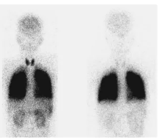
At the time of admission,the patient had jaundice (total bilirubin,4.5 mg/dl),spider nevi,clubbed finger, and esophageal varices,but ascites and hepatic ence- phalopathy were absent. The Child‑Pugh score was 5,the Model for End‑Stage Li ver Disease(MELD) score was 14,and the United Network for Organ Sharing‑modified MELD s core was 24. Arterial blood gas analysis obtained the following results:pH, 7.503;arterial carbon dioxide pressure(PaCO ),33.2 mm Hg;arterial oxygen pr essure(PaO ),57.5 mm Hg;arterial oxygen satur ation(SaO ),89.1% ;and alveolar‑arterial oxygen di fference(A‑aDO ),42 mm Hg. Respiratory function t esting obtained the fol- lowing results:the ratio of forced expiratory volume in 1 second to forced vital capacity,83.1% ;and the percent vital capacity,93. 3%. Pulmonary perfusion imaging using Tc‑macr oaggregated albumin (MAA)(Fig.1a)exhibited uptake in the brain and kidneys in addition to uptake in the lungs,suggesting an intrapulmonary shunt . The right‑to‑left(R‑L)
shunt ratio was calculated as 30.6% by comparing Tc‑MAA accumulation in both lungs with whole‑
body radioactivity. The results of chest radiography were unremarkable,and cont rast‑enhanced echocar- diography suggested delayed opacification in the left atrium and ventricle.
The patient underwent LRLT in November 2007 with an extended right lobe gr aft with the caudate
lobe donated by his younger brother. The graft weight was 662 g and the gr aft volume/recipient stan- dard liver volume ratio was 56.6%. The right hepatic vein was reconstructed wit h a third‑party homograft. The operation was uncomplicated and lasted 15 hours 34 minutes;the estimated bl ood loss was 4,600 mL.
The immunosuppressant agents administered were tacrolimus and methylpr ednisolone. Pathological examination of the resect ed liver specimen showed liver cirrhosis with veno‑occl usive lesions.
Postoperative recovery was favorable and with- out any major complications,and the patient was discharged on postoperative day 32 with normal liver function. The arterial bl ood gas saturation was measured sequentially af ter LRLT (Table 1)and showed gradual improvement in arterial oxygenation and a decrease in A‑aDO . A s ignificant improve-
ment of the R‑L shunt was also shown with pulmo- nary perfusion scanning(Fig.1b). All these vari- ables were normalized by 8 months after LRLT,and the patient has not needed home oxygen therapy since that time.
DISCUSSION
HPS is a pulmonary complication of chronic liver diseases. Although HPS commonl y occurs in patients with liver cirrhos is,some patients with non- cirrhotic portal hypertension and fulminant hepatic failure may be affected. Exer tional dyspnea is the most common symptom,and t he presence of spider nevi,cyanosis,digital clubbi ng,and severe hypoxemia suggests HPS. Diagnosti c criteria for HPS are as follows:PaO less than 80 mm Hg or A‑aDO more than 15 mm Hg while the patient is breathing room
T.GOCHO ,et al. Vol.57,No.1 40
Fig.1. Tc‑MAA lung scanning showing uptake in the brain and the kidneys. The R‑L shunt ratio im- proved from 30.6% preoperatively(Fig.1a)to 7.4% 8 months after LRLT ( Fig.1b).
Table 1.Sequential changes in arterial blood‑gas ten- sions and R‑L shunt
Pre‑op. Post‑op.day
14 24 89 178 245
pH 7.503 7.438 7.463 7.46 7.42 7.45 pCO (mmHg) 33.2 27. 9 29.3 26.7 35 30 pO (mmHg) 57.5 61. 8 62.1 51.8 85 102 pO (A‑a) (mmHg) 42 57.6 55.2 66.7 21 10.2 R‑L shunt (%) 30.6 33. 2 24.7 19.2 15.5 7.4
air,positive findings on contrast enhanced echocar- diography or abnormal uptake in the brain(>6%)on radioactive lung perfusion s canning,and portal hyper-
tension with or without cirrhosis.
The exact mechanism causing HPS is unclear, but IPVD is thought to be a pathologic hallmark.
IPVD can be caused by increased circulating levels of vasodilators,including nit ric oxide,prostaglandins,
progesterone,and estradiol,and by decreased circulat- ing levels of vasoconstrictors,including endothelin‑1 and serotonin. In the pres ent case,we measured such vasodilators and vasocons trictors in the serum to evaluate the etiology of HPS,but the levels of most were within their normal r anges preoperatively,and no significant changes were observed after LRLT. In an experimental model,int estinal endotoxin has been found to be one vasodilator that could cause HPS due to the failure of the damaged liver to clear it. The cause of HPS in the present case remains unclear,but multiple factors are thought t o be related to the development of HPS.
The prognosis is poor if HPS is treated medi- cally;the 5‑year survival rate of patients with HPS treated medically is 23%,but the survival rate after liver transplantation increas es to 76%,which is simi- lar to that after liver transplantation in patients without HPS. Therefor e,liver transplantation is the treatment of choice f or patients with HPS.
Because the long‑term survival rate for patients with HPS and a PaO of 60 mm Hg or less was reported to be worse even after liver t ransplantation,patients with HPS should undergo l iver transplantation before PaO deteriorates further. I n fact,the United Net- work for Organ Sharing gives extra MELD score points for HPS. In the pr esent case,the PaO was 57.5 mm Hg preoperativel y,and the patient was jud-
ged to be a good candidate for LRLT. The postoper- ative oxygenation gradually improved and had nor- malized by 8 months after LRLT,which is in agree- ment with previous studies describing the improve- ment in PaO occurring over months to years,depend- ing on the severity of hypoxemia. The mechanism for the improvement in oxygenat ion after liver trans-
plantation in patients with HPS has been reported to be the reversibility of diffus e dilatation of the pulmo- nary precapillary vessels. In the present case,such a mechanism seems reasonabl e.
CONCLUSION
The present case of HPS complicating cryptogenic cirrhosis was t reated successfully with liver transplantation,and mar ked improvements in oxygenation and R‑L shunt were obtained. Because hypoxemia is frequently pr ogressive and because a low PaO predicts poor sur vival in patients with HPS associated with advanced l iver cirrhosis,early liver transplantation should be cons idered for patients with a PaO of 60 mm Hg or les s.
REFERENCES
1. Kennedy TC,Knudson RJ. Exercise‑aggravated hypox- emia and orthodeoxia in cirrhosis. Chest 1977;72:
305‑9.
2. Schenk P,Fuhrmann V,Madl C,Funk G,Lehr S,Kandel O,et al. Hepatopulmonary s yndrome:prevalence and predictive value of various cut offs for arterial oxygena- tion and their clinical consequences. Gut 2002;51:
853‑9.
3. Rodriguez‑Roisin R,Krowka MJ. Hepatopulmonary syndrome;a liver‑induced l ung vascular disorder. N Engl J Med 2008;358:2378‑87.
4. Krowka MJ,Cortese DA. Hepatopulmonary syndrome:
an evolving perspective in the era of liver transplanta- tion. Hepatology 1990;11:138‑42.
5. Zhang HY,Han DW,Su AR,Zhang LT,Zhao ZF,Ji JQ, et al. Intestinal endotoxemia plays a central role in development of hepatopulmonar y syndrome in a cirr- hotic rat model induced by multiple pathogenic factors. World J Gastroenterol 2007;13:6386‑95.
6. Swanson KL,Wiesner RH,Krowka MJ. Natural his- tory of hepatopulmonary syndrome:impact of liver transplantation. Hepatology 2005;41:1122‑9.
7. Fallon MB,Mulligan DC,Gish RG,Krowka MJ. Mode for End‑Stage Liver Diseas e(MELD)exception for he- patopulmonary syndrome. Liver Transplant 2006;12:
S105‑7.
8. Krowka MJ,Cortese DA. Hepatopulmonary syndrome:
An evolving perspective in the era of liver transplanta- tion. Hepatology 1990;11:138‑42.
HPS Improved by LRLT
March,2010 41