Hirosaki Med.J.
67:92―101,2016ORIGINAL ARTICLE
MORPHOLOGICAL ANALYSIS OF THE RESIDUAL RECTAL MUCOSA IN 12 POSTOPERATIVE ULCERATIVE COLITIS PATIENTS
Satoko Morohashi
1),Hajime Morohashi
2),Tadashi Yoshizawa
1),Masanori Tanaka
3), Tatsuya Mikami
4),Takuji Kagiya
2),Takeshi Saito
2),Hideaki Hirai
1),
Toshihiro Haga
1),Yunyan Wu
1),Kenichi Hakamada
2),and Hiroshi Kijima
1)Abstract Background: There are no reports on the degree of inflammation in the residual rectal mucosa in asymptomatic UC patients after an operation for ileal pouch anal canal anastomosis (IACA).
Methods: We studied the residual rectal mucosal biopsies and rectal mucosa from the surgical specimens of 12 asymptomatic postoperative UC cases. We analyzed the degree of inflammation, including crypt architecture and mucus content in the rectal mucosa.
Results: Two pathological active cases (16.7%), four resolving cases (33.3%), and six remission cases (50%) were observed in the residual rectal mucosa. In terms of the degree of pathological inflammation in the residual rectal mucosa, one case (1/12, 8.3%) was upregulated, seven cases (7/12, 58.3%) were stable, and four cases (4/12, 33.3%)
were downregulated compared with the rectal mucosa of the surgical specimens.
Conclusions: We determined the natural history of the degree of inflammation on the residual rectal mucosa in postoperative UC patients. Eight cases (8/12, 66.6%) of asymptomatic UC patients were associated with prolonged inflammation after an IACA operation in the residual rectal mucosa compared with that in the rectal mucosa of the surgical specimens. Therefore, we could identify patients with prolonged inflammation in asymptomatic UC cases by a residual rectal mucosal biopsy.
Hirosaki Med.J. 67:92―101,2016
Key words: rectum; ulcerative colitis; residual rectal mucosa.
1) Department of Pathology and Bioscience, Hirosaki University Graduate School of Medicine
2) Department of Gastroenterological Surgery, Hirosaki University Graduate School of Medicine
3) Department of Pathology, Hirosaki Municipal Hospital
4) Department of Gastroenterology and Hematology, Hirosaki University Graduate School of Medicine
Correspondence: S. Morohashi
Received for publication, April 13, 2016 Accepted for publication, May 18, 2016
Introduction
Ulcerative colitis (UC) is a chronic inflam- matory bowel disease (IBD), in which the inflammation remains confined to the colon, and the rectum is involved in 95% of patients.
The inflammation is diffuse and continuous and largely confined to the mucosa
1). In Japan, an abdominal colectomy with ileal pouch- anal anastomosis (IPAA) and an ileal pouch anal-canal anastomosis (IACA) are standard methods for UC treatment. IPAA involves a hand-sewn method and anastomoses of the
ileo-anal pouch to the anus after removing the
mucosa of the anal canal more toward the oral
side than the dentate line
2). IACA is the method
used to staple the ileoanal pouch at the level of
the superior border of the anal canal without
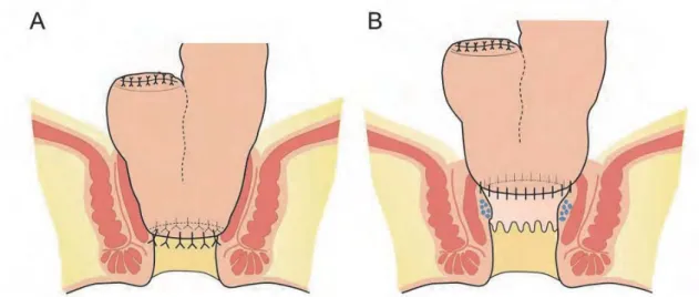
performing anorectal mucosectomy (Fig. 1)
3).
Compared to IPAA, IACA is more effective
in anastomosis and has the advantage of
avoiding the diverting stoma
4, 5). IACA has the
advantage of maintaining good postoperative
bowel function by ensuring that the anoderm
and the anal sphincter are kept secure. The
frequency of inflammation relapse from the
residual rectal mucosa is not high, but the possibility that colitis cancer occurs is reported to be 0%‒16%
6-9). There is an opinion that IPAA with an anorectal mucosectomy is desirable compared with IACA
10). In a clinical setting, the majority of UC patients do not experience symptoms and require no medication for long periods following the IACA. To the best of our knowledge, there are no published reports for evaluating the degree of inflammation in the residual rectal mucosa after the IACA operation.
The level of surveillance of the residual rectal mucosa for patients without symptoms is also unknown. Moreover, the natural course of healing of the residual rectal mucosa after the IACA procedure is not fully understood in asymptomatic UC patients. Here we studied the residual rectal mucosal biopsies and the rectal mucosa from the surgical specimens of 12 asymptomatic postoperative UC cases.
Material and methods
Patient samples
We performed a prospective analysis of the residual rectal mucosa from 12 UC patients
following the IACA operation between 1998 and 2015 at Hirosaki University Hospital (Hirosaki, Aomori Prefecture, Japan). These patients had no symptoms of UC and received no medication after the operation. There were two patients that exhibited symptoms of hematochezia as a consequence of ileal pouchitis. Biopsies were obtained from patients who consented to a biopsy from Hirosaki University Hospital.
All biopsies were performed from May to December, 2015. Informed consent was obtained from each patient regarding the use of clinical records and pathological specimens. The average time from the surgery to the residual rectal mucosa biopsy was 4198 ± 2075 (range:
132‒6418) days. The mean age of the patients was 33.6 ± 13.3 (range: 15‒55) years old at the time of the first operation. The mean age of the patients was 45.3 ± 16.3 (range: 24‒73) years old at the time of the biopsy. Of the patients, 10 were women and 2 were men. There were a total of 28 residual rectal mucosa biopsies, and the mean number of rectal mucosa biopsies per person was 2.3 specimens. We examined the rectal mucosa of a surgical specimen that was representative of the 12 UC patients.
Figure 1 A: The ileal pouch anal anastomosis (IPAA) operation is a hand-sewn method and anastomoses of the ileoanal pouch to the anus after removing the mucosa of the anal canal more toward the oral side than the dentate line. B: The ileal pouch anal canal anastomosis
(IACA) operation is a method in which the ileoanal pouch is stapled at the level of the anal canal superior border without performing an anorectal mucosectomy.
94
This study was approved by the ethics committees of Hirosaki University graduate School of Medicine.
Pathological examinations of the residual rectal mucosa biopsies and the rectal mucosa of surgical specimens
For the histopathological examination of the residual rectal mucosa biopsies and rectal mucosa, the surgical specimens were routinely formalin-fixed, paraffin embedded, thinly sectioned (4
ȝm), and stained with hematoxylin and eosin. We chose the rectal mucosa of the surgical specimens at the end of the anal side because it was available for epithelial evaluation without the formation of deep ulcers. Moreover,
we also analyzed the crypt architecture, mucus content in the residual rectal mucosa, a n d t h e r e c t a l m u c o s a o f t h e s u r g i c a l specimens. Furthermore, we studied the level of crypt, mucosal surface, and lamina propria inflammation in the residual rectal mucosa and the mucosa of the surgical specimens using the diagnostic criteria described by Tanaka et al.
11). Crypt architecture
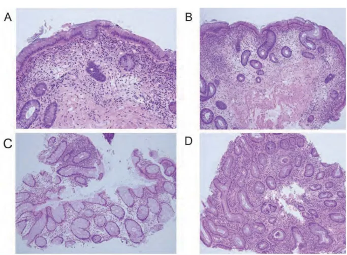
Crypt atrophy was recognized by the generally increased distance of the more than one crypt diameter and/or a general increase in the distance between crypts and the muscular mucosa in the well-oriented slides of an adequate-sized biopsy (Fig. 2-A). Crypt distortion was diagnosed if there were two or
Figure 2 A: Crypt atrophy, there was an increased distance of more than one crypt diameter between crypts and a general increase in the distance between the crypts and the muscular mucosa. B: Crypt distortion, there were two or more branched crypts that exhibited non-parallelism. C: Mucin depletion minimal (absent) case, there was no mucin depletion. Goblet cells were clearly evident. D: Mucin depletion severe case, goblet cell mucin was almost completely exhausted in all crypts as a diffuse change.
more branched crypts exhibiting non-parallelism
(Fig. 2-B). An equivocal change was recorded as absent.
Mucus content
Mucin depletion was divided into none or minimal (0: absent) , 1+, and 2+ (Fig. 2-C, D) . Mucin depletion was judged to be 2+ when the goblet cell mucin was almost completely exhausted in at least two crypts. Mucin depletion was not judged in the areas near the lymphoid follicles because it is often observed in this region of normal mucosa. In this study, we judged mucin depletion if minimal (0) as absent, and 1+
and 2+ as existing.
&U\SWLQÀDPPDWLRQ
Cryptitis and crypt abscesses were defined as either present or absent (Fig. 3-A). Distal
Paneth cell metaplasia was recorded either present or absent (Fig. 3-B).
0XFRVDOVXUIDFHLQÀDPPDWLRQ
Intraepithelial neutrophil infiltration was divided into three degrees as follows: 1) 0, there was no neutrophil infiltration within 100 surface epithelial cells; 2) 1+, 1‒4 infiltrating neutrophils within 100 surface epithelial cells; and 3) 2+,
≧5 infiltrating neutrophils within 100 surface epithelial cells. The extent of intraepithelial lymphocyte infiltration was divided into three degrees as follows: 1) 0, 0‒4 infiltrating lymphocytes within 100 surface epithelial cells;
2) 1+, 5‒29 infiltrating lymphocytes within 100 surface epithelial cells; and 3) 2+, ≧30 infiltrating lymphocytes within 100 surface epithelial cells.
Figure 3 A: Cryptitis, a few infiltrating neutrophils were seen in the crypt (arrow). B: Distal Paneth cell metaplasia, red granules were observed in the crypt cytoplasm of the residual rectal mucosa. C: severe
(2+) mononuclear cells, plasma cells were clustered together in the residual rectal mucosa. D: “Minimal
(0)” mononuclear cell infiltration, plasma cells, and/or lymphocytes infiltrated within the normal line.
96
/DPLQDSURSULDLQÀDPPDWLRQ
Basal lymphoid aggregation was defined as either present or absent. The infiltration of mononuclear cells (lymphocytes and plasma cells) was judged to be “severe (2+)” when they were clustered together (Fig. 3-C).
“Minimal (0)” infiltration was recorded as absent (Fig. 3-D). Mild to moderate (1+) was between severe (2+) and absent (0). Focal and diffuse mononuclear cell infiltrations were judged in this study. We considered which of lymphocytic infiltration, the plasma cell infiltration was superior. Basal plasmacytosis was arbitrarily recorded as present when at least three plasma cells per crypt diameter were found below the base of the crypt
11). Neutrophil infiltration in the lamina propria was divided into three degrees as follows: 1) 0, there was no apparent neutrophil infiltration;
2) 1+, 1‒3 infiltrating neutrophils; and 3) 2+, ≧ 4 infiltrating neutrophils within a high power field ( × 400). Eosinophil infiltration within the lamina propria was divided into three degrees as follows: 1) 0, 0‒19 infiltrating eosinophils; 2)
1+, 20‒59 infiltrating eosinophils; and 3) 2+, ≧ 60 infiltrating eosinophils within a high power field (× 400). The neutrophils and eosinophils in the capillaries of the lamina propria were omitted. Granuloma, giant cells, and apoptosis were defined as either present or absent.
3DWKRORJLFDOLQÀDPPDWRU\VWDJHRIWKHUHFWDOPXFRVD
We divided the stage of pathological inflammation of the rectal mucosa into three categories: 1) remission; 2) resolving; and 3)
active stages. The active stage was defined as high levels of diffuse plasma and mononuclear cell infiltration (2+). The resolving stage was defined as moderately diffuse plasma and mononuclear cell infiltration (1+). The remission stage consisted of cellular infiltration below the active and resolving stages.
Dysplasia and p53 immunohistochemistry of the rectal mucosa
We studied dysplasia and p53 expression in the rectal mucosa of surgical specimens and the residual rectal mucosa following IACA. For the histological examination, residual rectal mucosa specimens and the rectal mucosa of the surgical specimens were routinely fixed in formalin, embedded in paraffin, and thinly-sectioned (4
ȝm) . The sections were mounted on silane-coated glass slides. Immunohistochemical examination was performed on deparaffinized sections using the standard avidin-biotin-peroxidase complex method with an automated immunostainer
(Benchmark XT, Ventana Medical System, Tucson, AZ, USA) . We used an antibody for p53
(diluted antibody, clone DO-7; Dako) .
Results
Crypt architecture in the residual rectal mucosa and rectal mucosa of surgical specimens
The residual rectal mucosa of 12 postoperative UC patients exhibited crypt atrophy in three
(25%) cases, crypt distortion in nine (75%)
cases, and mucin depletion in nine (75%) cases.
The rectal mucosa of the surgical specimens for the 12 UC patients demonstrated crypt atrophy in eight (66.7%) cases, crypt distortion in eleven
(91.7%) cases, and mucin depletion in nine (75%)
cases (Table 1) .
&U\SWLQÀDPPDWLRQLQWKHUHVLGXDOUHFWDOPXFRVDDQG
rectal mucosa of surgical specimens
Crypt inflammation of the residual rectal mucosa was as follows: cryptitis in five (41.7%)
cases, crypt abscess in no (0%) cases, and distal Paneth cell metaplasia in eight (67%)
cases of the 12 postoperative UC patients.
Crypt inflammation in the rectal mucosa of the surgical specimens was as follows: cryptitis in six (50%) cases, crypt abscesses in two (16.7%)
cases, and distal Paneth cell metaplasia in three
(25%) cases of the 12 UC patients (Table 1).
Mucosal surface inflammation in the residual rectal mucosa and the rectal mucosa of surgical specimens Intraepithelial neutrophil infiltration of the residual rectal mucosa was as follows: 0 in six cases (50%), 1+ in three cases (25%), and 2+ in three cases (25%). Intraepithelial lymphocyte infiltration of the residual rectal mucosa was as follows: 0 in nine cases (75%), 1+ in three cases (25%), and 2+ in no cases (0%). The level of intraepithelial neutrophil infiltration in the rectal mucosa of the surgical specimens was as follows: 0 in nine cases (75%), 1+ in two cases
(16.7%), and 2+ in one case (8.3%). The extent of intraepithelial lymphocyte infiltration in the rectal mucosa of the surgical specimens was as follows: 0 in all 12 cases (100%) and none for 1+
or 2+ (0%) (Table 2).
Lamina propria inflammation in the residual rectal mucosa and rectal mucosa of surgical specimens Basal lymphoid aggregation was identified in nine (75%) cases in the residual rectal mucosa.
Mononuclear cell infiltration of the residual rectal mucosa was as follows: 0 in two cases
(16.7%), 1+ in eight cases (66.7%), and 2+ in two cases (16.7%). Neutrophil infiltration in the residual rectal mucosa was as follows: 0 in six cases (50%), 1+ in five cases (41.7%), and 2+
in one case (8.3%). Eosinophilic infiltration of the residual rectal mucosa was as follows: 0 in five cases (41.7%), 1+ in four cases (33.3%), and 2+ in three cases (25%). Basal plasmacytosis, granuloma, and giant cells were absent in all of
the residual rectal mucosa specimens. Apoptosis presented in three (25%) cases in the residual rectal mucosa. Basal lymphoid aggregation was evident in seven (58.3%) cases in the rectal mucosa of the surgical specimens. Mononuclear cell infiltration in the rectal mucosa of the surgical specimens was as follows: 0 in two cases (16.7%), 1+ in six cases (50%), and 2+ in four cases (33.3%). Neutrophil infiltration in the rectal mucosa of the surgical specimens was as follows: 0 in eleven cases (91.7%), 1+ in one case
(8.3%), and 2+ in no cases (0%). Eosinophilic infiltration in the rectal mucosa of the surgical specimens was as follows: 0 in three cases (25%), 1+ in seven cases (58.3%), and 2+ in two cases
(16.7%). Basal plasmacytosis was present in 11 (91.7%) cases in the rectal mucosa of the surgical specimens. Granuloma, giant cells, and apoptosis were absent in the rectal mucosa of all surgical specimens (Table 2).
3DWKRORJLFDOLQÀDPPDWRU\VWDJHLQWKHUHVLGXDOUHFWDO
mucosa and rectal mucosa of the surgical specimens of 12 UC patients
An active stage of inflammation was identified in two (16.7%) cases, the resolving stage in four
(33.3%) cases, and the remission stage in six
(50%) cases in the residual rectal mucosa of 12 postoperative UC patients. In addition, an active stage of inflammation was observed in four
(33.3%) cases, a resolving stage in four (33.3%)
cases, and the remission stage in four (33.3%)
cases in the rectal mucosa of the surgical specimens from the 12 UC patients (Table 3) .
Table 1. Crypt architecture of the rectal mucosa Residual Rectal Mucosa
n = 12 (100%)
Rectal Mucosa of Surgical Specimens n = 12 (100%)
Atrophy 3 (25%) 8 (66.7%)
Distortion 9 (75%) 11 (91.7%)
Mucin depletion 9 (75%) 9 (75%)
98
Dysplasia and p53 immunohistochemistry of the rectal mucosa
Dysplasia was not observed in all of the UC residual rectal mucosa and the rectal mucosa of the surgical specimens. The expression of p53
was weak and sporadic or focal/nested for all of the UC residual rectal mucosa and the rectal mucosa of the surgical specimens.
Table 3. Pathological inflammatory stage of the rectal mucosa Pathological Stage of the
Residual Rectal Mucosa
Pathological Stage of the Rectal Mucosa at IACA
Operation
Case 1 remission remission
Case 2 remission resolving
Case 3 resolving resolving
Case 4 remission remission
Case 5 remission active
Case 6 resolving active
Case 7 resolving active
Case 8 remission remission
Case 9 active resolving
Case 10 remission remission
Case 11 resolving resolving
Case 12 active active
IACA: ileal pouch anal canal anastomosis
Table 2. Crypt, mucosal surface, and lamina propria inflammation of the rectal mucosa
Residual Rectal Mucosa n = 12 (100%)
Rectal Mucosa of the Surgical Specimens
n = 12 (100%)
Crypt Inflammation
Cryptitis 5 (41.7%) 6 (50%)
Crypt Abscess 0 (0%) 2 (16.7%)
Distal Paneth Cell Metaplasia 8 (67%) 3 (25%)
Intraepithelial Neutrophil Infiltration 0 6 (50%) 9 (75%)
1+ 3 (25%) 2 (16.7%)
2+ 3 (25%) 1 (8.3%)
Intraepithelial Lymphocyte Infiltration 0 9 (75%) 12 (100%)
1+ 3 (25%) 0 (0%)
2+ 0 (0%) 0 (0%)
Lamina Propria Inflammation
Basal Lymphoid Aggregation 9 (75%) 7 (58.3%)
Mononuclear Cell Infiltration 0 2 (16.7%) 2 (16.7%)
1+ 8 (66.7%) 6 (50%)
2+ 2 (16.7%) 4 (33.3%)
Neutrophil Infiltration 0 6 (50%) 11 (91.7%)
1+ 5 (41.7%) 1 (8.3%)
2+ 1 (8.3%) 0 (0%)
Eosinophil Infiltration 0 5 (41.7%) 3 (25%)
1+ 4 (33.3%) 7 (58.3%)
2+ 3 (25%) 2 (16.7%)
Basal Plasmacytosis 0 (0%) 11 (91.7%)
Granuloma 0 (0%) 0 (0%)
Giant Cells 0 (0%) 0 (0%)
Apoptosis 3 (25%) 0 (0%)
Discussion
In this study, we revealed the natural history of the residual rectal mucosa in postoperative asymptomatic UC patients. Crypt distortion, mucin depletion, distal Paneth metaplasia, basal lymphoid aggregation, mononuclear cell infiltration, and eosinophil infiltration of the lamina propria were observed in more than half of the 12 UC patientsʼ residual rectal mucosa. In contrast, crypt abscesses, basal plasmacytosis, granuloma, giant cells, and cryptitis were not observed in the residual rectal mucosa of the postoperative UC patients. Using our analytical method, we identified two cases in the pathological active stage, four cases in the pathological resolving stage, and six cases in the pathological remission stage from the residual rectal mucosa of the postoperative UC patients without clinical manifestation. In terms of the level of mononuclear cell infiltration in the residual rectal mucosa, four cases (4/12, 33.3%) were downregulated, seven cases (7/12, 58.3%) were stable, and one case (1/12, 8.3%)
was upregulated in the pathological stage, compared with that in the rectal mucosa of the IACA operation samples. From these results, most of the postoperative UC patients might have prolonged inflammation in the form of mononuclear infiltration.
The reproducible grading scale used for histological assessment was previously reported by Geboes et al.
12)and Riley et al .
13). The Geobes scale is comprised of seven different parameters:
1) architectural distortion, 2) density of the mononuclear infiltrate, 3) density of eosinophils in the lamina propria, 4) density of neutrophils in the lamina propria, 5) presence of neutrophils in the crypts, 6) crypt destruction, and 7)
mucosal damage. The Geboes score ranges from 0 (normal mucosa) to 5.4 (severe inflammation, including ulcers and granulated tissue). The original Riley scale consists of six histological
features scored on a four-point scale: 1) acute inflammatory cell infiltrates, 2) crypt abscess, 3)
mucin depletion, 4) surface epithelial integrity, 5) chronic inflammatory cell infiltrate, and 6) crypt architectural irregularities. These histologic and endoscopic scores were correlated with each other for the assessment of UC activity by Bart L et al.
14), and architectural distortion and the density of the mononuclear infiltrate were observed in the low grade of a Mayo endoscopic score. Therefore, mononuclear infiltration can become a strong indicator for the low Mayo endoscopic score in their data. In the present study, we analyzed asymptomatic UC patients equivalent to a Mayo endoscopic score of 0 or 1. Therefore, we focused on the mononuclear infiltration after we assessed the degree of inflammation in the residual rectal mucosa.
Here we report that there was no dysplasia in the residual rectal mucosa and the rectal mucosa of the surgical specimens. A strong and diffuse expression of p53 was not observed in the residual rectal mucosa or the rectal mucosa of the surgical specimens. Many cases that were assessed >10 years after IACA were included in this study. Therefore, these findings indicate that there is a possibility of prolonged inflammation in asymptomatic patients following IACA. Severity of inflammation is a risk factor for colorectal neoplasia in ulcerative colitis
15). It is demanded that we have to perform a biopsy of the residual rectal mucosa in an occasion for patients without clinical manifestations.
We speculate that the residual rectal mucosa is reduced following the IACA operation, providing a potential reason why there were no symptoms even though prolonged inflammation was present.
There are some limitations of the current
study. There were very few medical cases
analyzed in this study. Therefore, we could not
perform a statistical analysis and indicated the
100
trends for the natural course only in UC patients.
In the future, this study should be performed with a larger number of cases. This study analyzed several different parameters, but the methods consisted of practical measurements.
Thus, parameter measurement of inflammation in asymptomatic UC patients is required for surveillance in the future.
Our study serves as a reference for the degree of inflammation in the residual rectal mucosa of postoperative UC patients without symptoms. Therefore, it is possible to identify individuals with prolonged inflammation in postoperative asymptomatic UC patients.
Acknowledgments
This study was supported by Grants-in-Aid for Science from the Ministry of Education, Culture, Sports, Science, and Technology of Japan; a Grant for Hirosaki University Institutional Research;
and the Fund for the Promotion of International Scientific Research.
References
1) Amy Noffsinger M, Cecilia fenoglil-Preiser M , D i p e n M a r u M , N o r m a n G i l i n s k y M . Gastrointestinal Disease. In: Donald West King M, editor. ATLAS OF NONTUMOR PATHOLOGY Washington, DC: American Resistry of Pathology Washington, DC in collaboration with the Armed Forces Institute of Pathology; 2007.p.700-28.
2) Utsunomiya J, Iwama T, Imajo M, Matsuo S, Sawai S, Yaegashi K, Hirayama R. Total colectomy, mucosal proctectomy, and ileoanal anastomosis. Dis Colon Rectum. 1980;23:459-66.
3) Heald RJ, Allen DR. Stapled ileo-anal anastomosis:
a technique to avoid mucosal proctectomy in the ileal pouch operation. Br J Surg. 1986;73:571-2.
4) Remzi FH, Fazio VW, Gorgun E, Ooi BS, Hammel J, Preen M, Church JM, et al. The outcome after restorative proctocolectomy with or without defunctioning ileostomy. Dis Colon Rectum.
2006;49:470-7.
5) Weston-Petrides GK, Lovegrove RE, Tilney HS, Heriot AG, Nicholls RJ, Mortensen NJ, Fazio VW, et al. Comparison of outcomes after restorative proctocolectomy with or without defunctioning ileostomy. Arch Surg. 2008;143:406-12.
6) Tsunoda A, Talbot IC, Nicholls RJ. Incidence of dysplasia in the anorectal mucosa in patients having restorative proctocolectomy. Br J Surg.
1990;77:506-8.
7) O'Riordain MG, Fazio VW, Lavery IC, Remzi F, Fabbri N, Meneu J, Goldblum J, et al.
Incidence and natural history of dysplasia of the anal transitional zone after ileal pouch-anal anastomosis: results of a five-year to ten-year follow-up. Dis Colon Rectum. 2000;43:1660-5.
8) Hyman N. Rectal cancer as a complication of stapled IPAA. Inflamm Bowel Dis. 2002;8:43-5.
9) Kariv R, Remzi FH, Lian L, Bennett AE, Kiran RP, Kariv Y, Fazio VW, et al. Preoperative colorectal neoplasia increases risk for pouch neoplasia in patients with restorative proctocolectomy.
Gastroenterology. 2010;139:806-12, 12 e1-2.
10) Sagayama K, Ikeuchi H, Nishigami T, Nakano H, Uchino M, Nakamura M, Noda M, et al. Incidence of and risk factors for dysplasia in mucosectomy area in ulcerative colitis patients undergoing restorative proctocolectomy. Int J Colorectal Dis.
2007;22:439-43.
11) Tanaka M, Riddell RH, Saito H, Soma Y, Hidaka H, Kudo H. Morphologic criteria applicable to biopsy specimens for effective distinction of inflammatory bowel disease from other forms of colitis and of Crohn's disease from ulcerative colitis. Scand J Gastroenterol. 1999;34:55-67.
12) Geboes K, Riddell R, Ost A, Jensfelt B, Persson T, Lofberg R. A reproducible grading scale for histological assessment of inflammation in ulcerative colitis. Gut. 2000;47:404-9.
13) Riley SA, Mani V, Goodman MJ, Dutt S, Herd ME.
Microscopic activity in ulcerative colitis: what does it mean? Gut. 1991;32:174-8.
14) Lemmens B, Arijs I, Van Assche G, Sagaert X, Geboes K, Ferrante M, Rutgeerts P, et al.
Correlation between the endoscopic and histologic score in assessing the activity of ulcerative colitis.
Inflamm Bowel Dis. 2013;19:1194-201.
15) Rutter M, Saunders B, Wilkinson K, Rumbles S, Schofield G, Kamm M, Williams C, et al. Severity
of inflammation is a risk factor for colorectal neoplasia in ulcerative colitis. Gastroenterology.
2004;126:451-9.