Fukushima Medical University
福島県立医科大学 学術機関リポジトリ
This document is downloaded at: 2021-11-08T00:35:17Z
Title The investigation of ALDH4A1 expression in the postmortem brains from patients with schizophrenia-genetic
neuropathology( 本文 ) Author(s) 長岡, 敦子
Citation
Issue Date 2019-03-22
URL http://ir.fmu.ac.jp/dspace/handle/123456789/979
Rights © The Author(s)
DOI
Text Version ETD
学 位 論 文
The investigation of ALDH4A1 expression in the postmortem brains from patients with schizophrenia
- genetic neuropathology
(統合失調症死後脳における ALDH4A1 に ついてのジェネティックニューロパソロジー)
福島県立医科大学大学院医学研究科 神経精神医学分野
長岡 敦子
1
論 文 内 容 要 旨(和文)
The investigation of ALDH4A1 expression in the postmortem brains from patients with schizophrenia-genetic neuropathology
(統合失調症死後脳におけるALDH4A1についてのジェネティックニューロパソロジー)
統合失調症は人口の1%弱が罹患する比較的頻度の高い精神疾患であるが、未だ分子メ カニズムは解明されていない。統合失調症の発症には遺伝的な関与が疑われており遺伝 学的研究が盛んに行われてきたが、統合失調症発症そのものを表現型とした場合、研究 間で一致した見解が得られてこなかったため、脳画像所見や神経生理学的所見などを中 間表現型として遺伝子との関連を検討する研究が現在主流となっている。我々は、統合 失調症死後脳における分子発現を中間表現型として、統合失調症発症に関わる遺伝子多 型との関連を解析し、統合失調症発症のメカニズムを解明するための研究を行ってきた。
我々は新規の技術による前頭前皮質のプロテオーム解析(2DICAL法)によって統合失 調症においてAldehyde dehydrogenase 4 family member A1(ALDH4A1)が著明に 上昇していることを見出した。ALDH4A1はプロリンが代謝されてグルタミン酸に至る 経路の中の酵素の一つであり、統合失調症の末梢単核球における遺伝子発現の増加や統 合失調症死後脳脳梁におけるタンパク発現量の増加が報告されている。本研究ではより 定量性の高いタンパク質測定法であるELISA法を用い、統合失調症の病態において重 要であると考えられている前頭前野、上側頭回について、統合失調症、双極性障害、健 常対照例、計64例の死後脳のALDH4A1の発現量を測定した。また同じサンプルセッ トの一部における ALDH4A1 の発現量とプロリン代謝経路に関わる分子の遺伝子多型 (SNPs)との関連を解析した。その結果、前頭前野と上側頭回で統合失調症群及び双極性 障害において ALDH4A1 が有意に上昇していたことが分かった。また前頭前野の ALDH4A1発現量は、プロリン代謝に関わる分子であるプロリダーゼの遺伝子のSNPs
(rs33823、rs153508)とP5Cシンセターゼの遺伝子のSNPs(rs10882639)とそれ ぞれ有意に相関することも分かった。
統合失調症の主要な病態仮説の1つに、NMDA受容体の機能低下が統合失調症の陽性 症状および陰性症状を引き起こすというグルタミン酸仮説があるが、なぜNMDA受容 体機能低下が引き起こされるかは解明されていない。統合失調症患者においてコラーゲ ン分解しプロリンを合成するプロリダーゼ活性が亢進していること、統合失調症患者の 血漿中でプロリンが上昇していることが報告されており、今回見出した統合失調症患者 死後脳における ALDH4A1 発現上昇の所見を合わせて考えると、統合失調症患者にお いてプロリンの合成、分解が亢進した結果グルタミン酸産生過剰を招き、二次的に NMDA 受容体の機能低下につながる可能性が示唆された。この知見はドパミン仮説に 基づく既存の抗精神病薬とは異なる新規の観点からの創薬につながる可能性がある。
2
Abstract
The molecular mechanisms underlying schizophrenia remain largely unclear. Until now,
we have consistently investigated the functional correlations between schizophrenia risk
alleles and the molecular expression profiles of postmortem brain samples from patients
with schizophrenia. Recently, we identified multiple proteins significantly altered in the
postmortem prefrontal cortex (PFC) of patients with schizophrenia using a novel liquid
chromatography tandem mass spectrometry (LC-MS/MS) method. Among these proteins,
aldehyde dehydrogenase 4 family member A1 (ALDH4A1) was especially elevated in
patients with schizophrenia. However, few studies have focused on the association
between ALDH4A1 expression and schizophrenia. Therefore, in this study, we
investigated levels of ALDH4A1 protein expression in the prefrontal cortex (PFC) and
superior temporal gyrus (STG) in postmortem brains from 24 patients with schizophrenia,
8 patients with bipolar disorder, and 32 controls using enzyme-linked immunosorbent
assay (ELISA). Moreover, we explored the associations between ALDH4A1 expression
and genetic variants in enzymes associated with proline metabolism, including
ALDH4A1 [schizophrenia (n=22), bipolar disorder (n=6), controls (n=11)]. We found
that ALDH4A1 levels were significantly elevated in both the PFC and STG in patients
with schizophrenia and bipolar disorder. Furthermore, ALDH4A1 expression levels in the
3
PFC were significantly associated with the following three single-nucleotide
polymorphisms (SNPs): rs10882639, rs33823, rs153508. These findings indicate that
altered expression of ALDH4A1 may reflect the potential molecular mechanisms
underlying the pathogenesis of schizophrenia and bipolar disorder, and may aid in the
development of novel drug therapies.
4
Introduction
The lifetime prevalence of schizophrenia is 0.30–0.66%, occurring at a rate of 10.2–22.0
per 100,000 each year [1]. In addition to positive symptoms such as auditory
hallucinations, patients with schizophrenia experience negative symptoms including a
lack of interest in social interaction, lack of motivation, anhedonia, and thought disorders
[2]. Cognitive impairment, such as decline of executive function, selective attention, and
working memory, may also be present [3]. Although many individuals experience these
symptoms, the molecular mechanisms underlying schizophrenia remain largely unclear.
Various neuroimaging, neurophysiological, neuropathological, and genetic
methods are used to investigate the brains of patients with schizophrenia. Among them,
postmortem investigations have become increasingly important, as they have the potential
to elucidate molecular phenotypes and region-specific alterations that may be related to
the pathophysiology of schizophrenia. Furthermore, given the recent evidence regarding
somatic mutations in the brain, such studies may further our understanding of how genetic
structures including rare mutations affect the molecular signatures of brain functions.
Due to the strongly associated genetic factors, numerous studies have utilized
genetic methods to investigate the mechanisms underlying schizophrenia. However, the
results of these genetic association studies are inconsistent. Researchers have relied on
5
neurobiological characteristics to determine endophenotypes (intermediate phenotypes).
Recent studies have investigated the functional correlations between schizophrenia risk
alleles and intermediate phenotypes using neuroimaging, as well as cognitive and
neurophysiological indicators. Kleinman et al. proposed a novel strategy known as
"genetic neuropathology" for identifying the ultimate intermediate phenotype based on
the expression of mRNA or protein in the postmortem brain [4]. They hypothesized that
insights into mechanisms underlying psychiatric diseases and identification of novel
therapeutic targets can be obtained by assessing the association between genetic variation
and the relevant molecular phenotypes in the postmortem brain [4]. Indeed, recent
postmortem brain studies [5] [6] [7] [8], including those by us [9] [10], have adopted this
strategy, providing unique insight into the genetic and molecular mechanisms underlying
schizophrenia.
Previously, we identified multiple proteins significantly altered in the
postmortem prefrontal cortex (PFC) of patients with schizophrenia using a novel liquid
chromatography tandem mass spectrometry (LC-MS/MS) method. Among these proteins,
aldehyde dehydrogenase 4 family member A1 (ALDH4A1) was especially elevated in
the brains of patients with schizophrenia [11]. ALDH4A1 is an enzyme that
dehydrogenizes glutamic-γ-semi-aldehyde—a metabolite in the conversion of proline to
6
glutamate. Two previous reports have focused on the association between ALDH4A1 and
schizophrenia. A previous microarray study demonstrated that expression of the
ALDH4A1 gene is increased in the peripheral blood cells of patients with schizophrenia
[12]. In addition, a postmortem nano-LC-MS/MS study reported increased ALDH4A1
expression in the corpus callosum of patients with schizophrenia [13].
While one previous study has examined the expression of ALDH4A1 in the
postmortem corpus callosum of patients with schizophrenia, no studies have focused on
other brain regions. Previous researchers have focused heavily on the PFC and superior
temporal gyrus (STG) in postmortem studies of schizophrenia [14], as negative symptoms
and cognitive deficiencies are thought to reflect frontal lobe dysfunction among affected
patients [15]. Indeed, functional neuroimaging studies have revealed that patients with
schizophrenia exhibit decreased blood flow in the frontal regions, relative to general
cerebral perfusion. One report noted that functional neuroimaging findings and
neuropsychological performance reflect prefrontal dysfunction in patients with
schizophrenia [16]. Heschl’s gyrus (HG), which is located in the STG, plays an important
role in auditory perception [17]. A functional MRI study by Dierks et al. revealed that HG
activation is associated with auditory hallucinations in patients with schizophrenia [18].
The STG is the major generator of mismatch negativity (MMN), which occurs via the
7
comparison between sound deviations and the neural trace of preceding sounds stored in
auditory sensory memory [19]. Because NMDA antagonists attenuate the magnitude of
MMN, N-methyl-D-aspartate (NMDA) receptor transmission is presumed to generate
MMN [20]. Indeed, some studies have reported dysfunction of glutamate systems in the
STG among patients with schizophrenia. For example, a postmortem study by Le Corre
et al. reported increased mRNA expression of the NMDA receptor NR1 subunit splice
variant in the STG of patients with schizophrenia [21]. In addition, ligand binding studies
have reported increased NMDA receptor density in the STG of patients with
schizophrenia [22]. Kasai et al. reported that schizophrenia is associated with attenuated
of MMN, as well as progressive decreases in gray matter volume in HG [23] [24] [25].
Taken together, these findings suggest that the STG is involved in auditory hallucinations
in patients with schizophrenia. As previously mentioned, the PFC and STG are thought
to be strongly related to the pathology of schizophrenia. Thus, we investigated the
expression of ALDH4A1 in the PFC and STG of patients with schizophrenia.
To further clarify the pathology of schizophrenia, we investigated the functional
correlations between schizophrenia risk alleles and molecular expression profiles in the
postmortem brains of patients with schizophrenia, which we hypothesized would reflect
intermediate phenotypes. We increased the number of samples relative to that used in our
8
previous study and measured postmortem levels of ALDH4A1 expression in the PFC and
STG of 24 patients with schizophrenia, 8 patients with bipolar disorder, and 32 healthy
controls using enzyme-linked immunosorbent assay (ELISA). We then analyzed the
relationship between ALDH4A1 expression levels and gene polymorphisms (SNPs) of
proline metabolism-containing molecules (PRODH, ALDH4A1, ALDH18A1, PYCR1
OAT, PEPD).
9
Materials and methods
Human brain samples
Postmortem brain samples from patients with schizophrenia, bipolar disorder, and
controls were obtained from the Fukushima Brain Bank and the Brain Research Institute
at the University of Niigata, as previously described [9] [26] [27]. The present study was
approved by the Ethics Committee of Fukushima Medical University and Niigata
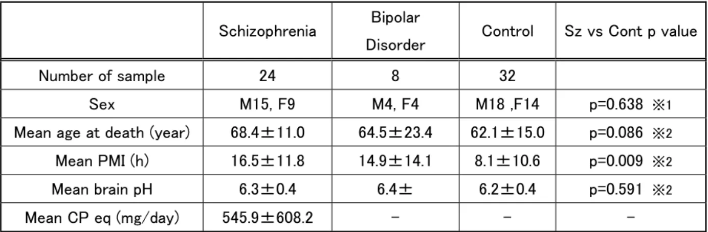
University, and was conducted in accordance with the Declaration of Helsinki. Detailed
demographic characteristics for the 24 patients with schizophrenia, eight patients with
bipolar disorder, and 32 controls is provided in Table 1. The Diagnostic and Statistical
Manual of Mental Disorders (DSM-IV) was used to diagnose both schizophrenia and
bipolar disorder. For each patient with schizophrenia, the daily dosage of antipsychotics
within 3 months before death is indicated as the chlorpromazine equivalent dose (mg/day)
(Table 1).
Protein expression analysis via ELISA
Sections (approximately 100 mg) of gray matter were selected from Brodmann area 10
(BA10) (i.e., PFC) and BA22 (i.e., STG) in the frozen brains. The tissues were suspended
in lysis buffer (N-PER Neuronal Protein Extraction Reagent, ThermoFisher, Japan)
10
supplemented with protease inhibitors (Protease Inhibitor Cocktail Set III, Calbiochem,
Japan) and phosphatase inhibitors (PhosSTOP, Roche, Japan), following which they
underwent sonication for 50 seconds (30% duty cycle) at an output level of 5 (Ultrasonic
processor W385, Astrason Heat Systms-Ultrasonics, NY, USA). The samples were the
centrifuged (40,000 × g for 30 min), and the total protein concentrations of the
supernatants were measured via a bicinchoninic acid assay (Protein Assay BCA Kit, Fuji
Film, Tokyo, Japan) using bovine serum albumin (BSA) as a control. Levels of
ALDH4A1 expression were determined using an ELISA kit (OKEH02446, Aviva
Systems Biology, USA), in accordance with the manufacturer’s protocol. Levels of
ALDH4A1 expression were normalized against the total protein concentration.
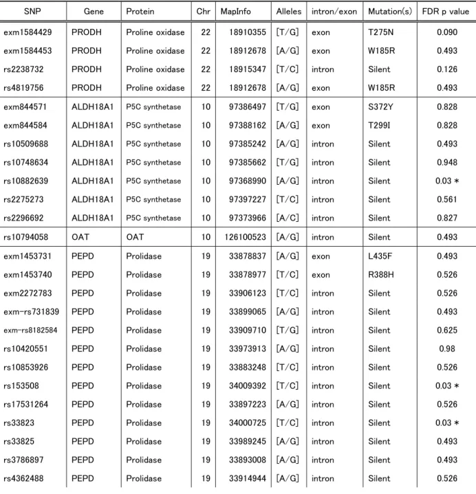
SNP genotyping
SNP data were determined in previous studies [10]. In brief, genomic DNA was extracted
from the frozen cerebellum or occipital cortex. Genotyping was performed using
HumanCoreExome -24 v1.0 Beadchip (Illumina, Tokyo, Japan) on an iScan system
(Illumina). In the present study, we examined 30 SNPs associated with proline
metabolism (Table 2). For analyses of the associations between SNPs and protein
expression, we excluded SNPs with call rates <99% and minor allele frequencies <5%.
11
Statistical analysis
Analyses of covariance (ANCOVA) were used to compare levels of protein (ALDH4A1)
expression among the groups (schizophrenia, bipolar disorder, and control). Diagnosis
and genotype were set as independent variables, while brain pH and postmortem interval
(PMI) were set as covariates. There were two missing values for PMI and three for brain
pH. Each missing value was represented using the average value. Fisher’s least significant
difference (LSD) post hoc comparisons were performed to evaluate group differences. In
addition, Spearman's rank correlation coefficients were used to assess the association
between protein expression and the following: age at death, PMI, brain pH, and estimated
total dosage of neuroleptic drugs. T-tests were used to assess the association between sex
and levels of ALDH4A1 expression. A p value <0.05 was considered significant in these
tests. For SNP analysis, the false discovery rate (FDR) was used to control for multiple
comparisons [28]. We reported a corrected p value with an FDR at q = 0.05 in the SNP
study. Statistica (version 10.0, Statsoft Inc., Tulsa, OH) was used for ANCOVA, and
Sigma Plot software (version 11, Systat Software, Inc., CA) was used for all tests that did
not involve FDR and ANCOVA values. The association study between gene
polymorphisms and schizophrenia was conducted using SNPStats
(http://bioinfo.iconcologia.net/SNPstats).
12
Results
We examined levels of ALDH4A1 expression in the PFC and STG of patients with
schizophrenia, bipolar disorder, and controls via ELISA. Our ANCOVA analysis revealed
that levels of ALDH4A1 expression in the PFC were significantly different between
schizophrenia, bipolar disorder and controls [F (2,57) =3.59, p<0.05)]. ALDH4A1 levels
in PFC were higher in patients with schizophrenia (p<0.05) and bipolar disorder (p<0.05)
than in controls (Figure 1a). Similarly, ANCOVA analysis revealed that levels of
ALDH4A1 expression in the STG were significantly different between schizophrenia,
bipolar disorder and controls [F (2,58) =10.36, p<0.05]. ALDH4A1 levels in STG were
higher in patients with schizophrenia (p<0.05) and bipolar disorder (p<0.05) than in
controls (Figure 1b).
We then examined the associations between ALDH4A1 expression and sex, age
at death, PMI, and brain pH using Pearson correlation coefficients (Figure 2). In patients
with schizophrenia, we also examined associations with the daily dosage of
antipsychotics in the 3 months prior to death. No significant effects of sex, age at death,
or antipsychotic dose were observed on ALDH4A1 expression in any brain region
examined. However, we observed a significant association between levels of ALDH4A1
expression in the PFC and PMI (p<0.05, r=0.459)/brain pH (p<0.005, r=0.265). We also
13
observed a significant association between levels of ALDH4A1 expression in the STG
and brain pH (p<0.005, r=0.291). Therefore, brain pH and PMI were set as covariates, as
described in Materials and Methods section.
We examined the effects of SNPs in molecules associated with proline
metabolism on levels of ALDH4A1 expression in patients with schizophrenia (n=22),
bipolar disorder (n=6), and controls (n=11). In our analysis of 111 SNPs of proline
metabolism-associated molecules (PRODH, ALDH4A1, ALDH18A1, PYCR1 OAT,
PEPD), 30 SNPs were retrieved for analysis using the criteria described in the Materials
and Methods section. We then associations between these 30 SNPs and levels of
ALDH4A1 expression in the PFC and STG. In the PFC, we observed significant
associations between ALDH4A1 expression and three SNPs for ALDH18A1
(rs10882639) and PEPD (rs33823, rs153508) (Figure 3). The rs10882639 SNP was
associated with an allele dose-dependent effect: Individuals homozygous for the A allele
and heterozygous AG individuals at rs10882639 exhibited significantly higher levels of
ALDH4A1 expression than homozygous G individuals. The rs33823 SNP was also
associated with allele dose-dependent effects: Individuals homozygous for the G allele
and heterozygous AG individuals at rs33823 exhibited significantly higher levels of
ALDH4A1 expression than homozygous A individuals. Furthermore, we also observed a
14
significant effect of SNP rs153508 on the expression of ALDH4A1: Individuals
homozygous for the G allele and heterozygous for AG at rs153508 exhibited significantly
higher levels of ALDH4A1 expression than homozygous A individuals. In the STG, we
observed no associations between ALDH4A1 expression and SNPs.
15
Discussion
The present study is the first postmortem investigation to examine levels of ALDH4A1
expression in the PFC and STG in patients with schizophrenia. We further examined the
relationship between levels of ALDH4A1 expression and genetic polymorphisms
involved in proline metabolism. Our results indicated that ALDH4A1 levels were
significantly increased in the PFC and STG of patients with schizophrenia or bipolar
disorder.
ALDH4A1 is among the enzymes involved in the metabolism of proline to
glutamate, which is the most downstream component of the pathway (Figure 4).
ALDH4A1 is expressed in all regions in the brain and is somewhat strongly expressed in
the hippocampus and caudate nucleus (THE HUMAN PROTEIN ATLAS
https://www.proteinatlas.org/). It is ubiquitously present in various cells and is an enzyme
located downstream of the pathway that metabolizes proline to glutamate in mitochondria
(Figure 4). The glutamate produced is converted to α-ketoglutarate and exits from the
mitochondria to the cytoplasm via an α-ketoglutarate-malate carrier (OGC) [29].
Aspartate aminotransferases(AST), is an enzyme that converts α-ketoglutarate into
glutamate, is present in the cytoplasm. Thus, higher expression of ALDH4A1 in
schizophrenia may result in increased glutamate and α-ketoglutarate levels in
mitochondria, inducing cytoplasmic glutamate and glutamine upregulation in the
16
astrocyte and presynaptic glutamate increase, thereby leading to increased glutamate
release into the synaptic cleft.
The relationship between schizophrenia and glutamate has attracted attention.
The glutamate hypothesis, one of the major pathological hypotheses of schizophrenia,
was proposed because overuse of phencyclidine (PCP), an antagonist of the NMDA
receptor, causes hypofunction of the NMDA receptor, resulting in positive and negative
symptoms [30]. It is known that in anti-NMDA receptor encephalitis, which exhibits
symptoms similar to those of schizophrenia, an autoantibody of the NMDA receptor
causes internalization of the NMDA receptor and thus a decrease of NMDA receptor
function [31].Neurotoxicity by excessive release of glutamate is thought to occur because
of disinhibition of glutamate nerve terminals caused by NMDA receptor hypofunction
[32]. However, the mechanisms underlying NMDA receptor hypofunction remain to be
elucidated. It was reported that NMDA receptors are internalized when activated by
glutamate [33] and that the internalization of NMDARs not only reduced the number of
cell surface NMDARs but also inhibited the activity of the non-internalized surface
NMDARs [34]. Due to the increase in glutamate it is possible that downregulation of
NMDA receptors by internalization or phosphorylation of NMDA receptors may occur.
Glutamate can be synthesized via four pathways. The first involves synthesis from α-
17
ketoglutarate by the action of an aminotransferase, the second from glutamate
dehydrogenase via the addition of free ammonia to α-ketoglutarate, the third via the
synthesis of glutamine to glutaminase, and the fourth via proline metabolism.Glutamate
that is used as a transmitter is generally synthesized from glutamine at the presynaptic
terminal and is stored in synaptic vesicles. Furthermore, it is known to be involved in the
pathology of schizophrenia, and several studies have reported the relationship between
schizophrenia and glutamate. Glutamate release induced by non-competitive NMDA
receptor agonists has been reported to increase prefrontal gray matter in model animals,
resulting in schizophreniform-like behaviors [35].Postmortem brain studies have shown
that glutamate levels were lower in the schizophrenia than in the control groups [36], and
that glutamate was higher in the schizophrenia group in the prefrontal region [37].
Furthermore, no significant difference between glutamate levels in the caudate nucleus,
nucleus accumbens, frontal cortex, amygdala, thalamus of was observed between the
schizophrenia and control groups [38]. Thus, no consensus on schizophrenia and
glutamate levels has been obtained in postmortem brain studies. Furthermore, a
metabolome study revealed an elevated serum glutamate levels in schizophrenia
patients[39]; however, glutamate levels in the cerebrospinal fluid were observed to be
lower in schizophrenic patients [40] in one study and in a few others, no difference in
18
these levels were observed compared with those in the control group [41][42][36]. Hence,
these results are inconsistent.A magnetic resonance spectroscopy (MRS) study revealed
that patients with acute schizophrenia had high levels of glutamine and patients with
chronic schizophrenia had low levels of glutamate and glutamine [43][44]. In addition,
glutamine levels were high in the cingulate gyrus and thalamus in first episode
schizophrenia patients, but glutamate and glutamine levels were low in the cingulate
gyrus in patients with schizophrenia in the chronic stage after treatment [45-47]. In studies
comparing results before and after drug treatment, glutamine levels were higher than
normal in the anterior cingulate gyrus and thalamus of untreated schizophrenic patients,
and were significantly reduced in the thalamus after 30 months of antipsychotic treatment
[48]. Moreover, higher levels of glutamate were observed in the striatum of antipsychotic-
naïve patients with first-episodic psychosis than in controls. After antipsychotic treatment
for 4 weeks, glutamate levels were not significantly different in the striatum [49]. These
results suggest that hyperactivity of glutamate mediated neurotransmission occurs in the
acute phase in schizophrenia, but these levels normalize following treatment. Conversely,
glutamate neurotransmission decreases in the chronic phase.
The relationship between schizophrenia and glutamate receptors have been
examined in previous studies. Glutamate receptors include ionotropic receptors (NMDA
19
receptor, α-amino-3-hydroxy-5-methyl-4-isoxazolepropionic acid (AMPA) receptor,
kainate receptor) and metabotropic receptors and their association with schizophrenia has
been studied. NMDA receptors have several subunits [GluN1 (NR1), GluN2A-2D
(NR2A-2D), GluN3A-3B (NR3A-3B)]. Postmortem brain studies have reported
decreased NR1 mRNA expression and elevated NR2B mRNA expression in the
hippocampus [50] and increased NR3A mRNA expression in the dorsolateral prefrontal
cortex (DLPFC) of schizophrenia patients [51]; however, another study reported that
expression of NR1 and NR3A proteins in the DLPFC was not significantly different
between the schizophrenia and control group [52]. Thus, consistent results were not
obtained. Neuroimaging studies reported decreased ligand binding of NMDA receptors
in the hippocampus and frontal lobe in untreated schizophrenia patients[53]. The AMPA
receptor has four subunits, namely GluA1-A4 (GluR1-GluR4). Postmortem brain studies
have investigated the binding of selective ligands and changes in the expression of each
subunit in patients with schizophrenia, and in the medial temporal lobe including the
hippocampus, ligand binding and a decrease in the expression of GluA1 and GluA2 in
schizophrenia patients was observed; [50, 54] however, these results lack reproducibility.
Changes in ligand binding in the prefrontal cortex were unclear[54], but a decrease in
GluA2 mRNA expression was reported[55][56]. The kainate receptor is an ion channel
20
formed by five subunits GluK1-GluK5. Collaborative research using our postmortem
brain showed decreased GluK4 expression in the prefrontal cortex of schizophrenia
patients. Among the 8 types of metabotropic glutamate receptors, mGlu1-mGlu8, mGlu2
and mGlu3 agonists improve positive and negative symptoms of schizophrenia [57].
Studies on the postmortem brains have shown the unchanged expression pattern and
amount of mGlu 2/3 protein[58] and decreased mGlu2 and unchanged mGlu3 mRNA
levels [59] in the prefrontal cortex of schizophrenia patients. Increased mGlu1 mRNA
and protein expression has been reported in the prefrontal cortex of schizophrenia[60]
[61], but the changes in the prefrontal cortex and hippocampus of mGlu5 mRNA have not
been elucidated[62]. Experiments in animals, have shown that the glutamate release in
the frontal lobe increases in rats administered with phencyclidine (PCP), and mGlu 2/3
agonists that suppress this phenomenon improve abnormal behaviors [63].
Developmental studies using mGlu 2/3 agonists for treating acute schizophrenia had
reached the advanced stages, but the therapeutic effects were somewhat lower than
olanzapine[57], and have hence been discontinued.
Previous studies have demonstrated that some neuropsychiatric disorders appear
to be related to hyperprolinemia [64] [65]. Several studies have suggested that glutamate
excitotoxicity may underlie neurological dysfunction in patients with hyperprolinemia
21
[66] [67] [68]. Previous reports have indicated that proline decreases Na+, K+, ATPase,
and acetylcholinesterase activity and induces oxidative stress in the rat brain [69] [70],
and that plasma proline levels are increased in patients with schizophrenia [39] [71].
Additional studies have reported that hyperprolinemic patients with schizophrenia are
significantly older at the first psychiatric hospitalization, suggestive of later onset.
However, hospital stays are approximately 46% longer in patients with hyperprolinemia
than in those without, suggesting that proline may be associated with the clinical picture
of schizophrenia [72]. Previous studies reporting that the onset of schizophrenia in
patients with hyperprolinemia is significantly delayed are of great interest. Considering
that proline is an agonist of the NMDA receptor GluN1 glycine site [73] and that it
enhances NMDA receptor function [74], the onset of schizophrenia may have been
delayed in these patients. Deficiencies in proline oxidase, an enzyme that converts proline
to D-1-pyrroline-5-carboxylate (P5C), can also cause hyperprolinemia [75]. The PRODH
gene encoding proline oxidase is located on chromosome 22 (22q 11.2). Carriers with chr
22 q 11. 2 deficiency exhibit a morbidity rate of 20-30% for schizophrenia, and this
deletion is among the most well-known risk factors for schizophrenia [76] [77] [78].
These findings indicate that hyperprolinemia may be related to clinical symptoms of
22q11 deletion syndrome, including the onset of schizophrenia. However, if proline
22
oxidase is deficient or decreased in a patient with the 22q11.2 deletion syndrome, the
efficiency of the metabolic cascade from proline to glutamine is decreases, so that even
if hyperprolinemia occurs, glutamate excess cannot occur. Factors other than glutamate
excess seem to be involved in schizophrenia onset in 22q11.2 deletion syndrome patients.
For example, the deleted region of 22q11.2 deletion syndrome includes schizophrenia
related genes, such as COMT, which codes an important enzyme in monoamine
catabolism; ZDHHC8, which codes a palmitoyl transferase enzyme; and DGCR8, a key
enzyme in the microRNA synthetic pathway[79]. One previous study reported that serum
prolidase activity was significantly higher in the schizophrenia group than in the control
group [80], in accordance with the finding that proline production is upregulated in
patients with schizophrenia [81]. Other studies have demonstrated that prolidase activity
in the production of proline from collagen is elevated in patients with schizophrenia,
along with plasma proline concentrations.
Conversely, that an increase in ALDH4A1 occurred secondarily in response to
the energy demand by neurons (e.g., overactivity of STG when auditory hallucinations
are active) generated in schizophrenic conditions cannot be ruled out. However, in the
PFC, cerebral blood flow reduction is observed in schizophrenia, and brain hypoactivity
is assumed and energy demand is expected to decline; however, ALDH4A1 increased
23
in the PFC. Thus, the increase in ALDH4A1 is not only due to the increase in energy
demand.
Based on the accumulated evidence, we hypothesized that glutamate production
is enhanced via increases in the expression of enzymes involved in degradation such as
prolidase and ALDH4A1, and that the pathway from proline to glutamate is accelerated
in patients with schizophrenia. Furthermore, glutamate production may be excessive,
which may in turn decrease NMDA receptor function. Thus, inhibiting the function of
ALDH4A1 and preventing excessive glutamate production may aid in the treatment of
schizophrenia. Disulfiram and cyanamide, which are currently used in the treatment of
alcoholism, also exert an inhibitory effect on ALDH4A1. However, inhibition of
ALDH4A1 may cause hyperprolinemia and other adverse effects. Some studies have
reported that disulfiram treatment for alcoholism does not alter Positive and Negative
Syndrome Scale (PANSS) scores in patients with schizophrenia [82]. Nonetheless, further
studies are required in order to determine the potential application of ALDH4A1
inhibition in the treatment of schizophrenia.
Our findings also indicated that ALDH4A1 expression was also elevated in the
PFC and STG of patients with bipolar disorder. Previous mass spectrometry studies have
analyzed the postmortem brains of patients diagnosed with mental illness (schizophrenia,
24
bipolar disorder, depression) in relation to those of healthy controls. Such studies have
examined changes in amino acid levels and glucose metabolism, revealing that
psychiatric disorders are associated with increases in proline levels [83]. An additional
study reported that serum prolidase activity was significantly higher in patients with
bipolar disorder than in healthy controls [84]. A meta-analysis of glutamate concentration
measurement via 1H-MRS further reported that brain glutamate concentrations are
increased in patients with bipolar disorder [85]. These findings indicate that schizophrenia
and bipolar disorder may share the tendency toward increased proline metabolism and
glutamic acid production. Although the results of our study support this notion, our
sample included only a small number of patients with bipolar disorder. Thus, our findings
should be interpreted with caution.
In the present study, we observed that ALDH4A1 expression levels were
significantly associated with the following three SNPs: rs10882639, rs33823, and
rs153508. The rs10882639 SNP is a genetic polymorphism of Δ-1-pyrroline-5-
carboxylane (P5C) synthetase, while rs33823 and rs153508 are genetic polymorphisms
of prolidase. P5C synthetase is an enzyme that functions inversely with ALDH4A1,
decomposing glutamic acid into glutamic-γ-semi-aldehyde. Prolidase is an enzyme that
generates proline from collagen. We could not confirm that ach gene of the 3 SNPs
25
identified in this study had a cis-acting effect on mRNA expression using Brain Cloud
(http://braincloud.jhmi.edu/). All three SNPs reflecting ALDH4A1 expression in this
study were intronic (iSNPs), located in the intergenic region (IGR) of the DNA sequence.
Although the IGR is a subset of noncoding DNA, recent studies have indicated that iSNPs
play key roles in regulating mRNA transcription rate, splicing, and maturation [86, 87].
Changes in the nucleotide sequence within the intron can affect splicing, as the sequence
is recognized by various splicing-related factors within introns of heterologous nuclear
RNA (hnRNA; alternative term for pre-mRNA). Most schizophrenia-associated SNPs are
thought to be iSNPs, and several studies have reported the role of iSNPs in psychiatric
disorders [88]. Such findings suggest that the three SNPs identified in the present study
play undetermined roles in schizophrenia.
It may be possible to predict levels of ALDH4A1 expression in patients with
schizophrenia by examining SNPs of P5C synthetase and prolidase in peripheral tissues
(i.e., blood, saliva). Furthermore, patients with schizophrenia exhibiting increased
ALDH4A1 expression may benefit from ALDH4A1 inhibition. As only one ALDH4A1
SNP associated with one genotype was observed in the present study, further studies are
required to determine whether other SNPs can be observed in patients with schizophrenia.
Future studies should also examine expression levels and gene polymorphisms for proline
26
and other molecules involved in proline metabolism.
The present study possesses several limitations of note. First, confounding
effects of disease-related factors including medication may have affected the expression
of ALDH4A1. Although we did not observe any effects of antipsychotic drugs on the
levels of ALDH4A1 expression in this study, additional animal studies are necessary to
investigate the effects of chronic antipsychotic administration on the expression of these
proteins in the PFC and STG. Second, our study population was relatively small,
particularly for a genetic study. Therefore, our findings must be verified via postmortem
examination in a larger brain cohort. Third, the age of each sample was slightly advanced,
and there was considerable variation in PMI. Therefore, we corrected the results via
statistical methods.Fourth, ALDH4A1 is also expressed in regions, such as the thalamus,
nucleus accumbens, and striatum, in which dopamine system abnormality is assumed,
other than the PFC and STG assessed in this study, and may be involved in schizophrenia
pathology; however ALDH4A1 expression in these regions were not measured in this
study. Further studies conducted will include these analyses. Fifth, the protein (P5C
synthetase, Prolidase) encoded by the three SNPs (rs 10882639, rs 33823, and rs 153508)
involved in ALDH4A1 expression were not measured in this study. However, our future
study measures the expression of these proteins and investigates the significance of the
27
trans-acting effect on ALDH4A1 expression. We also measure other proteins involved in
proline metabolism.
In summary, the findings of the present study demonstrate that ALDH4A1
expression is elevated relative to control levels in the postmortem brains of patients with
schizophrenia and bipolar disorder. Furthermore, such increases were accompanied by
genetic polymorphisms in molecules associated with proline metabolism. Such results
provide new insight into the potential pathogenic mechanisms underlying schizophrenia
and bipolar disorder.
28
Figure legends
Figure 1. Expression of ALDH4A1 in the diagnostic groups. Mean expression of
ALDH4A1 in the PFC (a) and STG (b) in patients with schizophrenia, patients with
bipolar disorder, and control participants. ELISA was used to measure protein expression.
Error bars indicate the SE of the mean. P values presented over a bar indicate that the
difference between these groups was significant based on a post hoc Fisher’s LSD test.
PFC, prefrontal cortex; STG, superior temporal gyrus; ELISA, enzyme-linked
immunosorbent assay; SE, standard error; LSD, least significant difference, Sz,
schizophrenia; BP: bipolar disorder; Cont, controls.
Figure 2. Association between levels of ALDH4A1 expression and each confounding
factor.
PFC, prefrontal cortex; STG, superior temporal gyrus; PMI, postmortem interval; CPeq,
chlorpromazine equivalent.
Figure 3. The effects of SNPs in molecules associated with proline metabolism on
levels of ALDH4A1 expression. There was a significant association between ALDH4A1
expression in the PFC and rs10882639 (a), rs33823 (b), and rs153508 (c).
Error bars indicate the SE of the mean. Asterisks presented over a bar indicate that the
29
difference between these groups was significant based on a post hoc Fisher’s LSD test.
SNP, single-nucleotide polymorphism; PFC, prefrontal cortex; SE, standard error; LSD,
least significant difference.
Figure 4. The proline metabolism system. The metabolism of proline involves two other
amino acids (glutamate and ornithine) and six enzymes [64].
P5C, Δ-1-pyrroline-5-carboxylane; TCA, tricarboxylic acid; OGC, α-ketoglutarate-
malate carrier; N, System N transporter (astrocytic from); A, System A transporter
(neuronal from); AST, aspartate aminotransferases.
30
Acknowledgments
I would like to express my deepest gratitude to Professor Hirooki Yabe for providing great
opportunities to study various aspects of psychiatry, and to the staff of the Postmortem
Brain Bank of Fukushima for Psychiatric Research in the Department of Neuropsychiatry
at the School of Medicine at Fukushima Medical University for their research support.
I am most grateful to Dr. Hiroyuki Nawa, Dr. Akiyoshi Kakita, and Dr. Akari Takeshima
of the Brain Research Institute of Niigata University for their assistance in obtaining the
brain samples. I also wish to thank Dr. Yasuto Kunii, Dr. Mizuki Hino, Dr. Akira Wada,
Dr. Junya Matsumoto, and Dr Ryuta Izumi. In addition, I thank Ms. Chiaki Watanabe and
Hiromi Onuma of the Postmortem Brain Bank of Fukushima for Psychiatric Research for
their contribution to sample collection and coordination. I wish to express special thanks
to the families of the deceased for the donations of brain tissue, and for their time and
effort devoted to the consent process and interviews. I also wish to express my gratitude
to my colleagues in the Department of Neuropsychiatry at the School of Medicine at
Fukushima Medical University.
Last, but not least, special thanks to my family for their support and warm encouragement.
31
References
1. McGrath, J., et al., Schizophrenia: a concise overview of incidence, prevalence, and mortality. Epidemiol Rev, 2008. 30: p. 67-76.
2. Holder, S.D. and A. Wayhs, Schizophrenia. Am Fam Physician, 2014. 90(11): p. 775- 82.
3. Weickert, T.W., et al., Cognitive impairments in patients with schizophrenia displaying preserved and compromised intellect. Arch Gen Psychiatry, 2000. 57(9): p.
907-13.
4. Kleinman, J.E., et al., Genetic neuropathology of schizophrenia: new approaches to an old question and new uses for postmortem human brains. Biol Psychiatry, 2011.
69(2): p. 140-5.
5. Dickinson, D., et al., Differential effects of common variants in SCN2A on general cognitive ability, brain physiology, and messenger RNA expression in schizophrenia cases and control individuals. JAMA Psychiatry, 2014. 71(6): p. 647-56.
6. Morita, Y., et al., Characteristics of the cation cotransporter NKCC1 in human brain:
alternate transcripts, expression in development, and potential relationships to brain function and schizophrenia. J Neurosci, 2014. 34(14): p. 4929-40.
7. O'Donovan, S.M., et al., Glutamate transporter splice variant expression in an enriched pyramidal cell population in schizophrenia. Transl Psychiatry, 2015. 5: p.
e579.
8. Ohi, K., et al., DEGS2 polymorphism associated with cognition in schizophrenia is associated with gene expression in brain. Transl Psychiatry, 2015. 5: p. e550.
9. Kunii, Y., et al., Elevated postmortem striatal t-DARPP expression in schizophrenia and associations with DRD2/ANKK1 polymorphism. Prog Neuropsychopharmacol Biol Psychiatry, 2014. 53: p. 123-8.
10. Hino, M., et al., Decreased VEGFR2 expression and increased phosphorylated Akt1 in the prefrontal cortex of individuals with schizophrenia. J Psychiatr Res, 2016. 82:
p. 100-8.
11. Hirayama-Kurogi, M., et al., Downregulation of GNA13-ERK network in prefrontal cortex of schizophrenia brain identified by combined focused and targeted quantitative proteomics. J Proteomics, 2017. 158: p. 31-42.
12. Sun, L., et al., Gene expression profiling in peripheral blood mononuclear cells of early-onset schizophrenia. Genom Data, 2015. 5: p. 169-70.
13. Saia-Cereda, V.M., et al., Proteomics of the corpus callosum unravel pivotal players in the dysfunction of cell signaling, structure, and myelination in schizophrenia
32
brains. Eur Arch Psychiatry Clin Neurosci, 2015. 265(7): p. 601-12.
14. Shenton, M.E., et al., A review of MRI findings in schizophrenia. Schizophr Res, 2001.
49(1-2): p. 1-52.
15. Semkovska, M., M.A. Bedard, and E. Stip, [Hypofrontality and negative symptoms in schizophrenia: synthesis of anatomic and neuropsychological knowledge and ecological perspectives]. Encephale, 2001. 27(5): p. 405-15.
16. de la Torre, J.C., M. Barrios, and C. Junque, Frontal lobe alterations in schizophrenia: neuroimaging and neuropsychological findings. Eur Arch Psychiatry Clin Neurosci, 2005. 255(4): p. 236-44.
17. Binder, J., The new neuroanatomy of speech perception. Brain, 2000. 123 Pt 12: p.
2371-2.
18. Dierks, T., et al., Activation of Heschl's gyrus during auditory hallucinations. Neuron, 1999. 22(3): p. 615-21.
19. Naatanen, R., et al., The mismatch negativity (MMN) in basic research of central auditory processing: a review. Clin Neurophysiol, 2007. 118(12): p. 2544-90.
20. Javitt, D.C., et al., Role of cortical N-methyl-D-aspartate receptors in auditory sensory memory and mismatch negativity generation: implications for schizophrenia.
Proc Natl Acad Sci U S A, 1996. 93(21): p. 11962-7.
21. Le Corre, S., et al., Increased levels of expression of an NMDARI splice variant in the superior temporal gyrus in schizophrenia. Neuroreport, 2000. 11(5): p. 983-6.
22. Nudmamud, S. and G.P. Reynolds, Increased density of glutamate/N-methyl-D- aspartate receptors in superior temporal cortex in schizophrenia. Neurosci Lett, 2001.
304(1-2): p. 9-12.
23. Kasai, K., et al., Mismatch negativity and N2b attenuation as an indicator for dysfunction of the preattentive and controlled processing for deviance detection in schizophrenia: a topographic event-related potential study. Schizophr Res, 1999.
35(2): p. 141-56.
24. Kasai, K., et al., Impaired cortical network for preattentive detection of change in speech sounds in schizophrenia: a high-resolution event-related potential study. Am J Psychiatry, 2002. 159(4): p. 546-53.
25. Kasai, K., et al., Progressive decrease of left Heschl gyrus and planum temporale gray matter volume in first-episode schizophrenia: a longitudinal magnetic resonance imaging study. Arch Gen Psychiatry, 2003. 60(8): p. 766-75.
26. Sakai, M., et al., Assessment of copy number variations in the brain genome of schizophrenia patients. Mol Cytogenet, 2015. 8: p. 46.
27. Matsumoto, J., et al., Abnormal phospholipids distribution in the prefrontal cortex
33
from a patient with schizophrenia revealed by matrix-assisted laser desorption/ionization imaging mass spectrometry. Anal Bioanal Chem, 2011. 400(7):
p. 1933-43.
28. Benjamini, Y., et al., Controlling the false discovery rate in behavior genetics research.
Behav Brain Res, 2001. 125(1-2): p. 279-84.
29. Satrustegui, J., et al., Role of aralar, the mitochondrial transporter of aspartate- glutamate, in brain N-acetylaspartate formation and Ca(2+) signaling in neuronal mitochondria. J Neurosci Res, 2007. 85(15): p. 3359-66.
30. Coyle, J.T., The glutamatergic dysfunction hypothesis for schizophrenia. Harv Rev Psychiatry, 1996. 3(5): p. 241-53.
31. Graus, F., et al., A clinical approach to diagnosis of autoimmune encephalitis. Lancet Neurol, 2016. 15(4): p. 391-404.
32. Olney, J.W., et al., NMDA antagonist neurotoxicity: mechanism and prevention.
Science, 1991. 254(5037): p. 1515-8.
33. Piguel, N.H., et al., Scribble1/AP2 complex coordinates NMDA receptor endocytic recycling. Cell Rep, 2014. 9(2): p. 712-27.
34. Fang, X.Q., et al., Regulated internalization of NMDA receptors drives PKD1- mediated suppression of the activity of residual cell-surface NMDA receptors. Mol Brain, 2015. 8(1): p. 75.
35. Moghaddam, B., et al., Activation of glutamatergic neurotransmission by ketamine:
a novel step in the pathway from NMDA receptor blockade to dopaminergic and cognitive disruptions associated with the prefrontal cortex. J Neurosci, 1997. 17(8):
p. 2921-7.
36. Tsai, G., et al., Abnormal excitatory neurotransmitter metabolism in schizophrenic brains. Arch Gen Psychiatry, 1995. 52(10): p. 829-36.
37. Kutay, F.Z., et al., Free amino acid level determinations in normal and schizophrenic brain. Prog Neuropsychopharmacol Biol Psychiatry, 1989. 13(1-2): p. 119-26.
38. Korpi, E.R., et al., Neurotransmitter amino acids in post-mortem brains of chronic schizophrenic patients. Psychiatry Res, 1987. 22(4): p. 291-301.
39. Oresic, M., et al., Metabolome in schizophrenia and other psychotic disorders: a general population-based study. Genome Med, 2011. 3(3): p. 19.
40. Kim, J.S., et al., Low cerebrospinal fluid glutamate in schizophrenic patients and a new hypothesis on schizophrenia. Neurosci Lett, 1980. 20(3): p. 379-82.
41. Do, K.Q., et al., gamma-Glutamylglutamine and taurine concentrations are decreased in the cerebrospinal fluid of drug-naive patients with schizophrenic disorders. J Neurochem, 1995. 65(6): p. 2652-62.
34
42. Perry, T.L., Normal cerebrospinal fluid and brain glutamate levels in schizophrenia do not support the hypothesis of glutamatergic neuronal dysfunction. Neurosci Lett, 1982. 28(1): p. 81-5.
43. Bartha, R., et al., Measurement of glutamate and glutamine in the medial prefrontal cortex of never-treated schizophrenic patients and healthy controls by proton magnetic resonance spectroscopy. Arch Gen Psychiatry, 1997. 54(10): p. 959-65.
44. Ohrmann, P., et al., Evidence for glutamatergic neuronal dysfunction in the prefrontal cortex in chronic but not in first-episode patients with schizophrenia: a proton magnetic resonance spectroscopy study. Schizophr Res, 2005. 73(2-3): p. 153- 7.
45. Theberge, J., et al., Glutamate and glutamine measured with 4.0 T proton MRS in never-treated patients with schizophrenia and healthy volunteers. Am J Psychiatry, 2002. 159(11): p. 1944-6.
46. Theberge, J., et al., Glutamate and glutamine in the anterior cingulate and thalamus of medicated patients with chronic schizophrenia and healthy comparison subjects measured with 4.0-T proton MRS. Am J Psychiatry, 2003. 160(12): p. 2231-3.
47. Tayoshi, S., et al., Metabolite changes and gender differences in schizophrenia using 3-Tesla proton magnetic resonance spectroscopy (1H-MRS). Schizophr Res, 2009.
108(1-3): p. 69-77.
48. Theberge, J., et al., Longitudinal grey-matter and glutamatergic losses in first- episode schizophrenia. Br J Psychiatry, 2007. 191: p. 325-34.
49. de la Fuente-Sandoval, C., et al., Glutamate levels in the associative striatum before and after 4 weeks of antipsychotic treatment in first-episode psychosis: a longitudinal proton magnetic resonance spectroscopy study. JAMA Psychiatry, 2013. 70(10): p.
1057-66.
50. Gao, X.M., et al., Ionotropic glutamate receptors and expression of N-methyl-D- aspartate receptor subunits in subregions of human hippocampus: effects of schizophrenia. Am J Psychiatry, 2000. 157(7): p. 1141-9.
51. Mueller, H.T. and J.H. Meador-Woodruff, NR3A NMDA receptor subunit mRNA expression in schizophrenia, depression and bipolar disorder. Schizophr Res, 2004.
71(2-3): p. 361-70.
52. Henson, M.A., et al., Developmental regulation of the NMDA receptor subunits, NR3A and NR1, in human prefrontal cortex. Cereb Cortex, 2008. 18(11): p. 2560-73.
53. Pilowsky, L.S., et al., First in vivo evidence of an NMDA receptor deficit in medication-free schizophrenic patients. Mol Psychiatry, 2006. 11(2): p. 118-9.
54. Meador-Woodruff, J.H. and D.J. Healy, Glutamate receptor expression in
35
schizophrenic brain. Brain Res Brain Res Rev, 2000. 31(2-3): p. 288-94.
55. Vawter, M.P., et al., Microarray analysis of gene expression in the prefrontal cortex in schizophrenia: a preliminary study. Schizophr Res, 2002. 58(1): p. 11-20.
56. Beneyto, M. and J.H. Meador-Woodruff, Lamina-specific abnormalities of AMPA receptor trafficking and signaling molecule transcripts in the prefrontal cortex in schizophrenia. Synapse, 2006. 60(8): p. 585-98.
57. Patil, S.T., et al., Activation of mGlu2/3 receptors as a new approach to treat schizophrenia: a randomized Phase 2 clinical trial. Nat Med, 2007. 13(9): p. 1102-7.
58. Crook, J.M., et al., Comparative analysis of group II metabotropic glutamate receptor immunoreactivity in Brodmann's area 46 of the dorsolateral prefrontal cortex from patients with schizophrenia and normal subjects. Mol Psychiatry, 2002. 7(2): p. 157- 64.
59. Gonzalez-Maeso, J., et al., Identification of a serotonin/glutamate receptor complex implicated in psychosis. Nature, 2008. 452(7183): p. 93-7.
60. Gupta, D.S., et al., Metabotropic glutamate receptor protein expression in the prefrontal cortex and striatum in schizophrenia. Synapse, 2005. 57(3): p. 123-31.
61. Volk, D.W., S.M. Eggan, and D.A. Lewis, Alterations in metabotropic glutamate receptor 1alpha and regulator of G protein signaling 4 in the prefrontal cortex in schizophrenia. Am J Psychiatry, 2010. 167(12): p. 1489-98.
62. Ohnuma, T., et al., Expression of the human excitatory amino acid transporter 2 and metabotropic glutamate receptors 3 and 5 in the prefrontal cortex from normal individuals and patients with schizophrenia. Brain Res Mol Brain Res, 1998. 56(1-2):
p. 207-17.
63. Moghaddam, B. and B.W. Adams, Reversal of phencyclidine effects by a group II metabotropic glutamate receptor agonist in rats. Science, 1998. 281(5381): p. 1349- 52.
64. Mitsubuchi, H., et al., Biochemical and clinical features of hereditary hyperprolinemia. Pediatr Int, 2014. 56(4): p. 492-6.
65. Raux, G., et al., Involvement of hyperprolinemia in cognitive and psychiatric features of the 22q11 deletion syndrome. Hum Mol Genet, 2007. 16(1): p. 83-91.
66. Nadler, J.V., A. Wang, and A. Hakim, Toxicity of L-proline toward rat hippocampal neurons. Brain Res, 1988. 456(1): p. 168-72.
67. Cohen, S.M. and J.V. Nadler, Proline-induced potentiation of glutamate transmission.
Brain Res, 1997. 761(2): p. 271-82.
68. Delwing, D., et al., In vivo and in vitro effects of proline on some parameters of oxidative stress in rat brain. Brain Res, 2003. 991(1-2): p. 180-6.