Forward genetic screen for Caenorhabditis
elegans mutants with a progressive decline in adult locomotor function
Author Kazuto Kawamura
Degree Conferral Date
2019‑03‑31
Degree Doctor of Philosophy Degree Referral
Number
38005甲第30号 Copyright
Information
(C) 2018 The Author.
URL http://doi.org/10.15102/1394.00000766
1
Okinawa Institute of Science and Technology Graduate University
Thesis submitted for the degree
Doctor of Philosophy
Forward genetic screen for Caenorhabditis elegans mutants with a progressive decline
in adult locomotor function
by
Kazuto Kawamura
Supervisor: Ichiro Maruyama
January 2019
2 1. Abstract
Some inherited mutations can cause a delayed onset of toxic symptoms. These types of mutations can be the underlying cause of age-related diseases or more prevalent age- related functional impairments. The isolation of mutants and identification of mutations in model organisms that show delayed onset of toxic symptoms may provide insights into evolutionarily conserved genetic regulators that function during the maintenance of functional capacities. In this study, we carried out an unbiased forward genetic screen for Caenorhabditis elegans mutants that show progressive loss of locomotor function during adulthood. After screening 3,352 F2 worms from 1,000 haploid genomes, we isolated five mutant strains that progressively lose their ability to complete a locomotor assay. For one of the mutant strains, a nonsense mutation in Elongator Complex Protein Component 2 (elpc-2) causes the progressive decline in locomotor function. Other C. elegans mutants with
mutations in subunits of the Elongator complex also showed progressive declines in adult locomotor function. The Elongator complex may play a critical role during the maintenance of adult locomotor function in C. elegans. The screening procedure, isolated mutants, and the Elongator complex are valuable tools to further explore how locomotor healthspan is
maintained in animals.
3 Acknowledgements
I am grateful for the gracious support of many people who helped me during my PhD.
Ichi Maruyama, my PhD advisor, has always encouraged me to pursue an independent project, and has provided an environment where I could learn to think as an independent scientist. He has allowed me to pursue my own interests while providing guidance at critical decision-points to keep the project on track. I thank him for giving me such a generous opportunity.
Bernd Kuhn and Davie Van Vactor have always been generous with their time to provide advice on coursework and research as thesis committee members. Their kind support and advice has helped me grow as a researcher.
I am grateful to Monica Driscoll for traveling from the other side of the world to examine my thesis proposal. The discussion that took place during the thesis proposal has served as a compass while moving the project forward. I thank Rick Morimoto and Alex Parker for taking the time and effort to examine my thesis and providing valuable guidance and advice for my future.
I thank the Maruyama Unit members, especially Takashi Murayama and Eiichiro Saita, for being supportive throughout my PhD. Takashi handed me my first plate of Caenorhabditis elegans worms and taught me from how to pick a worm from one plate to another, to injecting DNA fragments into the miniscule gonad of the worm. Eiichiro has always been supportive and helped me with setting up the video recording system to measure the locomotor function of worms.
None of my experiments would have been possible without the administrative support of Hitomi Ohtaki. She has always been a positive presence, and has handled all of the
administrative hoops and hurdles for me to obtain materials to carry out this thesis work.
4
I am grateful for the support of the OIST DNA sequencing section and Imaging section, especially Hiroki Goto and Koji Koizumi, for technical support as well as advice for my thesis project.
My first in-depth research experience came about because Takayuki Harada took a chance on me to work as a technician in his lab. This research experience motivated me to continue research and apply for a PhD. The encouragement and lessons that I learned while in the Harada Lab has remained with me through my PhD experience. Kazuhiko Namekata, Xiaoli Guo, Hayaki Watanabe, and Daiji Kittaka were great examples of researchers that I aspire to emulate.
I thank the Japan Society for the Promotion of Science and the Okinawa Institute of Science and Technology for providing funding for my PhD. The OIST graduate school staff has always been supportive from arranging my travel for the admissions workshop to coordinating the administrative support throughout my PhD.
Working on this thesis has been a growth experience for me. Sometimes I felt stuck or felt that I had chosen the wrong path in life. Through those times, I am happy to have had the support of my friends, especially Masakazu Igarashi, Tosif Ahamed, Tsai-Ming Luo, Ray Xin Lee, Yi-Jyun Luo, Yoriko Yamamura, Keisuke Ishihara, Alex Roertgen, Russ Hennessy, and Steve Asti.
Finally, I thank my family, Shoji, Mutsuko, and Yuko for their continued support.
They have encouraged me to try new things and work hard toward my goals while enjoying life. My extended family, especially Ken, Kuniko, Toru, Jeremy, and Masatoshi have helped me with encouragement and understanding. My wife, Aya, has been a constant source of support. Without her support, I would not have been able to create this thesis.
5 List of Abbreviations
AD Alzheimer’s disease
ALS amyotrophic lateral sclerosis
BC backcrossed
bp base pair
EDTA Ethylenediaminetetraacetic acid ELP Elongator complex protein ELPC Elongator complex protein EMS ethyl methanesulfonate EtOH ethyl alcohol
FUDR floxuridine
fALS familial amyotrophic lateral sclerosis HD Huntington’s disease
mcm methoxycarbonylmethyl ncm carbamoylmethyl
NGM nematode growth medium
RT room temperature
SNP single nucleotide polymorphism SMA spinal muscular atrophy
WT wild type
6 Table of Contents
1. Abstract ... 2
2. Problem Statement and Aims... 12
3. Chapter 1: Isolation of C. elegans mutants with progressive decline in adult locomotor function ... 13
3.1. Introduction ... 13
3.1.1 Inherited mutations can cause delayed onset of disease symptoms ... 13
3.1.2 Disease-related genes can be identified from forward genetic screens ... 16
3.1.3 C. elegans has been used successfully in forward genetic screens for complex phenotypes ... 19
3.1.4 C. elegans is used as a model for general mechanisms of neuromuscular aging and neuromuscular disease ... 20
3.1.5 Limitations in using C. elegans as the animal model ... 23
3.2 Materials and Methods ... 24
3.3 Results ... 26
3.3.1 “Edge Assay” can test locomotor function of hundreds of worms ... 26
3.3.2 Time points for distinguishing mutants with progressive locomotor decline from wild-type worms using the Edge Assay ... 30
3.3.3 Mutagenesis and screening procedure ... 32
3.3.4 Isolated mutants ... 33
3.4. Discussion ... 41
3.4.1 A new assay to measure C. elegans locomotor function ... 41
3.4.2 A new screening procedure to search for mutants with delayed onset of disease symptoms ... 42
3.4.3 Isolation of five mutants with progressive deficits in completing the Edge Assay . 43 3.4.4 Slight defects in development may cause strong deficits during adulthood ... 44
3.4.5 Genetic bases of lifespan and locomotor healthspan may not completely overlap . 44 4. Chapter 2: Identification of the causative mutation site in the isolated mutants ... 46
7
4.1 Introduction ... 46
4.1.1 Strategies to identify the causative mutation site in mutants isolated from forward genetic screens ... 46
4.1.2 Strategies to confirm the causative mutation site ... 50
4.2 Materials and Methods ... 53
4.3. Results ... 58
4.3.1 Whole genome sequencing of ix243 mutant strain ... 58
4.3.2 Wild-type version of elpc-2 rescues progressive loss of locomotor function in ix243 worms ... 62
4.3.3 Other C. elegans Elongator mutants show progressive loss of locomotor function 64 4.3.4 No additive effects are seen in Elongator double mutants ... 66
4.3.5 Expression pattern of elpc-2 transcriptional reporter ... 69
4.3.6 Whole genome sequencing of ix241 mutant strain ... 70
4.3.7 ix241(5x BC) #23 strain carries dys-1 mutation but maintains locomotor function 74 4.4 Discussion ... 79
4.4.1 Elongator Complex is important for maintaining adult locomotor function in C. elegans ... 79
4.4.2 ELP1 is implicated in familial dysautonomia... 80
4.4.3 ELP2 is involved in inherited neurodevelopmental disease ... 81
4.4.4 ELP3 is implicated in amyotrophic lateral sclerosis ... 82
4.4.5 ELP4 is implicated in rolandic epilepsy ... 83
4.4.6 ELP6 causes larval lethality in Drosophila melanogaster ... 83
4.4.7 Differences and commonalities among ELP-related diseases ... 84
5. Chapter 3: Characterization of the elpc-2 gene ... 86
5.1 Introduction ... 86
5.1.1 DAF-16 and HSF-1 are transcription factors that regulate aging in C. elegans ... 86
5.1.2 Insulin-signaling pathway involvement in locomotor healthspan and aging ... 86
5.1.3 Heat-shock factor involvement in locomotor healthspan and aging ... 88
8
5.1.4 tRNA modifications are involved in optimal protein translataion ... 88
5.2 Materials and Methods ... 89
5.3. Results ... 91
5.3.1 Adult locomotor function of daf-16(mu86) and hsf-1(sy441) worms ... 91
5.3.2 Genetic interaction of elpc-2(ix243) with hsf-1(sy441) ... 92
5.3.3 Genetic interaction of elpc-2(ix243) with tut-1(tm1297) ... 94
5.3.4 Induction of heat shock response is increased in elpc-2(ix243) worms ... 96
5.3.5 Increased heat shock response in aged elpc-2(ix243) worms ... 97
5.4. Discussion ... 98
5.4.1. elpc-2 and hsf-1 mutations cause progressive locomotor decline by an overlapping mechanism ... 98
5.4.2. tRNA modifications may be involved in maintenance of locomotor healthspan . 100 6. Concluding remarks ... 103
7. References ... 106
9 List of figures
Figure 3.1 Mechanisms underlying immediate and delayed toxicity………...13
Figure 3.2 Photos of Edge assay………..28
Figure 3.3 Edge Assay completion rates………..29
Figure 3.4 Edge Assay completion rates of known mutants………31
Figure 3.5 Schematic description of forward genetic screen………...32
Figure 3.6 Edge Assay completion rates of isolated mutants………..34
Figure 3.7 Maximum velocities of WT, ix239, ix240, and ix242 worms………35
Figure 3.8 ix241 and ix243 strains show progressive decline in locomotor function after four backcrosses………...36
Figure 3.9 Lifespan of WT, ix243, and ix241 worms………..37
Figure 3.10 Maximum velocity of WT and ix243 worms from adult day 1 to 10…………...39
Figure 3.11 Percent change in lifespan and locomotor healthspan………..40
Figure 3.12 Genetic factors that affect lifespan and healthspan may not completely overlap..45
Figure 4.1 Schematic diagram of Hawaiian variant mapping strategy………47
Figure 4.2 EMS variant density mapping by backcrossing……….………….48
Figure 4.3 EMS variant density mapping by bulk segregation………49
Figure 4.4 Mutation identification strategy for ix243 strain………59
Figure 4.5 Mutation frequencies of ix243 mutant strain………..60
Figure 4.6 Candidate mutation site………..…62
Figure 4.7 elpc-2 rescues progressive loss of locomotor function in ix243 worms………...63
Figure 4.8 The Elongator complex is required to maintain locomotor function…………..…65
Figure 4.9 elpc-3 mutation does not cause additive effects in elpc-2 mutant………..67
Figure 4.10 elpc-1 mutation does not cause additive effects in elpc-2 mutant….…….……...68
Figure 4.11 Expression pattern of elpc-2 transcriptional GFP fusion……….69
10
Figure 4.12 Mutation frequency of ix241 mutant strain………...70
Figure 4.13 Confirmation of dys-1 mutation site in ix241 worms………..74
Figure 4.14 Locomotor function of ix241 worms after the fifth backcross………....75
Figure 4.15 Amino acid alignment of C. elegans ELPC-2 and human ELP2………....82
Figure 5.1 Locomotor function of known aging mutants………...92
Figure 5.2 Locomotor function of elpc-2(ix243);hsf-1(sy441) double mutant worms……..93
Figure 5.3 Locomotor function of elpc-2(ix243);tut-1(tm1297) double mutant worms……95
Figure 5.4 hsp16.2p::GFP reporter expression after 30C 3-h heat shock………..97
Figure 5.5 Induction of heat shock response in non-heat shocked, aged animals…………..98
Figure 5.6 Working model of Elongator, TUT-1 and HSF-1 involvement in locomotor healthspan………102
Figure 6.1 Mutation-environment interactions………..105
11 List of tables
Table 3.1 Summary of types of inherited mutations and resulting phenotype……….14
Table 3.2 Reverse genetic disease models of neuromuscular disease in C. elegans………....22
Table 3.3 Number of viable mutants obtained from screen……….33
Table 3.4 Lifespan measurements of WT (N2), ix243, and ix241 worms………...38
Table 3.5 Development times of isolated mutant strains……….41
Table 4.1 Advantages and disadvantages of mutation identification strategies………...46
Table 4.2 Advantages and disadvantages of mutation confirmation strategies………...51
Table 4.3 Remaining mutations in ix243 mutant strains………..61
Table 4.4 Description of mutation types………..62
Table 4.5 Remaining mutations in ix241 mutant strains………...72
Table 4.6 Description of mutation types………..73
Table 4.7 Candidate mutation sites involved in maintained locomotor function in ix241(5x BC) #23 strain: Assuming loss of a loss-of-function mutation………...76
Table 4.8 Description of mutation types………...77
Table 4.9 Candidate mutation sites involved in maintained locomotor function in ix241(5x BC) #23 strain: Assuming gain of function mutation……….78
Table 4.10 Description of mutation types………..79
Table 5.1 Development times of tut-1(tm1297) and elpc-2(ix243);tut-1(tm1297) mutants..96
12 2. Problem Statement and Aims
What type of inherited mutation can cause delayed onset of disease symptoms? In inherited cases of adult-onset diseases such as Alzheimer’s Disease (AD) or amyotrophic lateral sclerosis (ALS), the causative mutation is present from birth, but obvious disease symptoms occur later in life. In theory, delayed onset of disease symptoms can occur from mutations that cause a slow, cumulative form of toxicity or mutations in which its toxicity is triggered by changes related to aging or adulthood. The delayed onset of disease symptoms takes many years to study in humans. Therefore, the isolation of mutant model organisms that show delayed onset of disease symptoms may be a more practical approach to explore how an inherited mutation can cause adult-onset disease.
In this thesis, we addressed the following three aims:
Aim 1 (Chapter 1): Isolation of Caenorhabdidtis elegans mutants with progressive decline in adult locomotor function
Aim 2 (Chapter 2): Identification of the responsible gene for the progressive decline in locomotor function for the isolated mutants
Aim 3 (Chapter 3): Characterization of the elpc-2 mutation
The accomplishment of these aims can provide new mutants to study the mechanisms of how an inherited mutation can lead to delayed onset of disease. The identified genes from the mutants may provide insights into the network of genes that are necessary for maintaining locomotor healthspan in C. elegans. These genes may also play a role in modifying the progression or age-of-onset of adult-onset diseases.
13
3. Chapter 1: Isolation of C. elegans mutants with progressive decline in adult locomotor function
3.1. Introduction
3.1.1 Inherited mutations can cause delayed onset of disease symptoms
As human life expectancy increases, there will be a greater need to understand the mechanisms of aging-related functional decline and aging-related disease. In some cases, mutations that are present from birth can cause a delayed onset of impairments that occur during normal aging or in adult-onset diseases.
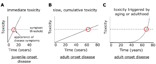
An inherited mutation with strong immediate toxic effects will lead to a juvenile-onset disease (Fig. 3.1A; Table 3.1). If the mutation causes a slow, cumulative form of toxicity, the mutation can cause a delayed onset of disease symptoms (Fig. 3.1B; Table 3.1). A mutation in which its toxic effects are triggered by changes related to aging or adulthood may also cause delayed onset of disease symptoms (Fig. 3.1C; Table 3.1).
Figure 3.1 Mechanisms underlying immediate and delayed toxicity
(A) Mutations with immediate toxic effects causes symptoms during youth and leads to juvenile-onset disease. (B) Mutations with slow, cumulative forms of toxicity can cause a delayed onset of disease symptoms and lead to adult-onset disease. (C) Mutations in which its toxic effects are triggered by changes related to aging or adulthood can cause a delayed onset of disease symptoms and lead to adult-onset disease.
14
From a pathophysiological perspective, it may be difficult to distinguish disease symptoms caused by gradual accumulation of toxicity versus those that are caused by a trigger related to aging (Fig. 3.1B vs. Fig. 3.1C). However, distinguishing between the two cases may be important for medical diagnosis and treatment. If the disease occurs from gradual accumulation of toxicity, there should be small damages that can be measured even during early life. In these cases, biochemical markers or imaging approaches may potentially detect the early signs of disease. However, in cases where a mutation’s toxic effects are triggered by changes related to aging, the disease will be very difficult to diagnose early in life. The use of genetic tests may be more prudent than biochemical or imaging approaches for diseases that are caused by mutations that are triggered by changes related to aging. In terms of treatment approaches, if a disease is of the age-related trigger type, certain aging pathways may be alternative therapeutic avenues along with targeting the disease mutation.
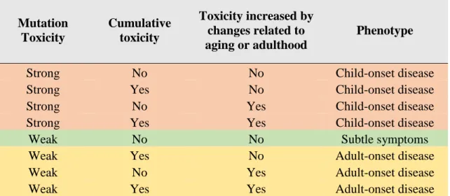
Table 3.1 Summary of types of inherited mutations and resulting phenotype
Mutation Toxicity
Cumulative toxicity
Toxicity increased by changes related to aging or adulthood
Phenotype
Strong No No Child-onset disease
Strong Yes No Child-onset disease
Strong No Yes Child-onset disease
Strong Yes Yes Child-onset disease
Weak No No Subtle symptoms
Weak Yes No Adult-onset disease
Weak No Yes Adult-onset disease
Weak Yes Yes Adult-onset disease
15
An example of a gain-of-function mutation that causes adult-onset symptoms is the polyglutamine repeat expansion that leads to Huntington’s disease (HD) (Ross et al., 1999).
Huntington’s disease is characterized by loss of motor control and cognitive deficits that begin generally between 35 and 44 years of age (Gil and Rego, 2008). HD is caused by an autosomal dominant polyglutamine repeat expansion in the Huntingtin (HTT) gene (Gil and Rego, 2008). The polyglutamine repeat varies from 6 to 39 repeats in unaffected individuals and from 36 to 180 repeats in HD patients (Rubinsztein et al., 1996). A greater number of repeats correlates with earlier onset of disease, with some juvenile-onset cases (Andresen et al., 2007). If age-related changes are required for the onset of HD, there should not be any juvenile forms of the disease. This suggests that aging is not necessarily required to trigger the toxic effects of long polyglutamine repeats and that a gradual accumulation of toxicity may lead to the symptom onset in HD. However, age-related changes may interact with both strong and weak polyglutamine toxicity to alter the severity of symptoms and disease
progression.
Loss-of-function mutations in molecular chaperones for protein folding can also cause adult-onset disease (Hartl et al., 2011). Chaperones are especially important for maintaining proper protein folding during adulthood (Labbadia and Morimoto, 2014a). Missense
mutations in the αB-crystallin gene (CRYAB), a molecular chaperone for myofilaments, can cause familial forms of late-onset myopathy (Reilich et al., 2010; Zobel et al., 2003). Patients with mutations in CRYAB develop muscle weakness often associated with the neck, trunk, cardiomyopathy, and cataracts (Reilich et al., 2010). The onset of symptoms is variable between patients, and reported cases have ranged from 32 years to 68 years (Reilich et al., 2010; Selcen and Engel, 2003; Zobel et al., 2003). The reduced chaperone activity of αB- crystallin causes disorganization of desmin myofilaments and leads to the aggregation of desmin and αB-crystallin in muscle cells (Zobel et al., 2003). In this case, CRYAB may be a
16
gene that is not required during development, but is required for the maintenance of muscle function during adulthood. Whether the accumulation of toxicity is gradual, or is triggered by an age-related change is still unknown.
Spinal muscular atrophy (SMA) is a disease characterized by degeneration of motor neurons in the spinal cord (Coovert et al., 1997). The most common cases are caused by loss- of-function mutations in the SMN1 gene, which leads to deficient SMN protein expression (Coovert et al., 1997). The disease is classified by the age of onset, which can range from infancy (Type 1) to adulthood (Type 4) (Coovert et al., 1997). SMN2 is a gene that is closely related to SMN1, and can also express the SMN protein, but at lower levels (D’Amico et al., 2011). Symptoms that are caused by loss of SMN1 are less severe for individuals with greater number of copies of the SMN2 gene (D’Amico et al., 2011). In this case, the number of copies of the protective SMN2 gene can explain the severity and age at onset of SMA.
However, an open question is how the neuroprotective role of SMN2 becomes diminished during adulthood in the late-onset cases of SMA. Transcription levels of SMN2 may decrease with age, and thus reduce the neuroprotective effect, or the built-up toxicity from loss of SMN1 may reach a threshold during adulthood that cannot be protected by SMN2.
3.1.2 Disease-related genes can be identified from forward genetic screens
Disease-related genes have been identified by studying human patients and animal models. Genetic sequencing of inherited cases of ALS led to the identification of ALS-
causing mutations in genes such as SOD1, TDP43, and C9ORF72 (Bruijn et al., 2004; Renton et al., 2011). An advantage of this approach is that the findings are directly relevant to human patients. However, evolutionarily conserved genes that are involved in aging-related
impairments may also be important for understanding the basic mechanisms of aging-related
17
impairments. Such genes can potentially be identified from genetic screens using animal models.
Genetic studies using model organisms can generally be divided into reverse genetic approaches or forward genetic approaches. Reverse genetic approaches start with a specific gene of interest and study the phenotypic and molecular pathways that are affected by the gene. On the other hand, forward genetic approaches start with a phenotype of interest and search for genes that are involved in the phenotype. A candidate-based RNAi screening approach can also test the potential effects of genes that may be involved in a specific
phenotype. However, in candidate-based screens, the selection of the candidate genes adds an element of bias in the genes that may be identified. RNAi approached are also limited in searching for phenotypes that result from decreased expression levels of a gene. Therefore, forward genetic approaches are suitable for searching for new genes involved in a phenotype in an unbiased way.
A classical forward genetic screen follows three steps: (1) Mutagenesis of animals, (2) Phenotypic screen to isolate mutants of a specific phenotype, and (3) Identification of the causative mutation site(s) in the isolated mutants. Further analysis of the causative mutation site may provide novel insights into how the mutation leads to the observed phenotype.
Findings from model organisms may not be directly relevant to human patients but may provide basic insights that lead to generalizable findings.
A forward genetic screen can be carried out on wild-type animals, or on transgenic or mutant animals that already demonstrate a specific phenotype. In the latter case, it is referred to as a modifier or suppressor screen, in which genes that affect a specific genetic pathway can be identified. A modifier screen in Drosophila melanogaster identified J proteins as suppressors of polyglutamine toxicity (Kazemi-Esfarjani and Benzer, 2000). Modifier screens
18
in C. elegans have also identified genetic modifiers of polyglutamine toxicity (van Ham et al., 2010; Silva et al., 2011). Inactivation of MOAG-4 suppressed the formation of
polyglutamine inclusion formations (van Ham et al., 2010). Silva and colleagues identified nine genes that suppress polyglutamine aggregation, improve the motility defect in C.
elegans, and improve temperature-sensitive misfolding of proteins (Silva et al., 2011). The nine genes are reported to be involved in the mitochondrial respiratory chain, tricarboxylic acid cycle, rRNA processing, and transcription (Silva et al., 2011).
A forward genetic screen for modifiers of TDP-43 toxicity in yeast identified the yeast ortholog of Ataxin-2 (Elden et al., 2010). Greater numbers of polyglutamine repeats in human ATXN2 was found to be linked to the incidence of ALS (Elden et al., 2010). A drug screening carried out in C. elegans has identified the neuroleptic drug pimozide has
protective effects in a pilot randomized double-blinded placebo-controlled randomized controlled trial for subjects with ALS (Patten et al., 2017). These studies indicate the potential of simple model organisms to provide insights into understanding and treating human diseases.
A forward genetic approach relies on a simple and reproducible screening procedure that can identify a specific phenotype of interest (Jorgensen and Mango, 2002). For an obvious phenotype such as abnormal body size, the screening procedure can be as simple as observing the mutants. However, screens for subtle phenotypes can be difficult to establish.
The methodological challenge in screening for progressive disease phenotypes is
distinguishing between degeneration that occurred during development versus after reaching adulthood. A paralytic phenotype that is observed in adulthood may have already been present begun during developmental stages. Adult-onset diseases are generally asymptomatic before adulthood, so the screening procedure must be able to separate developmentally
19
defective mutants from mutants that progressively lose their functional ability during adulthood.
3.1.3 C. elegans has been used successfully in forward genetic screens for complex phenotypes
Which animal model should be used for a forward genetic screen? The animal model used for mutagenesis must demonstrate the essential features of the target phenotype.
Additional considerations include lifespan, genome size, and ease of handling. For this study, the basic features of the target phenotype are physiological aging and a locomotor circuit controlled by motor neurons and muscles.
C. elegans is a small, 1.5-mm nematode which moves by generating sinusoidal waveforms by contracting and relaxing its body wall muscles. The C. elegans motor circuit resembles major aspects of the human motor circuit: both are comprised of the motor neuron, muscle, and neuromuscular junction (Von Stetina et al., 2005). C. elegans also undergoes aging, with a lifespan of about two weeks (Kenyon et al., 1993). These features make C.
elegans a suitable model to screen for mutants that show progressive declines in locomotor function. Sydney Brenner introduced C. elegans as an animal model and established its first forward genetic screen to identify genes involved in proper development and coordinated movement (Brenner, 1974). Many forward genetic screens have followed, providing the first examples of single gene mutations that regulate important biological processes such as apoptosis and lifespan (Friedman and Johnson, 1988; Hedgecock et al., 1983; Klass, 1983).
The C. elegans life cycle takes about three days from egg to adult and the mean adult lifespan lasts about two weeks. Despite this short lifespan, C. elegans undergo age-related changes such as neurodegeneration and sarcopenia that share signaling pathways with human
20
aging (Herndon et al., 2002; Toth et al., 2012). An unbiased forward genetic screen using C.
elegans led to the isolation of long-lived mutant strains and identified a single mutation in the age-1 gene that causes a 40% extension of mean lifespan (Friedman and Johnson, 1988;
Klass, 1983). This discovery led to the characterization of the evolutionarily conserved insulin signaling pathway in aging, and demonstrated that regulators of complex biological phenomena can be identified from forward genetic screens in C. elegans. Low levels of insulin signaling have been implicated in cases of exceptional human longevity (Milman et al., 2014).
Animal models such as C. elegans may be advantageous to identify genes involved in
complex disease processes due to their simple genetic architecture. In many cases, a family of human genes is represented by one gene in C. elegans. For example, the ErbB family of four genes in humans is represented by the single let-23 gene in C. elegans (Aroian et al., 1990). If a loss-of-function mutation occurs in one of the four ErbB genes in humans, the effects of the mutation may be masked by functional rescue from the other three related genes. However, a loss-of-function mutation in C. elegans let-23 will not be rescued due to the absence of homologs. Therefore, the functional contributions of some genes may be easier to identify in C. elegans. In some cases, mutations in genes that have many functions in higher organisms may cause lethality. These genes may have more simplified roles in C. elegans, leading to more subtle effects and allowing for their functional characterization.
3.1.4 C. elegans is used as a model for general mechanisms of neuromuscular aging and neuromuscular disease
C. elegans demonstrates signs of neuromuscular aging that are similar to humans.
Sarcopenia, the loss of muscle mass and strength with age, is observed both in humans and C.
21
elegans (Herndon et al., 2002). Genes that are involved in the progression of sarcopenia are largely unknown. Kashyap and colleagues conducted a candidate-based RNAi screen using C. elegans and identified several genes that significantly reduce the prolonged locomotor activity of the long-lived daf-2(e1370) strain (Kashyap et al., 2012; Kenyon et al., 1993).
Most of the genes that were identified were part of general cell maintenance pathways such as a vacuolar sorting protein, splicing factor, and fatty acid transport protein (Kashyap et al., 2012).
During aging, loss of motor neurons leads to the denervation of muscle fibers (Gonzalez-Freire et al., 2014). These muscle fibers can be re-innervated by nearby motor neurons, but age-related changes in the neuromuscular junction can prevent the re-innervation process (Gonzalez-Freire et al., 2014). Several mechanistic changes are implicated in the aged neuromuscular junction such as mitochondrial dysfunction, neurotransmitter
dysfunction from presynaptic nerve terminals, and chronic systemic inflammation (Gonzalez- Freire et al., 2014; Rudolf et al., 2014). Several studies have focused on changes that occur in the C. elegans neuromuscular junction as a result of aging (Liu et al., 2013; Mulcahy et al., 2012). Liu and colleagues found that the first signs of neuromuscular deterioration in C.
elegans is a deficit in synaptic vesicle fusion, followed by dysfunctions in neurotransmitter vesicle docking and priming (Liu et al., 2013). Muscle function defects are observed later in life (Liu et al., 2013). These findings implicate the neuromuscular junction as a critical location where interventions for neuromuscular deterioration may be most effective.
Reverse genetic models of various neuromuscular diseases have been developed in C.
elegans (Table 3.1). Various types of disease models for both muscle and motor neuron diseases demonstrate the wide applicability of C. elegans for locomotor function studies.
Disease phenotypes such as muscle degeneration, intracellular aggregate formation, and progressive paralysis have been described in C. elegans disease models (Table 3.1).
22
C. elegans models of ALS have been created by expressing human mutant SOD1 that has been implicated in familial ALS (fALS) (Oeda et al., 2001). C. elegans that express fALS-related mutant SOD1 show greater sensitivity to oxidative stress compared to C.
elegans that express the wild-type version of SOD1 (Oeda et al., 2001). C. elegans mutants that express fALS-related FUS or TDP-43 showed progressive declines in locomotor function (Liachko et al., 2010; Murakami et al., 2012; Vaccaro et al., 2012). Deletion of C. elegans alfa-1, the ortholog of human C9ORF72 implicated in fALS, leads to progressive loss of locomotor function and sensitivity to osmotic stress (Therrien et al., 2013). The toxic effects of fALS-related proteins in C. elegans demonstrate the evolutionarily conserved effects of disease-related mutations.
Disruption or knockdown of endogenous C. elegans genes such as dys-1, frh-1, or alfa-1 can cause progressive neuromuscular dysfunctions (Table 3.2). Therefore, targeting the phenotype of a progressive decline in locomotor function may uncover endogenous genes that work to maintain adult locomotor function in C. elegans.
Table 3.2 Reverse genetic disease models of neuromuscular disease in C. elegans
Disease Transgenic Model Phenotype Refs
Amyotrophic Lateral Sclerosis
• Expression of human familial mutation versions of SOD1, TDP43, or FUS
• Deletion of C. elegans alfa-1, ortholog of C9ORF72
Ÿ• Progressive locomotion defects
• Shortened lifespan in some cases
(Liachko et al., 2010;
Murakami et al., 2012;
Oeda et al., 2001;
Therrien et al. 2013;
Vaccaro et al. 2012) Duchenne Muscular
Dystrophy
Null mutation of C. elegans dys-1 with weak mutation in C. elegans MyoD homolog hlh-1
• Progressive muscle degeneration
• Progressive development of uncoordinated locomotion
(Gieseler et al., 2000) Friedreich’s Ataxia RNAi knockdown of C. elegans frh-1 • Shortened lifespan
• Lethargic behavior
(Vázquez-Manrique et al., 2006)
Inclusion Body Myositis Expression of human Aβ(1-42) • Progressive paralysis
• Intracellular aggregate formation (Link, 1995) Spinal Muscular
Atrophy Deletion of C. elegans smn-1 • Larval lethality
• Impaired locomotion (Briese et al., 2009)
23
Over 90% of ALS cases are caused by unknown factors and the causes of
polymyositis, a disease that causes adult-onset muscle weakness, are completely unknown (Bruijn et al., 2004; Dalakas and Hohlfeld, 2003). A recent study conducted whole-exome sequencing on 2869 ALS patients and 6405 controls and newly identified TBK1 as an ALS associated gene (Cirulli et al., 2015). However, TBK1 alone is unlikely to resolve all
unexplained ALS cases. Searching for new potential target genes using C. elegans may lead to novel insights that would not be found in human studies and studies using other animal models.
3.1.5 Limitations in using C. elegans as the animal model
Neuromuscular diseases can be classified by their pathogenic origin. The major origins are the muscle, motor neuron, or neuromuscular junction. Neuromuscular diseases can also be classified by age of onset in which some diseases begin during infancy, while others begin after reaching old age.
The features of the C. elegans motor circuit determine the types of human diseases it can model. The C. elegans motor circuit is made of 113 motor neurons and 95 muscle cells (Von Stetina et al., 2005; White et al., 1986). Seventy-five motor neurons are located in the ventral nerve cord (VNC) (Von Stetina et al., 2005; White et al., 1986). VNC motor neurons are divided into five classes: A, B, D, VC, and AS. The A, B, and D classes are further divided into DA, VA, DB, VB, DD, and VD based on whether they innervate dorsal (D) or ventral (V) muscles (White et al., 1986). The A, B, VC, and AS motor neurons are
cholinergic and excite muscle contraction while D motor neurons are GABAergic and inhibit muscle contraction in adulthood (Von Stetina et al., 2005; White et al., 1986).
24
Motor neurons in humans are also mainly cholinergic, demonstrating the evolutionary conservation of the motor circuit (Fambrough, 1979). However, C. elegans does not have an adaptive immune system and its neuronal axons are not surrounded by a myelin sheath. These differences preclude C. elegans from being used for specific neuromuscular diseases such as multiple sclerosis which is caused by loss of myelin (Glass et al., 2010).
3.2 Materials and Methods C. elegans strains
C. elegans Bristol strain N2 was used as wild-type worms. Worms were cultivated on NGM plates with Escherichia coli strain OP50. C. elegans strains CB408 unc-43 (e408), CB190 unc-54(e190), MT7929 unc-13(e51), and AM725 rmIs290 [unc-54p::Hsa-sod-1
(127X)::YFP] were obtained from the Caenorhabditis Genetic Center (University of Minnesota, MN, USA). All animals were maintained at 20oC.
Ethyl methanesulfonate (EMS) mutagenesis
Synchronized N2 young adult hermaphrodites were collected with 1 mL M9 buffer, and washed three times with 1 mL M9 buffer to remove bacteria. Animals were resuspended in 2 mL M9 buffer and mutagenized by adding 2 mL of 2x stock solution of 100 mM EMS to make a final concentration of 50 mM EMS. Animals were incubated in the 50mM EMS solution for 4 h. Animals were washed five times with 10 mL M9 buffer, then plated on OP50 seeded NGM plates.
Edge Assay
25
Edge Assay plates were prepared by pouring 16 mL of NGM agar into a circular 9 cm plate.
NGM plates were dried overnight at room temperature (RT) with the lid on, then kept at 4°C until use. On the day before the Edge Assay, a total of 100µL of E. coli suspension was spotted on four spots near the edge of the NGM plate. The tip of a 50 mL serological pipette was briefly placed over a flame to smoothen the tip. The NGM plate was placed on an inoculating turntable and the smoothened pipette tip was held against the E.coli drop. The plate was slowly rotated while holding the pipette tip still. The plate was rotated 360° to spread the E.coli around the edge of the whole plate. Plates were incubated overnight at RT and used the next day. Synchronized worms were collected and washed twice with M9 buffer containing 0.1% aqueous gelatin. Worms were placed on the center of an Edge Assay plate and excess M9 buffer was removed with the edge of a Kimwipe tissue. The number of worms that reached and did not reach the edge were counted at various time points to measure the Edge Assay completion rate.
Measurements of maximum speed and travel distance
Worms were synchronized by picking five adult day 1 worms onto an NGM plate with food, and allowed to lay eggs for 3 h. When the offspring reached adult day 1, 15 worms were picked randomly onto a 6 cm NGM plate without bacteria. After the worms moved away from the initial location with residual food, worms were again moved onto a different NGM plate without bacteria. The plate with worms was placed under a camera, and recorded for one min. Images were analyzed using ImageJ and wrMTrck software (plugin for ImageJ:
www.phage.dk/plugins) to produce maximum speed and total travel distance. Measurements were made with the lid on in a temperature-controlled room set at 20°C. At least three biological replicate plates of 15 worms each were measured for each strain. Representative locomotor tracks were also produced using wrMTrck software.
26 Lifespan Analysis
The lifespan of a population of worms was measured on NGM plates with food at 20°C.
Worms that did not move after gentle prodding to the head and tail were counted as dead.
Worms that were lost, died from an exploded vulva, or from the bag-of-worms phenotype were censored. For the ix243 strain, many worms died from the bag-of-worms phenotype.
Therefore, we measured lifespan on plates containing 25 µM floxuridine (FUDR), which is an inhibitor of germline proliferation. Worms were transferred from NGM plates to FUDR- containing plates after reaching the L4 stage.
Measurement of development time
The development time until adulthood was measured by allowing an adult day-1 worm to lay eggs for 1 h. The adult was then removed. The time until one of the offspring laid its first egg was measured as the development time.
3.3 Results
3.3.1 “Edge Assay” can test locomotor function of hundreds of worms
The first genetic screens that aimed to understand aging were focused on genetic or environmental interventions that affect lifespan. However, most scientists now agree that aging studies should place a stronger emphasis on “healthspan,” or the period of time that a person can maintain a healthy level of functional activity (Burch et al., 2014). Recent studies have suggested that the genetic bases of lifespan and healthspan may not completely overlap (Bansal et al., 2015; Iwasa et al., 2010; Tissenbaum, 2012). Some genes or environmental interventions may have effects on healthspan without large effects on lifespan and vice versa.
For example, lifelong spontaneous exercise was found to improve healthspan in mice without
27
any significant effects on lifespan (Garcia-Valles et al., 2013). A candidate-based genetic screen that searched for C. elegans worms with a prolonged swimming ability in adulthood found the epidermal growth factor signaling pathway to be involved in promoting locomotor healthspan without large effects on lifespan (Iwasa et al., 2010). Therefore, we also chose to focus on a healthspan-related phenotype: progressive loss of locomotor function during adulthood.
Loss of locomotor function is a sign of normal aging as well as a symptom of various age-related diseases. It is an evolutionarily conserved indicator of an animal’s healthspan from worms, flies, mice, and humans (Cesari et al., 2009; Grotewiel et al., 2005; Hahm et al., 2015; Justice et al., 2014). We decided to search for mutants that demonstrate normal
locomotor function during development and early adulthood, but lose their locomotor function much quicker than wild-type worms during adulthood. We addressed the issue of distinguishing mutants with developmental locomotor defects versus those with adult-onset locomotor deficits by establishing a sequential two-step screen in which we remove mutants that have strong developmental defects on the first day of adulthood and collect mutants that show locomotor deficits on the third and fifth days of adulthood. Our focus on locomotor function, a healthspan-related phenotype, aims to identify genes that are involved in the maintenance of healthspan.
In order to test the locomotor function of hundreds of worms at once, we established a procedure called the “Edge Assay.” The Edge Assay is carried out on a 9-cm circular agar plate with E. coli OP50 strain bacterial feed spread only on the outer edge (Fig. 3.2). Up to several hundred synchronized adult worms are collected with M9 buffer and placed on the center of the plate. Motile worms reach the E.coli and remain close to the edge but slow or paralyzed worms can be found remaining in the center of the plate (Fig. 3.2B). Almost all worms that reached the E. coli bacterial feed remained in the edge.
28 Figure 3.2 Photos of Edge assay
(Left) Photo of the Edge Assay after 5 min. (Center) Photo of the Edge Assay after 15 min.
(Right) Photo of the Edge Assay after 60 min.
The Edge Assay completion rate is calculated by counting the number of worms on the edge of the plate divided by the total number of worms (Fig. 3.3A). We measured the completion rates of adult wild-type worms for seven days at 5, 10, 15, 30, and 60 min time points (Fig. 3.3B). Over 90% of wild-type worms completed the Edge Assay in 15 min on the first day of adulthood. If given 60 min, over 90% of wild-type worms completed the Edge Assay during the first five days of adulthood (Fig. 3.3B).
29 Figure 3.3 Edge Assay completion rates
(A) Schematic diagram of Edge Assay and method to calculate Edge Assay completion rate.
(B) Completion rates of wild-type (WT) N2 C. elegans worms for the edge test from day 1 to day 7 of adulthood. Three biological replicate plates with approximately 100 worms per plate were assayed (n = 3). Error bars indicate 95% confidence intervals.
Large decreases in Edge Assay completion rate is observed from adult day 2 to 3 at the 10 and 15 min time points. In contrast, only minor decreases in the Edge Assay
completion rate is observed from adult day 1 to 5 at the 60 min time point (Fig. 3.3B).
Previous studies on the functional capacity of locomotor function and neuromuscular
function suggest declines that begin around adult day 5 to day 6 (Hahm et al., 2015; Liu et al., 2013). C. elegans chemotaxis towards benzaldehyde, an attractive chemical cue, was
30
significantly diminished beginning on the fourth day of adulthood (Leinwand et al., 2015).
Therefore, declines in Edge Assay completion rate that occurred on adult day 3 at the 10 and 15 min time points may indicate deficits in sensory perception or result from suboptimal search behavior. The adult day 3 worm may start to show deficits in sensing the food cue but still have fully functional neuromuscular activity.
3.3.2 Time points for distinguishing mutants with progressive locomotor decline from wild-type worms using the Edge Assay
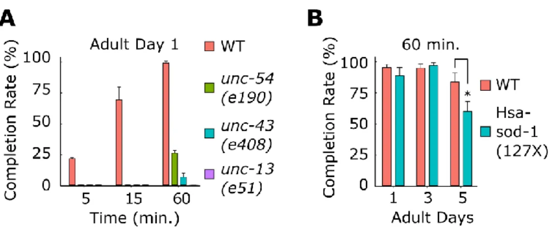
We tested whether the Edge Assay can distinguish wild-type worms from mutants with a progressive decline in locomotor function. We tested C. elegans mutant strains that have been previously isolated and are defective in the function of neurons (unc-13(e51), unc- 43(e408))) (Maruyama and Brenner, 1991; Reiner et al., 1999) or muscles (unc-54(e190)) (MacLeod et al., 1981) using the Edge Assay (Fig. 3.4A). On the first day of adulthood, all three mutant strains could not reach the edge in 15 min (Fig. 3.4A). After 60 min, 26% of unc-54(e190) mutants, 6.4% of unc-43(e408) mutants, and 0% of unc-13(e51) mutants reached the edge (Fig. 3.4A). In contrast, 91.3% of wild-type worms reached the edge in 15 min on the first day of adulthood and 99.6% reached the edge in 60 min (Fig. 1B). Therefore, carrying out the Edge Assay for 15 min on the first day of adulthood is a time point that can clearly distinguish wild-type worms from worms with strong developmental locomotor defects.
31
Figure 3.4 Edge Assay completion rates of known mutants
(A) Completion rates for WT and developmental motor deficit mutants (unc-54(e190), unc- 43(e408), (unc-13(e51)) on the first day of adulthood. (B) Completion rates of WT and previously reported adult-onset motor deficit mutant (Hsa-sod-1(127X)). Three biological replicate plates with approximately 100 worms per plate were assayed (n = 3). Error bars indicate 95% confidence intervals. *P < 0.05; Unpaired Student’s t test.
Next, we tested whether the Edge Assay can distinguish wild-type worms from worms that show progressive declines in locomotor function. A C. elegans model of amyotrophic lateral sclerosis that expresses a truncated version of human SOD1 (SOD1- G127insTGGGstop) was tested using the Edge Assay (Gidalevitz et al., 2009). The Hsa-sod- 1(127X) mutant showed a significant reduction in the Edge Assay completion rate on the fifth day of adulthood compared to wild-type worms (Fig. 3.4B). Therefore, carrying out the Edge Assay for 60 min on the fifth day of adulthood is a time point that can distinguish wild-type worms from worms that show progressive declines in locomotor function.
32 3.3.3 Mutagenesis and screening procedure
Taking into account the Edge Assay completion rates of mutants with developmental and progressive locomotor deficits, we established a forward genetic screening procedure to isolate mutants that show progressive declines in locomotor function during adulthood (Fig.
3.5). The screening procedure removes mutants with developmental locomotor defects and isolates mutants that show a locomotor deficit in adulthood.
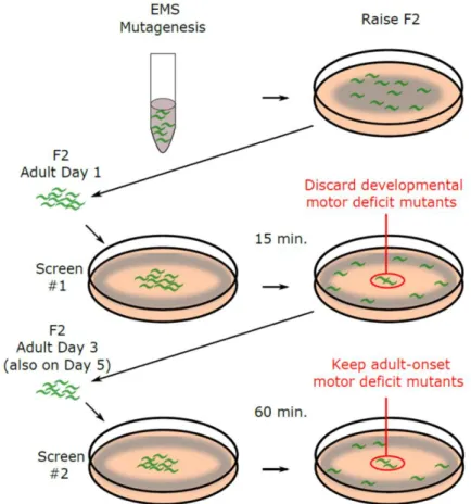
Figure 3.5 Schematic description of forward genetic screen
Schematic description of genetic screening procedure to isolate mutants that show a progressive decline in locomotor function.
We mutagenized worms using EMS and screened 3,352 F2 worms from 500 F1 worms (1,000 haploid genomes). We carried out a three-step screening procedure to isolate mutants with progressive locomotor deficits (Fig. 3.5). First, we carried out the Edge Assay
33
for F2 worms on the first day of adulthood to remove worms with developmental locomotor defects. Worms that were unable to reach the E. coli ring on the edge of the plate in 15 min were aspirated away. Second, we repeated the Edge Assay on the third day of adulthood using only the worms that were able to complete the Edge Assay on the first day. We
collected the worms that remained in the central region of the Edge Assay plate after 60 min as mutants that show a progressive loss of locomotor function on the third day of adulthood.
Third, we repeated the Edge Assay on the fifth day of adulthood using the worms that were able to complete the Edge Assay on the third day. We collected the worms that remained in the central region of the Edge Assay plate after 60 min as mutants that show a progressive loss of locomotor function on the fifth day of adulthood. We carried out two separate instances of the screening procedure and obtained 22 viable mutants (12 viable mutants on the third day of adulthood and 10 viable mutants on the fifth day of adulthood) (Table 3.3).
Table 3.3 Number of viable mutants obtained from screen
3.3.4 Isolated mutants
Five out of the 22 isolated mutants reproducibly showed progressive deficits in completing the Edge Assay (Fig. 3.6). Reductions in completing the Edge Assay may be caused by deficits in locomotor function, sensory perception, or search behavior. To
determine whether the isolated mutants have deficits in locomotor function, we measured the locomotor function of individual worms on an agar plate without food. For each strain, we
Day 3 Day 5 Day 3 Day 5 Total
Adult-onset mutants 13 23 17 17 70
Viable adult-onset mutants 3 8 9 2 22
EMS-A: 400 genomes EMS-B: 600 genomes
34
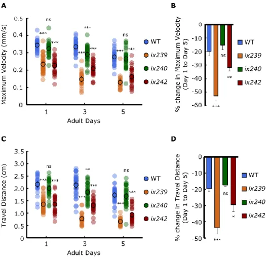
recorded three plates of 15 worms on the first, third, and fifth days of adulthood. The videos were analyzed using ImageJ and wrMTrck software (www.phage.dk/plugins) to produce the instantaneous maximum velocity that the worm reached within a 1-second interval and the total travel distance of the worm during the 1-min recording. The ix239 and ix242 mutant strains showed significantly greater reductions in maximum velocity and travel distance compared to wild-type worms (Fig. 3.7A–D). However, ix240 worms did not show a significant reduction in maximum velocity or travel distance from adult day 1 to 5 in
comparison to wild-type worms (Fig. 3.7A–D). ix240 worms may have progressive deficits in functions other than locomotor function, such as sensory perception or search behavior.
Figure 3.6 Edge Assay completion rates of isolated mutants
Edge Assay completion rates of isolated mutants for adult days 1, 3, and 5. Three biological replicate plates with approximately 100 worms per plate were assayed (n = 3). *P < 0.05;
**P < 0.01; Paired Student’s t test vs. adult day 1 completion rate.
35
Figure 3.7 Maximum velocities of WT, ix239, ix240, and ix242 worms
(A) Maximum velocity of WT, ix239, ix240, and ix242 worms. (B) Percent change in
maximum velocity of WT, ix239, ix240, and ix242 worms. For maximum velocity and travel distance experiments, n = 30–45 worms per strain for each day (10–15 worms from 3
biological replicate plates). For percent change in maximum velocity graphs, n = 3 biological replicate plates. *P < 0.05; **P < 0.01; ***P < 0.001; ns, not significant; One-way ANOVA with Dunnett’s post hoc test vs. WT.
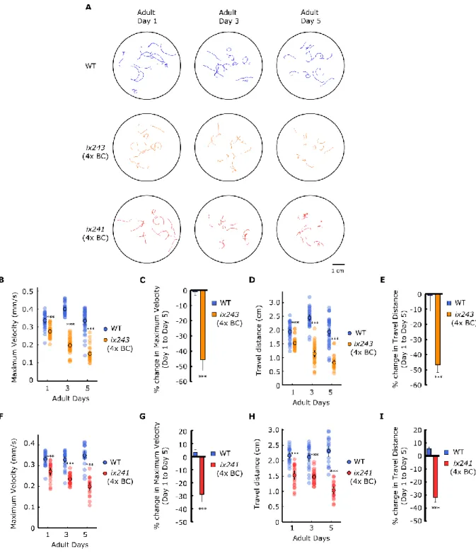
To check whether the progressive decline in locomotor function is maintained after backcrossing, ix241 and ix243 worms were crossed with the parental N2 strain. After each backcross, we checked for lines that still showed the progressive decline in locomotor function by measuring the maximum velocity and travel distance of individual worms. Both ix241 and ix243 worms showed significant reductions in both travel distance and maximum velocity after four backcrosses (Fig. 3.8A-I).
36
Figure 3.8 ix241 and ix243 strains show progressive decline in locomotor function after four backcrosses
(A) Representative tracks of WT, ix241, and ix243 worms during 1-min video recording. n = 10–15 tracks per plate (some worms were unable to be tracked for the full minute, and were removed from analysis). (B) Maximum velocities of WT and ix243 worms for adult days 1, 3, and 5. (C) Percent change in maximum velocity for ix243 worms from adult day 1 to 5. (D) Travel distances of WT and ix243 worms for adult days 1, 3, and 5. (E) Percent change in
37
travel distance for ix243 worms from adult day 1 to 5. (F) Maximum velocities of WT and ix241 worms for adult days 1, 3, and 5. (G) Percent change in maximum velocity for ix241 worms from adult day 1 to 5. (H) Travel distances of WT and ix241 worms for adult days 1, 3, and 5. (I) Percent change in travel distance for ix241 worms from adult day 1 to 5. For maximum velocity and travel distance experiments, n = 30–45 worms per strain for each day (10–15 worms from 3 biological replicate plates). For percent change in maximum velocity and travel distance graphs, n = 3 biological replicate plates. ***P < 0.001; Unpaired
Student’s t test.
To check whether the ix241 and ix243 mutant strains were simply aging faster than wild-type worms, we measured the lifespans of the ix241 and ix243 worms (Fig. 3.9A, B, Table 3.4). The lifespan of the ix241 worms was not significantly decreased compared to wild-type worms (Fig. 3.9B). The median lifespan of the ix243 worms was decreased by two days (Fig. 3.9A). For the lifespan measurement of ix243 worms, we used FUDR at low concentrations (25 µg/ml) for both ix243 worms and WT worms since the ix243 worms are slightly egg-laying deficient. ix243 worms can lay eggs, but many worms die from the bag- of-worms phenotype in late adulthood. Although both ix241 and ix243 worms showed very little movement in adulthood, they were still able to move enough to orient its head to new patches of bacterial feed. We did not observe instances where the bacteria became absent near the head of ix241 or ix243 worms.
38 Figure 3.9 Lifespan of WT, ix243, and ix241 worms
(A) Kaplan Meier survival curve of WT (n = 56) and ix243 (n = 89) worms. (B) Kaplan Meier survival curve of WT (n = 94) and ix241 (n = 77) worms. Log-rank test for lifespan comparisons.
Table 3.4 Lifespan measurements of WT (N2), ix243, and ix241 worms
Strain Median
Lifespan (adult days)
P value vs WT (Log-rank test)
Worms (Counted/Total)
FUDR
N2 18 56/90 25 µg/ml
ix243 (4x backcrossed)
16 P = 0.00078 89/90 25 µg/ml
N2 18 82/90 25 µg/ml
ix243 (4x backcrossed)
16 P < 0.0001 84/90 25 µg/ml
N2 18 87/90 25 ug/ml
ix243 (4x
backcrossed) 16 P < 0.0001 87/90 25 ug/ml
N2 17 94/120 0
ix241 (4x backcrossed)
16 P = 0.095 77/120 0
N2 14 66/90 0
ix241 (4x backcrossed)
15 P = 0.12 64/90 0
N2 12 67/90 0
ix241 (4x backcrossed)
14 P = 0.024 74/90 0
The ix241 worms do not live shorter than wild-type worms. Therefore, the progressive decline in locomotor function is not due to a shorter lifespan. The ix241 allele affects
39
locomotor healthspan, but not lifespan. This example suggests that the genetic bases of locomotor healthspan and lifespan may not completely overlap.
Figure 3.10 Maximum velocity of WT and ix243 worms from adult day 1 to 10
Maximum velocities of individual WT and ix243 worms from day 1 to day 10 of adulthood. n
= 30–45 worms per strain for each day (10–15 worms from 3 biological replicate plates).
The ix243 worms live two days shorter than wild-type worms. In order to compare the relative reductions in lifespan and locomotor healthspan, we quantified the reductions in lifespan and locomotor healthspan as compared to wild-type worms. The area under a healthspan curve has been suggested as a way to compare the relative changes in healthspan between individuals (Kaeberlein, 2018). We used this approach to quantify the reduction in lifespan and healthspan of ix243 worms relative to wild-type worms. We measured the maximum velocity of wild type and ix243 worms for 10 days and created a curve for locomotor healthspan (Fig. 3.10). We quantified the decrease in locomotor healthspan of ix243 worms relative to wild-type worms by comparing the areas under the decline in maximum velocity curves (Fig. 3.11A). We quantified the decrease in lifespan of ix243 worms relative to wild-type worms by comparing the areas under the survival curves (Fig.
40
3.11B). In the ix243 worms, there is an average 11.5 percent decrease in lifespan, while there is a significantly greater decrease of 18.5 percent for locomotor healthspan (Fig. 3.11C).
These results suggest that the ix243 allele affects locomotor healthspan more than lifespan.
The gene that is mutated in the ix243 allele may play a more critical role for locomotor
healthspan compared to lifespan. Other assays for healthspan-related phenotypes may provide insights into whether the ix243 allele specifically affects locomotor healthspan, or affects healthspan in general.
Figure 3.11 Percent change in lifespan and locomotor healthspan
(A) Calculation method for percent change in lifespan. (B) Calculation method for percent change in locomotor healthspan. (C) Percent change in lifespan and locomotor healthspan of ix243 worms compared to wild-type worms. n = 3 biological replicate plates. *P < 0.05;
Unpaired Student’s t test.
41
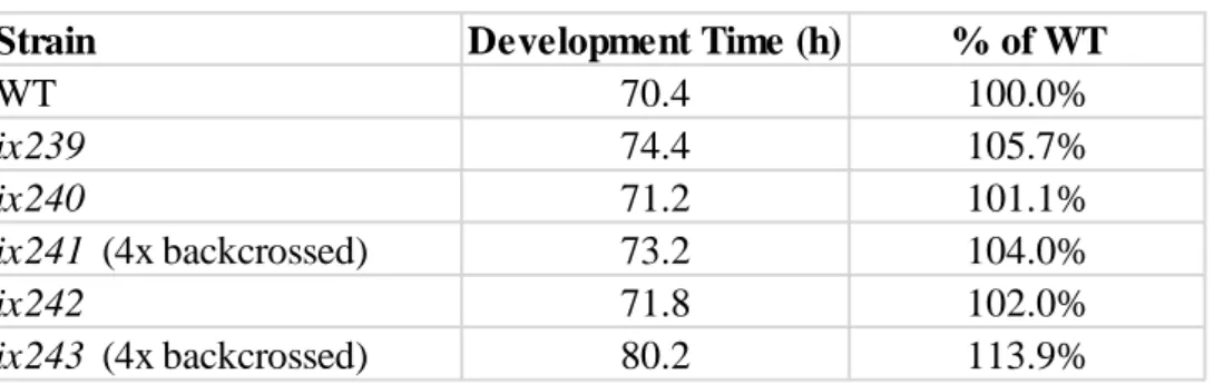
Four out of the five mutants that showed progressive deficits in completing the Edge Assay showed a progressive decline in locomotor function as measured by maximum velocity and travel distance. All four of those mutants showed slight but significant defects in
locomotor function on the first day of adulthood. In order to check the developmental effects on the mutants, we measured the time it takes for the isolated mutant strains to reach
development. Increases ranging from 1.1% to 13.9% were observed in our mutants, indicating that the induced mutations also have effects on development (Table 3.5).
Table 3.5 Development times of isolated mutant strains
Development time from egg to first egg-lay (n = 5 worms per strain).
3.4. Discussion
3.4.1 A new assay to measure C. elegans locomotor function
Here, we developed the “Edge Assay” to measure locomotor function of hundreds of worms at once. The Edge Assay can be used as an assay to compare the locomotor function of different populations of worms, or can be used to measure the decline in locomotor function of worms over time. We used E. coli bacterial feed on the edge of the plate as an attractant for the worms. Reductions in completion rate on the Edge Assay may represent deficits in locomotor function, sensory perception, or search behavior. Follow-up
measurements are necessary to determine which modality was affected in worms with reduced completion rates. The Edge Assay can be used as a preliminary test or screen to measure the locomotor function of a population of worms. The main advantage of the Edge
Strain Development Time (h) % of WT
WT 70.4 100.0%
ix239 74.4 105.7%
ix240 71.2 101.1%
ix241 (4x backcrossed) 73.2 104.0%
ix242 71.8 102.0%
ix243 (4x backcrossed) 80.2 113.9%
42
Assay is its simplicity and that it can be conducted in any lab that has the facilities to maintain laboratory strains of C. elegans.
3.4.2 A new screening procedure to search for mutants with delayed onset of disease symptoms
The main difficulty in searching for mutants with progressive declines in adult functional capacity is distinguishing between mutants that had a developmental defect versus mutants that show a progressive decline in functional capacity. We overcome this issue by removing mutants with strong developmental locomotor defects on the first day of adulthood.
Using only the mutant worms that completed the locomotor assay on the first day of adulthood, we collect mutants that show a loss of locomotor function on the third and fifth days of adulthood. To our knowledge, our screening procedure is the first unbiased screen using C. elegans that focuses on the progressive decline of adult locomotor function.
Many C. elegans models of neurodegenerative diseases show progressive deficits in locomotor function (Table 3.1). A C. elegans model of inclusion body myositis that expresses human beta amyloid, the protein that comprises the aggregates found in Alzheimer’s disease, is reported to undergo age-dependent paralysis (Wu et al., 2006). C. elegans worms that express green fluorescent protein (GFP) preceded by polyglutamine repeats show age- dependent accumulations of GFP aggregates (Morley et al., 2002). However, the accumulation of protein aggregations does not always correlate with the severity of
locomotor dysfunction (Silva et al., 2011). In those cases, locomotor function may serve as a better indicator of the severity of symptoms compared to protein aggregation levels.
43
Simple methods to test the locomotor function of many worms can accelerate the implementation of suppressor screens using previously created C. elegans models of neurodegenerative disease. For example, mutagenesis can be carried out on C. elegans mutants that carry human beta amyloid, and the Edge Assay can be used to isolate mutants that show improvements in locomotor function. The best age to carry out the screening procedure to enrich for mutants with a suppressed phenotype will depend on the severity of the locomotor deficit in each C. elegans disease model.
The E. coli bacterial lawn can be replaced with specific chemical cues to test the sensory functions of C. elegans. It may be possible to carry out forward genetic screens to isolate mutants with progressive loss of specific sensory functions by carrying out the Edge Assay with a repellent on the edge of the plate. First, mutagenized adult day 1 worms that remain in the center and are repelled by the repellent are collected. Those worms can be tested again on adult day 3 and day 5 for mutants that then cannot sense the repellent and move towards the edge of the plate. In addition to these examples, the Edge Assay may be a valuable assay for various types screening paradigms.
3.4.3 Isolation of five mutants with progressive deficits in completing the Edge Assay Typically, the frequency that a loss-of-function allele occurs for a particular gene following EMS is 1/2000 (Brenner, 1974; Jorgensen and Mango, 2002). This equals 10 null- mutations per genome since there are 20,000 genes. If we screen 2,000 genomes, we will have caused approximately one mutation in each gene. Since we screened 1,000 genomes, we caused loss-of-function mutations in approximately 10,000 genes. Therefore, there may be genes that are involved in our specific phenotype that were not mutagenized in our
screenings. Another screening of 1,000 genomes may identify more genes that are involved
44
in this phenotype. A saturation screen would theoretically test all 20,000 genes, so we can roughly estimate that our current screen is halfway towards saturation. However, even if a screen is saturated, genes may not be found if they have redundant functions or are small in size. Therefore, a saturation screen will not be able to test all C. elegans genes. Generally, a screen is considered saturated when multiple alleles of the same gene are identified.
3.4.4 Slight defects in development may cause strong deficits during adulthood Four out of the five mutants that we isolated in our screen showed a progressive decline in locomotor function as measured by maximum velocity and travel distance in 1 min on an agar plate without food. All four of those mutants showed slight but significant defects in locomotor function on the first day of adulthood, indicating signs of developmental neuromuscular deficits. Those four mutants also showed slight delays in development. The slight effects on development may be analogous to the early signs of dysfunction in brains of young adults with risk alleles for Alzheimer’s Disease (Mormino et al., 2016). Potential links between genes implicated in development and those implicated in aging or age-related impairments may provide clues for early diagnosis of late-onset diseases.
3.4.5 Genetic bases of lifespan and locomotor healthspan may not completely overlap In the ix241 mutant strain, a progressive decline in locomotor function was observed without a reduction in lifespan. In the ix243 mutant strain, there was a significantly greater negative effect on locomotor healthspan compared to lifespan. These results support the notion that the genetic bases of lifespan and locomotor healthspan may not completely overlap (Bansal et al., 2015; Tissenbaum, 2012). Although genetic regulators of lifespan in many cases may also affect healthspan, some genes may affect lifespan without largely
45
affecting locomotor healthspan and vice versa (Fig. 3.12). Therapeutic targets to improve healthspan should therefore target genes that have disproportionate effects on healthspan instead of genes that have the most robust effects on lifespan.
Figure 3.12 Genetic factors that affect lifespan and healthspan may not completely overlap
Conserved genetic regulators of specific aspects of healthspan may be easier to identify from model organisms compared to conserved genetic regulators of lifespan.
Lifespan is regulated by various causes of death, which may be very different from one organism to another. For example, one of the major causes of death in C. elegans is bacterial colonization of the gut (Podshivalova et al., 2017; Zhao et al., 2017). Pharyngeal swelling is caused by bacterial infection by E. coli, which is an innocuous food source until later stages of adulthood (Zhao et al., 2017). Since the leading cause of death in humans is heart disease, the improvement of heart function may be the most effective way to prolong lifespan in the human population (Heron, 2013).
In contrast, the genetic factors that affect specific domains of healthspan are functionally conserved. For example, locomotor healthspan in humans and C. elegans are maintained by the neuromuscular system in both species. Therefore, genetic factors related to healthspan that are found from model organisms may have a greater probability of having similar effects in humans.