1-3 皮膚の光物性計測に関する従来の研究
これまでに,皮膚の光物性は,主に医療分野での応用を目的として,その計測手法 の開発およびデータ計測に関する多くの研究が進められてきた[20]–[64].医療分野で
の実際の応用として,乳癌などの腫瘍の診断技術[38]や port wine stain(ぶどう酒様血
管腫)の治療及び評価・診断に向けた[6][9][10]光物性の研究が行われている.また, 近年では,皮膚に対する光線治療(シミやほくろの除去,美容整形,様々な皮膚疾患 の治療など)が急速に進歩しており,家庭用の治療機器も販売されるようになってき ているが,現状として,皮膚に対するそれらの治療メカニズムは未だ十分に明らかに なっておらず,治療成果が得られないだけでなく,医療トラブルを生じることも少な くないため,光線治療へ向けた光物性の研究も行われている[6][7][44][58]. 光物性を計測する方法には,主に,積分球を利用した方法や,DOSI(Diffuse Optical Spectroscopic Imaging),RSPM(Reflection Spatial Profile Measurement)などがある.積分
A および B を決めることで波長分布が求められる)こと[49]–[51]や,近赤外波長域の 吸収係数がヘモグロビン[52],水[53],脂肪[54]によって決められると仮定すること[55] で,レーザー光以外の波長における散乱係数および吸収係数を推定した. ss(l) = Al–B Cerussi ら[38]は,この方法を用いて,胸部の腫瘍および正常な皮膚を 58 人計測した. そして,腫瘍と正常な皮膚の光物性に特徴的に違う部分があることを明らかにし,非 侵襲での光物性計測が腫瘍の診断技術として応用できる可能性を示した. Tseng ら[39]は,上述したレーザー光の波長における計測や近似を用いた幅広い波 長域の補間ではなく,オプティカルスイッチや分光計を利用することで,波長 l = 500–1000 nm において連続的に光物性を計測することを可能にした.また,Tsengら
[40]は,この方法の実用性を確認するために,Fitzpatrick skin phototypes[56]が I–II, III– IV, V–VI のグループに分け,各 6 人ずつの計測を行った.また,Chao-kai ら[42]は,
今後の治療への基礎データとして,71 人のケロイド瘢痕と一般的な傷跡,傷跡のない 正常な皮膚の光物性を計測した.そして,ケロイド瘢痕の重症度の診断として,この 光物性計測を用いることが可能であること,治療による改善度合いを長期的に追跡す ることに有効であることを示した. 1-3-2 RSPMによる光物性計測 RSPM は空間的に一様でない光を照射し,その反射光の空間分布から光物性を推定 する方法である[44]–[48]. Dognitz ら[44]は,円状に照射部と非照射部を繰り返すように光を計測対象物に照 射することで,その反射光の分布とモンテカルロ法を用いたシミュレーションの結果 から逆解析によって散乱性媒質の光物性が推定可能であることを示した.さらに,こ の方法を用いて,l = 400, 500, 633, 700 nm の 4 つの波長における人の皮膚の光物性の 計測を行った. Saager ら[45]は,計測部に分光計を用いた新たな光学系のシステムにすることで, l = 430–1050 nm の幅広い波長域の光物性の計測を可能にした.この方法を用いて, Sagger ら[46]は,Fitzpatrick skin phototypes が I–VI の範囲に当たる 12 人の前腕内側・
1-4 本研究の目的および内容 1-3 で述べたように,近年,皮膚における光物性の計測方法に関して,特に非侵襲 での計測を中心として研究が進められている.しかしながら,1-1 で述べたように, 未だ皮膚の見え方と光物性との関係を明らかにすることができていない.これは,非 侵襲での皮膚の光物性に関する研究が医療分野の目的のみとなっており,化粧分野な ど他の様々な分野で応用する目的を持って多数の人の光物性計測した例がないこと や,実験室レベルでなく幅広い分野で使用されやすい装置の開発が行われていないこ とが一つの原因となっていると考えられる.生きた人の皮膚の見え方は,人種,部位, 状態によって異なるだけでなく,風呂上がりや日焼けによってもいつもとは異なり, その光物性も異なる.したがって,皮膚の見え方と光物性の関係を明らかにする上で, 多様な皮膚の評価・計測が不可欠であり,その場計測が可能な装置を用いて膨大なデ ータの蓄積をする必要がある.従って,迅速で簡便な計測が可能な,標準化された実 用機の開発が必要といえる.具体的には,装置性能を明確化し,装置の信頼性を示し た上で,可搬性や操作性に優れた光物性計測装置の実用機を開発することが必要であ る.そこで,本研究では,これらの要素を可能にした,標準化に向けた光物性計測装 置の開発することを第一の目的とした. RSPM は,DOSI と比較し,白色光源を用いた steady-state の計測のみから光物性の 計測が可能であり,光学系が単純化でき,短時間での計測も可能なため,本研究の目 的に適していると考えられる.また,D. J. Cuccia ら[48]は,この方法を用いて,高精 度に光物性の計測が可能であることを示しているが,その一方で,RSPM を利用した 計測方法は光学系の性能に依存して,光物性の推定に誤差を生じさせてしまう可能性 があることも述べられている.そこで,本研究では,光学設計ソフト Zemax(Zemax, LLC)を使用して開発する装置の光学性能を評価し,RSPM を利用した装置に生じる光 学系の性能に依存した誤差のシミュレーションを行った.これにより,計測に生じる 得る誤差を明確化した上で,計測精度の高い標準化に向けた光物性計測装置の実用機 を開発した(第 4 章–第 5 章).そして,開発した装置の有用性を確認するために, 水分量の変化に伴う光物性の変化を計測した(第6 章).
(a ≠ 0) である.区間(0,1)の乱数を⼀つ選ぶことによってこの式から天頂⾓z が決定される. またh については, が得られる.整理すると, である.この式から別に区間(0,1)の乱数 R を⼀つ選ぶことによってh が決定できる. このz , h を⽤いて,散乱後の光束の進⾏⽅向を⽰す単位ベクトルは,媒体を基準に した座標系で次のように表される. q’, f’⽅向に散乱された光束の挙動は,本節で述べてきた⽅法を引き続き繰り返すこと によって求められる. cosζ =Y2+Y2a2−1 2aY2 R= 1 2π 0 η
∫
dηη
= 2π
R sinθ' cosφ' sinθ' sinφ' cosθ' ⎛ ⎝ ⎜ ⎜ ⎜ ⎞ ⎠ ⎟ ⎟ ⎟ =cosθ cosφsinζ cosη − sinφsinζsinη + sinθ cosφ cosζ cosθsinφsinζ cosη + cosφsinζsinη + sinθsinφ cosζ
(=π)のとき とする. これらの式から, ここで とおく. よって , 以上より 積分して, となる.残りの2区間の場合も同様にそれぞれ ζ1≤ζ < ζ2 Φ(ζ) = B⋅ 1− b2 (1+ b2− 2 ⋅b ⋅ cosζ)3/2 S1= A⋅ 1− a 2 (1+ a2− 2 ⋅ a ⋅ cosζ)3/2 ⋅ sinζdζ ζ0 ζ1
∫
= A(1− a2) (1+ a2− 2a cosζ)−3/2sinζdζ ζ0 ζ1
∫
cosζ
= t dt dζ = −sinζ= A(1− a2) (1+ a2− 2at)−3/2sinζ ⋅ −dt
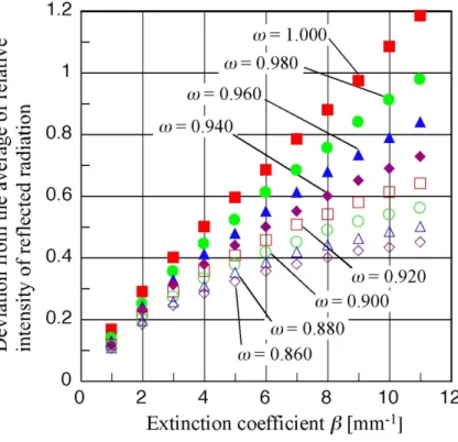
図 3-14.偏差と減衰係数の関係
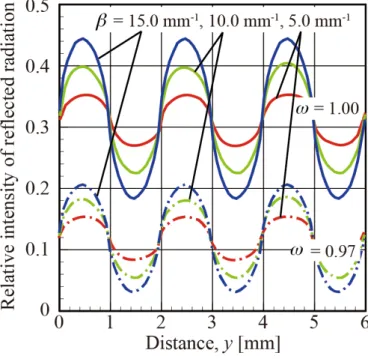
図 3-15.反射光強度の平均値とアルベドの関係
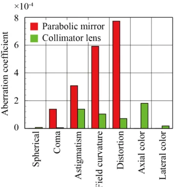
4-2 光学設計 結像光学系を組んだ時,どんなに理想的なレンズを⽤いたとしても,回折限界の原 理から,収差が⽣じることは避けられない.結像光学系に収差があるとき,本来点で あるものが広がりを持ったエネルギー分布として計測されてしまう.収差の種類によ って,その広がりに与える影響は異なるが,収差が⼤きいほどエネルギー分布はより 広がったものとなる.本推定法においては,その広がりが推定される光物性値に誤差 を⽣じさせる. 本章では,より⾼い光学系の性能を⽬指す光学設計の⼀例を⽰すとともに,光学系 の性能が推定される光物性に与える影響を数値解析により明らかにした. 4-2-1 光学性能の評価
4-2-2 計測装置に生じうる誤差のシミュレーション 光学性能の評価 4-2-1 節の光学設計に基づき,開発する計測装置に生じうる誤差を 収差が存在するときの光学系をシミュレーションにより求めた. 光学系に収差が存在しないとき,拡大投影される縞状の照射光は理想的に鋭角な分 布となる.そして,皮膚から射出された反射光分布は,正確に計測される.したがっ て,収差が存在しないときは,計測に誤差が生じることはなく,皮膚の持つ減衰係数 が正確に推定される.一方,光学系に収差が存在するとき,投影される縞状の光は波 形のような分布となる.そして,皮膚から射出された反射光分布は,収差がないとき に比べて,偏差が減った分布となって計測される.この偏差の減り方は,4-2-1 節で説 明した光学系の性能を表す MTF の値に依存して決まる.第 3 章で述べたように,減 衰係数は,反射光分布の偏差から推定するため,ここで誤差が生じてしまう. この偏差の減少をシミュレーションするために,ここでは線広がり関数(LSF:line spread function)[66]を用いることとした.LSF は理想的な線光源が,光学系を通過し た時にどの様に広がるかを表す関数である.また,LSF は MTF を逆フーリエ変換す ることによって得られる関数であるため,拡大投影部および分光計測部の MTF から それぞれのLSF を算出した.ここで,それぞれの LSF(拡大投影部 Lep(y)と分光計測 部 Lss(y))を図 4-4 に示す.これらの LSF を用いて,開発する装置の光学系での収差 の影響をシミュレーションした.そのシミュレーションの概要を図 4-5 に示す. まず,拡大投影される縞状の光の分布をUin(y)とすると,収差が光学系に存在した 時に投影される像は以下の式で表される. Uinpre(y) = Uin(y − τ )Lep(τ )dτ −∞ ∞
∫
. シミュレーションでは,このUinpre(y)を皮膚への照射光の分布として与えた. この時の皮膚内部での光伝播は第 3 章のモンテカルロ法による解析と同様に行い, 皮膚から射出される反射光の分布を求める.モンテカルロ法により得られた反射光の 分布をUrefl(y)とすると,光学系の収差の影響を考慮した場合に計測される反射光の分布Ureflpre(y)は以下の式で表される.
図 4-5.シミュレーションの流れ
図 5-5.富士フィルム製のシャープカットフィルタの透過率
図 5-7.白色板に SC64 を使用したときの撮影画像
5-2-3 計測データの例
まず,前腕内側部を計測している時の様⼦を図 5-8 に⽰す.次に,皮膚に縞状の光 を照射したときに撮影される画像の例を図 5-9 に示す.
図 5-9.皮膚の撮影画像
この得られた画像から,反射率 を算出したグラフの例を図 5-10 に示す.
図 5-10.算出された反射率
この反射率から,反射光の平均強度,および,振幅の大きさ(平均強度からの偏差の
図 5-11.平均強度と振幅の大きさ
ここで得られた実験結果と,第3 章の解析結果を合わせて,逆解析を行うことにより,
第6章 水分量変化に伴う光物性の変化
本装置の有用性を示すために,デモを行った.風呂上がりなど,水分を含み潤って いる皮膚はキレイに見えると言われる.皮膚の光物性に与えるこの潤いの影響を評価
した.そのため,開発した装置を用いて,27 歳の日本人男性の前腕内側の肌および水
に30 分間浸した後に計測した.
52 図 6-1.表皮水分計(Skin Moisture Sensor MY-808S, Scalar Corporation)
図 6-2.真皮水分計(MoistureMeter D Compact, Delfin Technologies Ltd.)
皮膚の表面近くを計測
皮膚の深い部分を計測
少し押当て,1秒程度で計測終了
そっと乗せて,1秒程度で計測終了
Skin Moisture Sensor MY-808S
水分量計測機器について
MoistureMeter D Compact
どちらの機器も計測原理は同じで 皮膚の水分量を比誘電率で測定しています.
プローブ①から発せられた信号は,表皮②や真皮③で
一部吸収されます.このとき皮膚の水分量が多いほど
プローブに戻ってくる信号が減少します.
この際の減少量から組織の比誘電率を計算します.
(従って,非侵襲での計測が行えます.)
2つ用いているのは,それぞれ先端のプローブの形が異なり,表皮②を計測するものと真皮③を計測
するものと,計測深さが異なるためです.
皮膚の表面近くを計測
皮膚の深い部分を計測
少し押当て,1秒程度で計測終了
そっと乗せて,1秒程度で計測終了
Skin Moisture Sensor MY-808S
図 6-3.水分量変化させるために前腕内側部を水に浸している時の様子 表 6-1. 水に前腕をつける前後の表皮および真皮の水分量 図 6-4.計測部分(赤い四角の中の 9 点を計測) State Moisture (%) Epidermis Dermis Before soaking in water 28.6 ± 1.1 46 ± 3
(a)
(b)
第7章 日本人の標準皮膚の計測
第 6 章の実験を通して,開発した本光物性計測装置の妥当性および実用性が示され た.そこで,今後の様々な応用に向けて,基準となる日本人の標準皮膚の光物性を計 測するために,開発した装置を用いて多人数の光物性計測を行った.本計測実験では, 芝浦工業大学の学生および職員を対象として,計198 名の日本人の皮膚の光物性を計 測した.に,計測対象者の年齢および男女別の人数を示す.本実験では,体の部位に よる光物性の違いも見るために,①手を軽く握った時の親指と人差し指の間,②手の 甲,③頬,④前腕内側,の4 か所の異なる部位(図 7-1)で計測を行った. 表 7-1. 計測人数Age Male Female
(a)
(c)
(e)
(g)
(h)
(a)
(b)
(a)
(b)
なっていたと言える.図 8-7 に前腕内側部の計測深さteff = 5.7 から幾何学厚さδ算出
したものを示す.この結果から,計測深さは波長によって異なるが約0.3 mm から 0.6
mm までの深さが関与していたこと分かる.ただし,厳密な計測深さを知るためには,
各波長での減衰係数の違いなどを考慮した有効光学厚さteff の解析を行う必要がある.
81
構成表
× MEASUREMENT INSTRUMENT for RADIATIVE PROPERTIES 0 (1) 装置完成図
× CCD BLOCK UNIT 1 (1) 組立図1
× CCC HOLDER 1-1 1
× CCD HOLDER COVER 1-2 1
× CCD CONNECT BLOCK 1-3 1
× GRATING HOLDER 1-4 1
× LENS BLOCK UNIT 2 (1) 組立図2
× LENS APERTURE HOLDER 2-1 1
× LENS APERTURE SPACER 2-2 1
× LENS CONNECT BLOCK 2-3 1
× TARGET BLOCK UNIT 3 (1) 組立図3
× WAVELENGTH APERTURE HOLDER 3-1 1
× WAVELENGTH APERTURE COVER 3-2 1
× CONNECT BLOCK 3-3 1
× TARGET CONNECT 3-4 1
× TARGET POLE 3-5 4
× TARGET SURFACE 3-6 1
× TARGET FILTER SPACER 3-7 2
× PROJECTION UNIT 4 (1) 組立図4
× PROJECTION CONNECT 4-1 1
× MASK PROJECTION CONNECT 4-2 1
× MASK HOLDER 4-3 1
× PIN 4-4 3
× LAMP BOX UNIT 5 (1) 組立図5
× LAMP BOX FRONT 5-1 1
× LAMP BOX BACK 5-2 1
× LAMP BOX TOP 5-3 1
× SOURCE BOX UNIT 6 (1) 組立図6
× LAMP FOUNDATION 6-1 1
× SOURCE BOX TOP 6-2 1
× SOURCE BOX FRONT 6-3 1
× SOURCE BOX BACK 6-4 1
× SOURCE BOX RIGHT 6-5 1
× SOURCE BOX LEFT 6-6 1
参考文献
[1] H. Nishimura, Y. Takasukam, and M. Yamamoto. "Optical Properties of Skin Gloss and Development of a» Mizumizushii «Look Makeup Foundation." International Journal of
Cosmetic Science 28.1 (2006): 70-70
[2] R. Ohtsuki, T. Shoji, and R. Hikima. "Appearance analysis of human skin with cosmetic foundation." Proc. SPIE 8292 (2012)
[3] C. Donner, and H. W. Jensen. "A Spectral BSSRDF for Shading Human Skin." Rendering
Techniques 2006 (2006): 409-418.
[4] T. Weyrich, W. Matusik, H. Pfister, et al. "Analysis of human faces using a measurement-based skin reflectance model." ACM Transactions on Graphics (TOG). Vol. 25. No. 3. ACM, 2006.
[5] Y. Takemae, T. Moriyama, and S. Ozawa. "The correspondence between physical features and subjective evaluation on skin image." Proc. Institute of Electronics, Information and
Communication Engineers (1998)
[6] W. Jia, G. Aguilar, W. Verkruysse, et al. "Improvement of port wine stain laser therapy by skin preheating prior to cryogen spray cooling: A numerical simulation." Lasers in
surgery and medicine 38.2 (2006): 155-162.
[7] S. Miller, and K. Mitra. "Simulation of the dependence of spatial fluence profiles on tissue optical properties." SPIE BiOS. International Society for Optics and Photonics, 2016. [8] J. Preissig, K. Hamilton, and R. Markus. "Current laser resurfacing technologies: a review
that delves beneath the surface." Seminars in plastic surgery. Vol. 26. No. 03. Thieme Medical Publishers, 2012.
[9] M. Figurová, V. Ledecký, M. Karasová, et al. "Histological assessment of a combined low-level laser/light-emitting diode therapy (685 nm/470 nm) for sutured skin incisions in a porcine model: a short report." Photomedicine and laser surgery 34.2 (2016): 53-55. [10] E. B. Podgoršak. Radiation physics for medical physicists. Berlin: Springer, 2006. [11] B. A. Gilchrest. "A review of skin ageing and its medical therapy." British Journal of
Dermatology 135.6 (1996): 867-875.
[12] T. Lister, P. Wright, and P. Chappell. "Spectrophotometers for the clinical assessment of port-wine stain skin lesions: a review." Lasers in medical science 25.3 (2010): 449-457. [13] T. Igarashi, K. Nishino, and S. K. Nayar. "The appearance of human skin: A
survey." Foundations and Trends® in Computer Graphics and Vision 3.1 (2007): 1-95. [14] R. R. Anderson, and J. A. Parrish. "The optics of human skin." Journal of investigative
considerations." The Journal of Medical Investigation 44.3-4 (1998): 121-126.
[16] J. R. Howell, M. P. Menguc, and R. Siegel. Thermal radiation heat transfer. CRC press, 2010.
[17] M. Q. Brewster. Thermal radiative transfer and properties. John Wiley & Sons, 1992. [18] R. Ohtsuki, T. Sakamaki, and S. Tominaga. "Analysis of skin surface roughness by visual
assessment and surface measurement." Optical Review 20.2 (2013): 94-101
[19] K. Yoshida, M. Miyaki, N. Ojima, and K. Iwata. "Relationship between microstructure of the skin surface and surface reflection based on geometric optics." Journal of
dermatological science 66.3 (2012): 225-232.
[20] C. R. Simpson, M. Kohl, M. Essenpreis, and M. Cope. "Near-infrared optical properties of ex vivo human skin and subcutaneous tissues measured using the Monte Carlo inversion technique." Physics in medicine and biology 43.9 (1998): 2465.
[21] R. Marchesini, A. Bertoni, S. Andreola, et al. "Extinction and absorption coefficients and scattering phase functions of human tissues in vitro." Applied Optics 28.12 (1989): 2318-2324.
[22] Y. Du, X. H. Hu, M. Cariveau, et al. "Optical properties of porcine skin dermis between 900 nm and 1500 nm." Physics in Medicine and biology 46.1 (2001): 167.
[23] E. Salomatina, B. Jiang, J. Novak, and A. N. Yaroslavsky. "Optical properties of normal and cancerous human skin in the visible and near-infrared spectral range." Journal of
biomedical optics 11.6 (2006): 064026-064026.
[24] T. L. Troy, and S. N. Thennadil. "Optical properties of human skin in the near infrared wavelength range of 1000 to 2200 nm." Journal of biomedical optics 6.2 (2001): 167-176.
[25] T. Lister, P. A. Wright, and P. H. Chappell. "Optical properties of human skin." Journal
of biomedical optics 17.9 (2012): 0909011-09090115.
[26] A. N. Bashkatov, E. A. Genina, V. I. Kochubey, and V. V. Tuchin. "Optical properties of human skin, subcutaneous and mucous tissues in the wavelength range from 400 to 2000 nm." Journal of Physics D: Applied Physics 38.15 (2005): 2543.
[27] S. L. Jacques. "Optical properties of biological tissues: a review." Physics in medicine
and biology 58.11 (2013): R37.
[28] R. H. Wilson, K. P. Nadeau, F. B. Jaworski, et al. "Review of short-wave infrared spectroscopy and imaging methods for biological tissue characterization." Journal of
biomedical optics 20.3 (2015): 030901-030901.
[30] S. J. Matcher, M. Cope, and D. T. Delpy. "In vivo measurements of the wavelength dependence of tissue-scattering coefficients between 760 and 900 nm measured with time-resolved spectroscopy." Applied Optics 36.1 (1997): 386-396.
[31] P. Taroni, A. Pifferi, A. Torricelli, et al. "In vivo absorption and scattering spectroscopy of biological tissues." Photochemical & Photobiological Sciences 2.2 (2003): 124-129. [32] H. W. Wang, T. C. Zhu, M. E. Putt, et al. "Broadband reflectance measurements of light
penetration, blood oxygenation, hemoglobin concentration, and drug concentration in human intraperitoneal tissues before and after photodynamic therapy." Journal of
biomedical optics 10.1 (2005): 014004-01400413.
[33] T. Svensson, S. Andersson-Engels, M. Einarsdóttír, and K. Svanberg. "In vivo optical characterization of human prostate tissue using near-infrared time-resolved spectroscopy." Journal of biomedical optics 12.1 (2007): 014022-014022.
[34] C. Albert. "Diffuse optical spectroscopic imaging." https://sites.google.com/site
/dosiatbli/home, (accessed 2017-02-13).
[35] B. J. Tromberg, L. O. Svaasand, T. T. Tsay, R. C. Haskell. "Properties of photon density waves in multiple-scattering media." Applied Optics 32.4 (1993): 607-616.
[36] F. Bevilacqua, A. J. Berger, A. E. Cerussi, et al. "Broadband absorption spectroscopy in turbid media by combined frequency-domain and steady-state methods." Applied
optics 39.34 (2000): 6498-6507.
[37] M. Bohnert, R. Walther, T. Roths, and J. Honekamp. "A Monte Carlo-based model for steady-state diffuse reflectance spectrometry in human skin: estimation of carbon monoxide concentration in livor mortis." International journal of legal medicine 119.6 (2005): 355-362.
[38] A. Cerussi, N. Shah, D. Hsiang, et al. "In vivo absorption, scattering, and physiologic properties of 58 malignant breast tumors determined by broadband diffuse optical spectroscopy." Journal of biomedical optics 11.4 (2006): 044005-044005.
[39] S. H. Tseng, A. Grant, and A. J. Durkin. "In vivo determination of skin near-infrared optical properties using diffuse optical spectroscopy." Journal of biomedical optics 13.1 (2008): 014016-014016.
[40] S. H. Tseng, P. Bargo, A. Durkin, and N. Kollias. "Chromophore concentrations, absorption and scattering properties of human skin in-vivo." Optics express 17.17 (2009): 14599-14617.
[42] C. K. Hsu, S. Y. Tzeng, C. C. Yang, et al. "Non-invasive evaluation of therapeutic response in keloid scar using diffuse reflectance spectroscopy." Biomedical optics
express 6.2 (2015): 390-404.
[43] C. H. Cheng-Hung, T. C. chou, C. K. Hsu, S. H. Tseng. "Broadband absorption and reduced scattering spectra of in-vivo skin can be noninvasively determined using δ-P 1 approximation based spectral analysis." Biomedical optics express 6.2 (2015): 443-456. [44] N. Dögnitz, and G. Wagnières. "Determination of tissue optical properties by steady-state
spatial frequency-domain reflectometry." Lasers in medical science 13.1 (1998): 55-65. [45] R. B. Saager, J. C. David, and A. J. Durkin. "Determination of optical properties of turbid
media spanning visible and near-infrared regimes via spatially modulated quantitative spectroscopy." Journal of biomedical optics 15.1 (2010): 017012-017012.
[46] R. B. Saager, M. Balu, V. Crosignani, et al. "In vivo measurements of cutaneous melanin across spatial scales: using multiphoton microscopy and spatial frequency domain spectroscopy." Journal of biomedical optics 20.6 (2015): 066005-066005.
[47] J. Yamada, Y. Arita, A. AN, et al. "Estimation of radiative properties for human skin by reflection profile measurement." Transactions of the Japan Society of Mechanical
Engineers Series B 74.745 (2008): 2034-2039.
[48] D. J. Cuccia, F. Bevilacqua, A. J. Durkin, et al. "Quantitation and mapping of tissue optical properties using modulated imaging." Journal of biomedical optics 14.2 (2009): 024012-024012.
[49] R. Graaff, J. G. Aarnoudse, J. R. Zijp, et al. "Reduced light-scattering properties for mixtures of spherical particles: a simple approximation derived from Mie calculations." Applied optics 31.10 (1992): 1370-1376.
[50] J. R. Mourant, T. Fuselier, J. Boyer, et al. "Predictions and measurements of scattering and absorption over broad wavelength ranges in tissue phantoms." Applied optics 36.4 (1997): 949-957.
[51] J. M. Schmitt, and G. Kumar. "Optical scattering properties of soft tissue: a discrete particle model." Applied Optics 37.13 (1998): 2788-2797.
[52] S. Wray, M. Cope, D. Delpy, et al. "Characterization of the near infrared absorption spectra of cytochrome aa3 and haemoglobin for the non-invasive monitoring of cerebral oxygenation." Biochimica et Biophysica Acta (BBA)-Bioenergetics 933.1 (1988): 184-192.
[53] L. Kou, D. Labrie, and P. Chylek, "Refractive indices of water and ice in the 0.65- to 2.5-mm spectral rangel." Applied Optics 32.19 (1993): 3531-3540.
dissertation Lund University, Lund, Sweden (1999).
[55] C. L. Tsai, J. C. Chen, and W. J. Wang. "Near-infrared absorption property of biological soft tissue constituents." Journal of Medical and Biological Engineering 21.1 (2001): 7-14.
[56] S. Astner, and R. R. Anderson. "Skin phototypes 2003." Journal of Investigative
Dermatology 122.2 (2004): xxx-xxxi.
[57] S. T. Flock, B. C. Wilson, and M. S. Patterson. "Total attenuation coefficients and scattering phase functions of tissues and phantom materials at 633 nm." Medical
Physics 14.5 (1987): 835-841.
[58] S. L. Jacques, C. A. Alter, and S. A. Prahl. "Angular dependence of HeNe laser light scattering by human dermis." Lasers Life Sci 1.4 (1987): 309-333.
[59] R. Marchesini, A. Bertoni, S. Andreola, et al. "Extinction and absorption coefficients and scattering phase functions of human tissues in vitro." Applied Optics 28.12 (1989): 2318-2324.
[60] J. R. Mourant, J. P. Freyer, A. H. Hielscher, et al. "Mechanisms of light scattering from biological cells relevant to noninvasive optical-tissue diagnostics." Applied optics 37.16 (1998): 3586-3593.
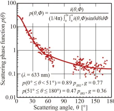
[61] K. Naito, J. Yamada, T. Ogawa, S. Takata. "Measurement of scattering phase function of human skin." Japan journal of Thermophysical properties 24.2 (2010): 101-108.
[62] Y. Lee, and K. Hwang. "Skin thickness of Korean adults." Surgical and radiologic
anatomy 24.3-4 (2002): 183-189.
[63] R. B. Saager, A. Truong, D. J. Cuccia, and A. J. Durkin. "Method for depth-resolved quantitation of optical properties in layered media using spatially modulated quantitative spectroscopy." Journal of biomedical optics 16.7 (2011): 077002-077002.
[64] S. J. Yeh, O. S. Khalil, C. F. Hanna, et al. "Temperature dependence of optical properties of in-vivo human skin." Proc. SPIE 4250 (2001)
[65] D. Yudovsky, J. Q. Nguyen, and A. J. Durkin. "In vivo spatial frequency domain spectroscopy of two layer media." Journal of Biomedical Optics 17.10 (2012)
[66] G. D. Boreman. Modulation transfer function in optical and electro-optical systems. Vol. 21. Bellingham, WA: SPIE press, 2001.
[67] International Standard ISO 12233:2000(E).
[68] A. K. Ahmad, H. H. Mohammed, and S. M. Khorsheed. "MTF Estimation for IR Optical Systems." relation 4.2: 040.
IEEE, 2011.
[70] P. Wissmann, S. B. Oh, and G. Barbastathis. "Simulation and optimization of volume holographic imaging systems in Zemax®." Optics express 16.10 (2008): 7516-7524. [71] J. Tortora, Gerard, and B. H. Derrickson. Principles of anatomy and physiology. John
Wiley & Sons, 2008.
[72] K. Nakamura, and J. Yamada. "Development of measurement system for bidirectional reflectance/transmittance using a paraboloidal mirror." Japan Journal of Thermophysical
properties 30.1 (2016): 18-26.
![図 3-6.Naito らの計測結果および近似式一覧 3-3-2 散乱位相関数を既知としたモンテカルロ法への適用 本研究では,散乱位相関数を除いた2つの光物性(アルベド w ,減衰係数 b ) を推定する.散乱位相関数も他の光物性と同時に推定することが可能であるが,すべ ての光物性を同時に逆解析することによって,複雑さを生み,推定誤差が増す.この ため,本解析では,培養皮膚(表皮モデル)について Naito ら [61] が計測した散乱位相 関数を既知のものとして与え,残り2つの光物性を推定する](https://thumb-ap.123doks.com/thumbv2/123deta/9766121.1850125/23.892.267.644.118.483/および式一覧モンテカルロ本研究アルベドがすべによっについて.webp)