Impact of Cardiac Progenitor Cells on Heart Failure and Survival in Single Ventricle Congenital Heart Disease
Toshikazu Sano, M.D.1, Daiki Ousaka, Ph.D.1, Takuya Goto, M.D.1, Shuta Ishigami,M.D., Ph.D.4, Kenta Hirai, M.D.2, Shingo Kasahara, M.D., Ph.D.1, Shinichi Ohtsuki, M.D., Ph.D.2, Shunji Sano, M.D., Ph.D.4, and Hidemasa Oh, M.D., Ph.D.3, *
Departments of Cardiovascular Surgery1 and Pediatrics2, Okayama University Graduate School of Medicine, Dentistry, and Pharmaceutical Sciences; Department of Regenerative Medicine3, Center for Innovative Clinical Medicine, Okayama University Hospital, Okayama, Japan;
Department of Pediatric Cardiothoracic Surgery4, University of California San Francisco, San Francisco, California, U.S.A.
Running Title: Outcomes after cell therapy in single ventricles
Key Words: single ventricle, stem cell, heart failure, congenital heart disease, diastolic function Abstract: 300 words
Text: 7,540 words
This work will be presented at the American Heart Association’s Council on Cardiovascular Disease in the Young (CVDY) and was selected to receive a 2017 CVDY Outstanding Research Award in Pediatric Cardiology.
*Correspondence:
Hidemasa Oh, M.D., Ph.D.
Department of Regenerative Medicine, Center for Innovative Clinical Medicine, Okayama University Hospital,
2-5-1 Shikata-cho, Kita-ku, Okayama 700-8558, Japan Tel.: +81-086-235-6506 Fax: +81-086-235-6505
E-mail: hidemasa@okayama-u.ac.jp
Abstract
Rationale- Intracoronary administration of cardiosphere-derived cells (CDCs) in patients with single ventricles resulted in a short-term improvement in cardiac function.
Objective- To test the hypothesis that CDC infusion is associated with improved cardiac function and reduced mortality in patients with heart failure.
Methods and Results- We evaluated the effectiveness of CDCs using an integrated cohort study in 101 patients with single ventricles, including 41 patients that received CDC infusion and 60 controls treated with staged palliation alone. Heart failure with preserved (HFpEF) or reduced ejection fraction (HFrEF) was stratified by the cardiac function after surgical reconstruction. The main outcome measure was to evaluate the magnitude of improvement in cardiac function and all-cause mortality at 2 years. Animal studies were conducted to clarify the underlying mechanisms of HFpEF and HFrEF phenotypes. At 2 years, CDC infusion increased ventricular function (stage 2: +8.4% ± 10.0% vs +1.6% ± 6.4%, P=0.03; stage 3: +7.9% ± 7.5% vs -1.1% ± 5.5%, P<0.001) compared with controls. In all available follow-up data, survival did not differ between the 2 groups (Log-rank P=0.225), whereas overall patients treated by CDCs had lower incidences of late failure (P=0.022), adverse events (P=0.013), and catheter intervention (P=0.005) compared with controls. CDC infusion was associated with a lower risk of adverse events (hazard ratio: 0.411 [95% confidence interval, 0.179 to 0.942], P=0.036). Notably, CDC infusion reduced mortality (P=0.038) and late complications (P<0.05) in patients with HFrEF but not with HFpEF. CDC-treated rats significantly reversed myocardial fibrosis with differential collagen deposition and inflammatory responses between the heart failure phenotypes.
Conclusions- CDC administration in patients with single ventricles showed favorable effects on
ventricular function and was associated with reduced late complications except for all-cause mortality after staged procedures. Patients with HFrEF but not HFpEF treated by CDCs resulted in significant improvement in clinical outcome.
Clinical Trial Registration: URL: ClinicalTrials.gov Unique Identifier: NCT01273857, NCT01829750.
Non-standard Abbreviations and Acronyms CDCs cardiosphere-derived cells
HFpEF heart failure with preserved ejection fraction HFrEF heart failure with reduced ejection fraction HLHS hypoplastic left heart syndrome
cMRI cardiac magnetic resonance imaging EDV end-diastolic volume
ESV end-systolic volume WAZ weight for age
ESPVR end-systolic pressure volume relationship EDPVR end-diastolic pressure-volume relationship Col collagen
IL interleukin
MMP matrix metalloproteinase NPPA atrial natriuretic peptide
Introduction
In patients with congenital heart disease, single ventricle heart defects have a high risk of mortality in the first year of life and often result in late complications developing during the staged palliative repair.1 Although single ventricle reconstruction trials have sought to determine predictors of poor outcome at 3 years in patients with single ventricle physiology on the basis of initial shunt types (Norwood procedure) and the timing of stage 2 palliation, a longitudinal cohort study in patients with Fontan (stage 3 procedure) circulation has shown that the risk of death or cardiac transplantation over 12 years was closely associated with poorer ventricular function and lower exercise performance after staged surgical procedures, rather than the type of shunt connection or ventricular morphology.2-4
The prevalence of heart failure in pediatric patients is increasing, despite improved treatment of structural heart disease.5 Heart failure presents as a diverse syndrome and is characterized by elevated intracardiac pressure or reduced cardiac output. Although functional deterioration and an increased risk of extracardiac comorbidities in patients with single systemic ventricles are thought to occur over time, little is known about the involvement of heart failure with preserved ejection fraction (HFpEF) in the progression to multiorgan systemic dysfunction.1 The epidemiology and pathophysiology of HFpEF in patients with single ventricle physiology remain elusive, largely due to disease-specific heterogeneity and attribution of underlying genetic factors, as well as technical variations in surgical procedures that may impact the outcome.6 To date, no definitive therapy has been shown to improve cardiac function under chronic volume or pressure overload that may predispose HFpEF patients to a worse prognosis.7
Stem cell therapy trials are ongoing in a variety of congenital heart diseases to investigate their
favorable effects on cardiac performance and functional health status.5 Our early phase 1/2 clinical trials using intracoronary administration of autologous cardiosphere-derived cells (CDCs) have shown reliable and safe results for the treatment of patients with single ventricle physiology.8,
9 These clinical trials had the limitation that only a few study participants were enrolled as a long- term control arm due to ethical considerations.10 As such, we conducted a retrospective cohort study to evaluate the therapeutic efficacy of CDC infusion in patients with single ventricle physiology at 2 years. Data from CDC-infused patients was integrated with that of a larger population who met the overarching criteria for inclusion and were treated by staged surgical palliation alone in the same study period as a comparative effective clinical research to yield previously unappreciated information.
Methods
Patient Population
The study protocols and the initial results of the TICAP phase 1 (Transcoronary Infusion of Cardiac Progenitor Cells in Patients with Single Ventricle Physiology) and PERSEUS phase 2 (Cardiac Progenitor Cell Infusion to Treat Univentricular Heart Disease) trials have been previously described.8-10 A total of 280 patients with single ventricle lesions constitutively underwent staged palliations between January 5, 2011 and January 30, 2015 (first patient enrolled for the TICAP study to the last patient assigned for the PERSEUS study). Among the study participants, 112 patients were excluded as they underwent stage 1 palliative procedures (Figure I in the online-only Data Supplement). Forty-one patients were prospectively assigned to receive CDC infusions in the TICAP and PERSEUS trials. Medical records of 60 patients were obtained
and analyzed as a control group who were eligible, but were treated by staged palliation alone.
The comparative arm was constitutively enrolled and included 7 control subjects, 20 single ventricle patients excluded for having non-hypoplastic left heart syndrome (HLHS) in the TICAP trial, 30 patients who were not able to participate in both trials because of the review process of the PERSESUS study protocol, 2 patients who withdrew consent, and 1 patient excluded due to endocarditis after surgery in the PERSEUS trial. No placebo infusions were given to control patients.
Study Design
The inclusion criteria were that patients were aged under 6 years old who were diagnosed with single ventricle lesions undergoing stage 2 or 3 palliation. Ventricular EF should be <60% during the initial screening through to the final enrollment. This study was composed of 2 prospective and 1 retrospective cohort studies to investigate ongoing long-term follow-up after CDC infusion.
The retrospective study protocol for collecting medical information from control patients was approved by the Ethics Committee of Okayama University and followed the guidelines on Clinical Research issued by the Ministry of Health, Labor, and Welfare, Japan. The study was conducted in accordance with the Declaration of Helsinki and by an opt-out method through the institutional website.
Follow-up and End Points
The pre-specified end point was the improvement of cardiac EF from baseline to 2 years of follow-up, as measured by echocardiography. Two-year clinical events analyzed included the composite of death of any cause and type, late failure, adverse events, and unplanned catheter intervention. Late failure was defined as late death (precluded death during the initial hospital
stay), protein-losing enteropathy, stage 2 or 3 procedure takedown, stage 2 procedure patients who were not able to proceed to stage 3, Ross classification 3 or 4 at follow-up, or rehospitalization due to heart failure requiring additional medication or surgery. Adverse events included death, reoperation, ventricular arrhythmia (tachycardia and pacemaker implantation), thromboembolic events, atrioventricular valve regurgitation ≥moderate at follow-up, and rehospitalization due to heart failure.
Blinded core laboratories assessed cardiac function measured by echocardiography, cardiac magnetic resonance imaging (cMRI), and ventriculography. For patients who died or underwent additional surgery or catheter-based re-intervention, the primary outcome measure was obtained from the most recent cardiac imaging study immediately before the adverse event. For long-term outcome analysis, patients and investigators were unblinded. However, data acquisition and categorization of events were performed by an independent data manager, as well as by an event review committee who were unware of the group assignment.
Heart Failure Classification
Cardiac function was analyzed by 2-dimmentional and color Doppler examinations and stored digitally for offline analysis by 2 pediatric cardiologists who were blinded to the group assignment. End-diastolic volume (EDV) and end-systolic volume (ESV) were obtained from the dominant ventricle by the monoplane-modified Simpson’s method through a transverse apical 4-chamber view as previously described.8 Patients with HFpEF were categorized according to the presence of heart failure symptoms and 50%≤ EF <60% measured by echocardiography, whereas heart failure with reduced EF (HFrEF) was defined as <50%. In cMRI, EF was calculated based on the quantification of single ventricle volume through short-
axis cine images and was categorized as 40% to less than 50% for HFpEF, whereas HFrEF was defined as <40%.
Statistical Analysis
As no previous cell therapy trials in single ventricle lesions have reported long-term evaluation of mortality and adverse events, clinical outcome measures in this study were descriptive and exploratory with no statistical hypothesis based on an estimated power calculation. Continuous variables are presented as the means and the standard deviation (SD) if normally distributed.
Comparisons between groups were made by Student’s t-tests, and changes from baseline within groups were evaluated by paired t-tests. If normality could not be established, differences between groups were tested using nonparametric Mann-Whitney U tests, and changes from baseline within groups were tested by nonparametric Wilcoxon signed-rank tests. Categorical variables were expressed as the number and percentage of observations and were analyzed with Fisher’s exact tests. Clinical outcome was assessed by Kaplan-Meier analyses, and Log-rank tests were applied to determine whether the distributions of time to the earliest event of death, late failure, adverse events, and catheter intervention during all available follow-ups differed between groups. The assumption of proportional hazards with respect to the clinical outcome after stage 2, 3, or combined stages was assessed by plotting the log-minus-log function over time. The model was adjusted for all the variables that were associated with an adverse clinical outcome in the crude analysis. Univariate regression analysis was performed to identify variables predictive of improvement in ventricular function. Nonlinearly associated variables were adjusted for stepwise selection for multivariate analysis. Univariate and multivariate stepwise Cox proportional hazard regression models were applied to assess the independent competing risks and hazard ratios (HR)
associated with the occurrence of adverse events. For comparison of 2 different categorical independent variables on the time interaction term within groups, 2-way analysis of variance (ANOVA) was used. EF calculated by echocardiography was compared with cMRI results by linear regression analysis. All statistical tests reported were two-sided, and values of P<0.05 were considered to be significant. All analyses were performed using the SPSS software (version 24, IBM Corporation, Armonk NY, USA). The statistical methods applied were critically reviewed and supervised by a medical biostatistician who had no role in data collection and interpretation.
Results
Our previous results from the TICAP phase 1 and PERSEUS phase 2 prospective studies in 48 patients have shown that intracoronary administration of CDCs may lead to global and regional functional improvement at 3 months by a significant reduction in myocardial elastance compared with patients treated by standard palliation alone (Figures II and III and Table I in the online-only Data Supplement). The major study limitations of these trials are the lack of a control arm for long-term comparison, and that a mixed study population undergoing different stages of surgical repair, either stage 2 or 3 procedures, were enrolled during the initial registry. Therefore, this cohort study was sought to determine the stage-specific clinical outcome after intracoronary CDC transfer in patients with single ventricles by direct comparison with the subjects treated by surgical palliation alone in larger population.
Baseline Characteristics
Between January 5, 2011 and January 30, 2015, 41 patients with single ventricle physiology were enrolled in either TICAP or PERSEUS trials to receive CDC infusions, and 60 control subjects,
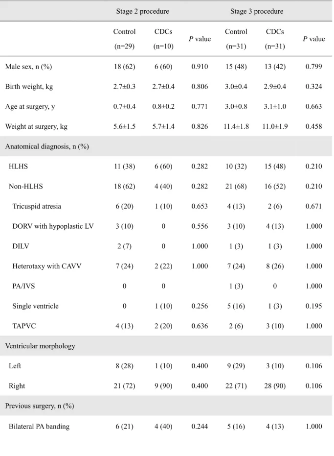
including 7 patients prospectively allocated as controls in the TICAP study, were treated using staged surgical repair without cell delivery. Of 101 patients, 39 subjects (39%) underwent stage 2 and 62 subjects (61%) underwent stage 3 palliation. Baseline characteristics of the CDC-treated patients and controls in each stage did not differ significantly (Table 1). Anatomical diagnosis, previous surgery received, and values of brain natriuretic peptide (BNP) were similar within the groups.
Response to CDC Infusion at 2 Years after Stage 2 or 3 Palliation
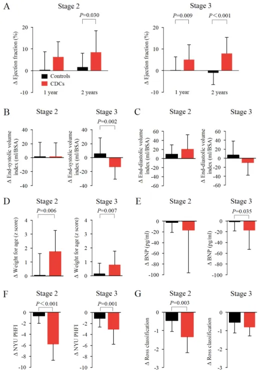
Cardiac function was assessed by echocardiography in 60 controls and 41 CDC-treated patients 1 month after staged palliation and compared with that at 2 years after treatment. The absolute changes in EF from baseline to 2-year follow-up showed significant improvements in CDC- treated patients compared with controls after each staged procedure (Figure 1A; stage 2: +8.4%
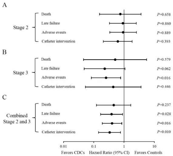
± 10.0% vs +1.6% ± 6.4%, P=0.03; stage 3: +7.9% ± 7.5% vs -1.1% ± 5.5%, P<0.001). Somatic growth and heart failure status were both markedly improved in CDC-treated patients compared with controls in both palliative stages (Figures 1D and 1F). A significant reduction in BNP levels was found in patients treated with CDCs after the stage 3 procedure (P=0.035, Figure 1E). A 2- year follow-up of clinical events was completed in 101 patients (100%). The median follow-up time was 38 months (interquartile range: 31 to 60 months). CDC infusions in combined stage 2 and 3 procedures significantly favored the pre-specified end points, including late failure, adverse events, and catheter intervention, as shown by the HR: 0.387 (95% confidence interval [CI], 0.167 to 0.9, P=0.028), 0.4 (95% CI, 0.19 to 0.844, P=0.016), and 0.333 (95% CI, 0.144 to 0.768, P=0.01), respectively (Figure 2C). In the patients that underwent stage 3 palliation, CDC infusion was shown to be significantly associated with a lower hazard of adverse events (HR, 0.253 [95%
CI, 0.083 to 0.771], P=0.016; Figure 2B).
Patient Outcome
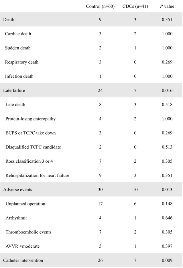
A total of 12 deaths (11.9%) occurred during the follow-up, including 9 (survival, 85%) in controls and 3 (survival, 92%) in the CDC-treated group (Table 2). There were no significant differences in all-cause mortality between the groups (P=0.351); however, patients that received CDC infusion showed significantly reduced incidences of late failure (P=0.016), adverse events (P=0.013), and unplanned catheter intervention (P=0.009) compared with controls (Table 2).
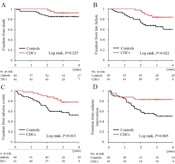
Although event-free survival at 2 years was significantly higher in CDC-treated patients compared with controls (100% vs 86.7%, P=0.02), overall survival did not differ between the 2 groups in all available follow-up data (Log-rank P=0.225, Figure 3A). None of the patients in this study received heart transplantation. Corresponding to the results shown in Table 2, patients treated by CDCs had significantly lower incidences of late failure (Log-rank P=0.022), adverse events (Log-rank P=0.013), and catheter intervention (Log-rank P=0.005) over time compared with controls (Figures 3B to 3D).
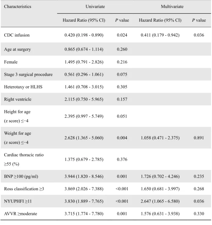
Competing Risks for Adverse Events and Factors Associated with Functional Improvement The prognostic relevance of adverse events was analyzed using univariate and multivariate Cox proportional hazard models. The strongest association was identified in patients that had a higher heart failure status, as defined by the New York University Pediatric Heart Failure Index (NYUPHI) ≥11, for which the risk was increased >2.6-fold (P=0.036, Table 3). Most importantly, CDC infusion in patients with single ventricles was significantly associated with a lower hazard ratio to suffer adverse events after staged palliation (HR: 0.411 [95% CI, 0.179 to 0.942], P=0.036; Table 3). In addition, multivariate regression analysis revealed baseline EF to have the
greatest prognostic impact on functional improvement after CDC infusion (P=0.015, Table II in the online-only Data Supplement).
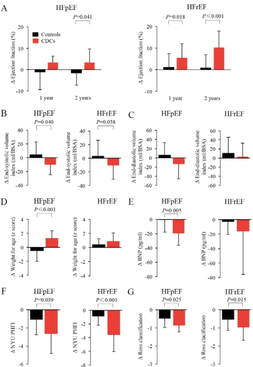
Outcomes in Patients According to the Subtypes of Heart Failure after CDC Application As there is still a poor understanding of how the grade of heart failure affects the clinical outcome after CDC treatment in patients with single ventricles, we sought to determine longitudinal changes in cardiac function and outcomes in patients with HFpEF and HFrEF. In the short-term results of TICAP and PERSEUS studies, cardiac function measured by cMRI in HFpEF patients receiving CDCs did not differ from controls (Figures IVA to IVC in the online-only Data Supplement). Although the therapeutic efficacy could only be observed in HFrEF patients at 3 months, there was a significant improvement in the circumferential global strain in both HFpEF (P=0.006) and HFrEF (P=0.02) patients 3 months after CDC infusion compared with controls (Figure IVD in the online-only Data Supplement). In the long-term study, baseline characteristics and medications of the patients did not differ between CDC-treated patients and controls in each categorized heart failure group (Table III in the online-only Data Supplement). At 2 years, patients treated by CDCs showed a significant functional improvement in both HFpEF and HFrEF groups compared with controls (HFpEF: +3.3% ± 6.5% vs -1.6% ± 5.7%, P=0.041; HFrEF: +10.3% ± 7.7% vs +0.9% ± 6.0%, P<0.001; Figure 4A). The improvement in cardiac function could be seen by a significant reduction in cardiac systolic volumes (Figure 4B). The absolute changes of weight for age corrected by z scores (WAZ) and BNP were greater only in the HFpEF group (WAZ: +1.3
± 1.1 vs -0.5 ± 1.5, P<0.001; BNP: -19.2 ± 17.3 pg/mL vs -0.2 ± 17.7 pg/mL, P=0.005; Figures 4D and 4E). Compared with controls, CDC treatment significantly decreased the heart failure status, assessed by NYUPHFI and the Ross classification, in both groups (Figures 4F and 4G).
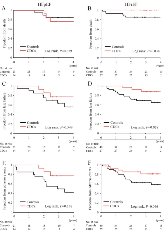
During follow-up, 3 CDC-treated patients with HFpEF died after 2 years. The overall survival in the HFpEF group was 75% for patients receiving CDCs vs 85% for controls (Log-rank P=0.679, Figure 5A), whereas no patients died in the HFrEF group treated by CDC infusion, as shown by a significantly lower all-cause mortality during follow-up (Log-rank P=0.038, Figure 5B). The improved outcome in patients with HFrEF receiving CDCs was further characterized by significantly reduced incidences of late failure (Log-rank P=0.028) and adverse events (Log-rank P=0.046), compared with those treated by staged palliation alone (Figures 5D and 5F). In the HFpEF cohort study, the benefits of CDC infusion for preventing late complications were not statistically evident during follow-up (Figures 5C and 5E).
Mechanisms of CDC-Mediated Cardiac Repair in Congenital Heart Failure
The pathophysiology of HFpEF in congenital heart disease, including single ventricles, is yet to be fully elucidated. A pulmonary artery banding was created to model right heart failure in rats in order to investigate the underlying mechanisms involved in cardiac repair followed by CDC therapy. At 4 weeks after banding, a pressure volume study was performed to define HFpEF based on an EF ≥40%. Compared with medium-treated rats, animals with HFpEF that received CDC infusions for 4 weeks had a significant increase in the dP/dt max (4414 ± 1859 mmHg/s vs 2796
± 1088 mmHg/s, P=0.03) and ventricular elastance measured by the slope of the end-systolic pressure volume relationship (ESPVR: 0.39 ± 0.15 mmHg*m2/mL vs 0.23 ± 0.18 mmHg*m2/mL, P<0.001) relative to the baseline (Table IV in the online-only Data Supplement). In medium- treated animals, cardiac function deterioration was manifested by a decrease in EF (P=0.006) with elevated end-diastolic pressure (P=0.004) and myocardial stiffness measured by the slope of the end-diastolic pressure-volume relationship (EDPVR, P=0.002), whereas these parameters were
relatively preserved in CDC-treated rats with HFpEF. In the HFrEF rat study, medium-treated animals showed significant ventricular dilatation in end-systole (P=0.03). The therapeutic effects of CDCs appeared more obvious in HFrEF rats, as shown by a significantly greater improvement in EF (43.3 ± 5.7% at 1 month vs 27.3 ± 7.7% at baseline, P<0.001) that coincided with a marked reduction in ESV (P=0.03) and an increase in cardiac output and stroke volume compared with the baseline (P<0.001, Table V in the online-only Data Supplement).
Histological evaluation revealed that right ventricular hypertrophy was observed 4 weeks after banding with remarkable fibrotic remodeling of the myocardium that was significantly attenuated by CDC infusion 4 weeks after treatment in both HFpEF and HFrEF rats (P<0.001, Figure V in the online-only Data Supplement). Compared with sham operated rats, transcripts of collagen depositions and inflammatory cytokines were upregulated in both HFpEF and HFrEF animals.
The gene expressions of collagen type 3 (Col3A1) and interleukin-6 (IL-6) were markedly suppressed by CDC infusion in both groups. Interestingly, collagen type 1 (Col1A2), matrix metalloproteinase type 2 (MMP2), and atrial natriuretic peptide (NPPA) expressions remained unchanged in HFrEF rats compared with a significant reduction seen in HFpEF rats (Figure VI in the online-only Data Supplement).
Discussion
The disproportionate share of mortality and morbidity in patients with single ventricles among congenital heart diseases may limit long-term observational studies to relatively small sample sizes with biased populations toward survivors at each stage.11, 12 In this complex context, variability in the underlying genetic causes may exist in the study population, and heterogeneity in the types of single ventricle may also influence maladaptive responses after surgical
reconstruction.13 Our results addressed several unanswered questions with regard to stage- specific responses to CDC therapy with potent clinical implications. Heterogeneity in the study population resulted from structural differences and phenotypic diversity in heart failure in patient and animal studies. A significant feature of this integrated cohort study was its evaluation of the clinical outcome, including all-cause mortality and late complications, by direct comparison with controls treated by standard surgical procedures alone. As such, the limitations of this study are its observational nature, which included neither prospective data collection, nor randomization.
The anatomic and morphological subtypes of single ventricles are critical determinants of the outcome. Predictors for functional improvement after CDC therapy were analyzed and it was revealed that HLHS or heterotaxy syndrome were not risk factors for a poor outcome (Table II in the online-only Data Supplement). However, disease variation in complex structural heart disease might exist and may not have been detected in the relatively small sample size of this study, thus affecting its practical therapeutic efficacy. Increasing the number of specific
univentricular heart diseases, such as HLHS, across multi-centers using trial registry rather than recruiting a diverse range of single ventricles in a single-center may address these limitations.
Another issue of the early TICAP and PERSEUS studies was the initial results obtained from the mixed study population treated by different staged palliations.14 Direct comparisons within the stratified staged-procedure in this cohort study may enable us to elucidate the CDC- mediated functional benefits and clinical outcome in a similar systemic circulation by ventricular remodeling after surgical reconstruction. In patients that underwent stage 2 palliation, survivors who were able to proceed to the stage 3 procedure in the CDC-treated group and controls at a 2-year follow-up did not differ (80% vs 90%, P=0.586). The functional improvement after 1 year in stage 2 patients may be affected by the inter-stage variance of patients proceeding to stage 3 palliation, leading to differential responses with altered relaxation properties and ventricular filling, as well as increased afterload in the absence of pulsatile flow in Fontan circulation.15
The heterogeneous population in this study was further explored by functional subtypes of heart failure. The results of predictor analysis suggested that cardiac function measurement at baseline may provide insight into the therapeutic responsiveness and implications of CDC treatment in single ventricles (Table II in the online-only Data Supplement). In all available follow-up data, all-cause mortality in control patients with HFrEF increased after stage
palliation, but not in those treated by CDCs, whereas overall survival declined after more than 1 year in the HFpEF group (Figure 5). This differential 1-year outcome between HFpEF and HFrEF was supported by the fact that lower cardiac function at baseline was associated with an increased risk of death or transplantation.2 In the HFpEF group, death occurred in 3 CDC- treated and 3 control patients. Among them, sudden cardiac death occurred in 1 case for each treatment. Death attributed to progression of heart failure occurred in 2 controls, while 2
patients that underwent stage 2 palliation died 2 years after CDC infusion due to circulatory failure caused by conduit obstruction after the stage 3 procedure. In the absence of compelling outcome data to support CDC therapy in patients with HFpEF, our finding of all-cause mortality in patients with HFpEF by 2 years is consistent with the favorable effects of CDC treatment, as seen by improved regional myocardial strain at 3 months resulting in enhanced global cardiac function at 2 years (Figure IV in the online-only Data Supplement and Figure 4).16
The role of HFpEF in adult heart disease has been extensively studied; however, its pathogenic and prognostic significance in single ventricles remain largely unknown.17, 18 No definitive therapy has been shown to have favorable effects on mortality in outcome trials in patients with HFpEF.7 Although medication trials in adults to reduce all-cause mortality by testing an angiotensin-converting enzyme inhibitor or mineralocorticoid receptor antagonist have shown moderately positive outcomes in a retrospectively-defined subgroup or by an adjusted analysis, no treatments have been shown to affect mortality in HFpEF patients with congenital heart disease.19, 20 The difficulty in treatment may be attributed to distinct
pathophysiological mechanisms that develop the heart failure into diverse HFpEF phenotypes.21 A possible mechanism found in our animal studies was the involvement of myocardial fibrosis through extracellular matrix modification and inflammatory responses via IL-6.22, 23 Whether resident fibroblasts or extracardiac progenitors were responsible for aberrant homeostatic control of collagen remains unknown.24, 25 Higher expression of Col3A1 in HFpEF compared with that in HFrEF without CDC treatment suggest that there might be a different degree of cardiac fibrosis composition through procollagen synthesis and secretion between HFpEF and HFrEF groups (8.3 ± 0.9 vs 4.4 ± 0.7, P=0.008; Figure VI in the online-only Data
Supplement).22 Although we did not detect any specific configuration of cardiac fibrosis in both types of heart failure models, collagen metabolism, involving collagen degradation after cell therapy, might differ between these distinct heart failure phenotypes.26 Recent advances in cMRI-derived T1 mapping may enable us to determine the composition of myocardial fibrosis, such as diffuse or focal depositions, in patients with single ventricles, as well as to monitor fractional changes after cell therapy in real-time.27, 28
Limitations
As with all observational cohort studies, our results may be biased, confounding, and inferior to those obtained from randomized controlled trials.29 This integrated comparative research was designed and conducted on the basis of the difficulty to recruit patients with rare congenital heart diseases, as well as the inability to treat control subjects by standardized surgery alone for long-term observation as a hierarchical randomized controlled trials due to ethical
considerations. Manufacturing CDCs should be conducted by institutional departments alongside tissue harvesting and subsequent cell transfer, this would limit the study to a single- center experience. Although the control data were obtained from patients treated by the usual palliative procedure during the same period as the TICAP and PERSEUS trials, the registry settings may have affected the results. The total sample size was not determined based on statistical power considerations. Subgroups for stage-specific and heart failure phenotype- dependent analyses were performed in small sample sizes, limiting their power to assess associations and the potential outcome. cMRI and catheter examinations were not performed in all control patients at 2 years of follow-up, to obtain myocardial strain analysis and pulmonary
vascular function. These data may provide additional prognostic value for heart failure, but could not be assessed in a portion of controls.16, 30 Instead, echocardiography was used to assess cardiac function without missing data for imputation. Along with the TICAP and PERSEUS protocols, we precluded patients with an EF ≥60%, limiting the sample size in HFpEF patients as relatively small to assess causality. Findings of improved mortality in CDC-treated HFrEF patients compared with controls were encouraging, but the observational period may be too short to infer the pragmatic effects of CDC treatment in the long-term. Our results failed to determine the mechanisms involved in late death occurring in HFpEF patients receiving CDCs.
Extracardiac comorbidities that may drive ventricular dysfunction through circulatory failure should be taken into account in this complex pathophysiology.
Despite the fact that pressure overload may not directly recapitulate the systemic
hemodynamics of single ventricular circulation, this experimental approach has been shown to assess reversible ventricular remodeling by attenuating myocardial fibrosis after stem cell injection in a porcine model of right heart failure.31 The definition of HFpEF in animal studies was determined based on a cardiac function of ≥40%, as measured by a catheter, and may differ from cohort studies that stratified the borderline as 50% by echocardiography.
Conclusions
In this integrated cohort study, intracoronary delivery of CDCs significantly improved ventricular function and reduced the incidences of late failure, adverse events, and unplanned catheter intervention in patients with single ventricles that underwent stage 2 or 3 palliation at 2 years of follow-up. Patients treated with CDCs had a lower hazard for the magnitude of
treatment effects on adverse events. Baseline cardiac function was an independent predictor of functional improvement after cell therapy. Although CDC infusion improved cardiac function and heart failure status in both HFpEF and HFrEF patients, all-cause mortality and late
complications were significantly reduced in HFrEF, but not in HFpEF subjects receiving CDCs.
Longer follow-up is underway to clarify the differential therapeutic responses underlying the phenotypic diversity in heart failure with single ventricle physiology.
Authors Contributions
HO was the chief investigator for the study, and designed and managed the study. TS, SI, and HO collected the electronic data and conducted the statistical analyses. SK and SS contributed to cardiac surgery. SO performed the catheter examination and cell infusion in patients. DO, TG, and KH performed animal experiments. TS and HO wrote the manuscript.
Acknowledgments
We thank all the patients and their parents who gave consent to participate in this study.
Source of Funding
This study was supported by grant from the Research Project for Practical Application of Regenerative Medicine (17bk0104052h0002) by the Japan Agency for Medical Research and Development (to H. Oh).
Role of the Funding Source
The study funding source had no role in study design, data collection, data analysis and interpretation, and preparation of the manuscript. All authors had full access to the data and its analysis. The corresponding author had final responsibility for the decision to submit for publication.
Disclosures
HO applied for a patent that was issued and licensed out to Japan Regenerative Medicine and
which provided no funding for this study. The authors have declared that no conflicts of interest exist.
Affiliations
From the Departments of Cardiovascular Surgery (TS, DO, TG, SK); Pediatrics (KH, SO), Okayama University Graduate School of Medicine, Dentistry, and Pharmaceutical Sciences, Japan; Regenerative Medicine, Center for Innovative Clinical Medicine, Okayama University Hospital, Japan (HO); and Pediatric Cardiothoracic Surgery, University of California San Francisco, U.S.A. (SI, SS).
References
1. Hinton RB and Ware SM. Heart Failure in Pediatric Patients With Congenital Heart Disease. Circ Res. 2017;120:978-994.
2. Newburger JW, Sleeper LA, Frommelt PC, Pearson GD, Mahle WT, Chen S, Dunbar- Masterson C, Mital S, Williams IA, Ghanayem NS, Goldberg CS, Jacobs JP, Krawczeski CD, Lewis AB, Pasquali SK, Pizarro C, Gruber PJ, Atz AM, Khaikin S, Gaynor JW, Ohye RG and Pediatric Heart Network I. Transplantation-free survival and interventions at 3 years in the single ventricle reconstruction trial. Circulation. 2014;129:2013-20.
3. Meza JM, Hickey E, McCrindle B, Blackstone E, Anderson B, Overman D, Kirklin JK, Karamlou T, Caldarone C, Kim R, DeCampli W, Jacobs M, Guleserian K, Jacobs J, Jaquiss R and Congenital Heart Surgeons' Society Timing of SPWG. The Optimal Timing of Stage-2- Palliation After the Norwood Operation. Ann Thorac Surg. 2017.
4. Atz AM, Zak V, Mahony L, Uzark K, D'Agincourt N, Goldberg DJ, Williams RV, Breitbart RE, Colan SD, Burns KM, Margossian R, Henderson HT, Korsin R, Marino BS, Daniels K, McCrindle BW and Pediatric Heart Network I. Longitudinal Outcomes of Patients With Single Ventricle After the Fontan Procedure. J Am Coll Cardiol. 2017;69:2735-2744.
5. Oh H. Cell Therapy Trials in Congenital Heart Disease. Circ Res. 2017;120:1353-1366.
6. Ohye RG, Schranz D and D'Udekem Y. Current Therapy for Hypoplastic Left Heart Syndrome and Related Single Ventricle Lesions. Circulation. 2016;134:1265-1279.
7. Metra M and Teerlink JR. Heart failure. Lancet. 2017.
8. Ishigami S, Ohtsuki S, Tarui S, Ousaka D, Eitoku T, Kondo M, Okuyama M, Kobayashi J, Baba K, Arai S, Kawabata T, Yoshizumi K, Tateishi A, Kuroko Y, Iwasaki T, Sato
S, Kasahara S, Sano S and Oh H. Intracoronary autologous cardiac progenitor cell transfer in patients with hypoplastic left heart syndrome: the TICAP prospective phase 1 controlled trial.
Circ Res. 2015;116:653-64.
9. Ishigami S, Ohtsuki S, Eitoku T, Ousaka D, Kondo M, Kurita Y, Hirai K, Fukushima Y, Baba K, Goto T, Horio N, Kobayashi J, Kuroko Y, Kotani Y, Arai S, Iwasaki T, Sato S, Kasahara S, Sano S and Oh H. Intracoronary Cardiac Progenitor Cells in Single Ventricle Physiology: The PERSEUS Randomized Phase 2 Trial. Circ Res. 2017.
10. Tarui S, Ishigami S, Ousaka D, Kasahara S, Ohtsuki S, Sano S and Oh H.
Transcoronary infusion of cardiac progenitor cells in hypoplastic left heart syndrome: Three-year follow-up of the Transcoronary Infusion of Cardiac Progenitor Cells in Patients With Single- Ventricle Physiology (TICAP) trial. J Thorac Cardiovasc Surg. 2015;150:1198-1207, 1208 e1-2.
11. Bouma BJ and Mulder BJ. Changing Landscape of Congenital Heart Disease. Circ Res.
2017;120:908-922.
12. Kaltman JR, Burns KM and Pearson GD. Perspective on Congenital Heart Disease Research. Circ Res. 2017;120:898-900.
13. Zaidi S and Brueckner M. Genetics and Genomics of Congenital Heart Disease. Circ Res. 2017;120:923-940.
14. Bittle GJ, Wehman B, Karathanasis SK and Kaushal S. Clinical Progress in Cell Therapy for Single Ventricle Congenital Heart Disease. Circ Res. 2017;120:1060-1062.
15. Veldtman GR, Opotowsky AR, Wittekind SG, Rychik J, Penny DJ, Fogel M, Marino BS and Gewillig M. Cardiovascular adaptation to the Fontan circulation. Congenit Heart Dis.
2017.
16. Kraigher-Krainer E, Shah AM, Gupta DK, Santos A, Claggett B, Pieske B, Zile MR, Voors AA, Lefkowitz MP, Packer M, McMurray JJ, Solomon SD and Investigators P. Impaired systolic function by strain imaging in heart failure with preserved ejection fraction. J Am Coll Cardiol. 2014;63:447-56.
17. Vedin O, Lam CSP, Koh AS, Benson L, Teng THK, Tay WT, Braun OO, Savarese G, Dahlstrom U and Lund LH. Significance of Ischemic Heart Disease in Patients With Heart Failure and Preserved, Midrange, and Reduced Ejection Fraction: A Nationwide Cohort Study. Circ Heart Fail. 2017;10.
18. Ohuchi H, Hayama Y, Negishi J, Noritake K, Iwasa T, Miyazaki A, Yamada O and Shiraishi I. Heart failure with preserved right ventricular ejection fraction in postoperative adults with congenital heart disease: A subtype of severe right ventricular pathophysiology. Int J Cardiol.
2016;212:223-31.
19. Lund LH, Benson L, Dahlstrom U and Edner M. Association between use of renin- angiotensin system antagonists and mortality in patients with heart failure and preserved ejection fraction. JAMA. 2012;308:2108-17.
20. Shah AM, Claggett B, Sweitzer NK, Shah SJ, Deswal A, Anand IS, Fleg JL, Pitt B, Pfeffer MA and Solomon SD. Prognostic Importance of Changes in Cardiac Structure and Function in Heart Failure With Preserved Ejection Fraction and the Impact of Spironolactone.
Circ Heart Fail. 2015;8:1052-8.
21. Shah SJ, Kitzman DW, Borlaug BA, van Heerebeek L, Zile MR, Kass DA and Paulus WJ. Phenotype-Specific Treatment of Heart Failure With Preserved Ejection Fraction: A Multiorgan Roadmap. Circulation. 2016;134:73-90.
22. Spinale FG and Zile MR. Integrating the myocardial matrix into heart failure recognition and management. Circ Res. 2013;113:725-38.
23. Seiler M, Bowen TS, Rolim N, Dieterlen MT, Werner S, Hoshi T, Fischer T, Mangner N, Linke A, Schuler G, Halle M, Wisloff U and Adams V. Skeletal Muscle Alterations Are Exacerbated in Heart Failure With Reduced Compared With Preserved Ejection Fraction:
Mediated by Circulating Cytokines? Circ Heart Fail. 2016;9.
24. Snider P, Standley KN, Wang J, Azhar M, Doetschman T and Conway SJ. Origin of cardiac fibroblasts and the role of periostin. Circ Res. 2009;105:934-47.
25. Moore-Morris T, Guimaraes-Camboa N, Banerjee I, Zambon AC, Kisseleva T, Velayoudon A, Stallcup WB, Gu Y, Dalton ND, Cedenilla M, Gomez-Amaro R, Zhou B, Brenner DA, Peterson KL, Chen J and Evans SM. Resident fibroblast lineages mediate pressure overload- induced cardiac fibrosis. J Clin Invest. 2014;124:2921-34.
26. Zile MR, Baicu CF, Ikonomidis JS, Stroud RE, Nietert PJ, Bradshaw AD, Slater R, Palmer BM, Van Buren P, Meyer M, Redfield MM, Bull DA, Granzier HL and LeWinter MM.
Myocardial stiffness in patients with heart failure and a preserved ejection fraction: contributions of collagen and titin. Circulation. 2015;131:1247-59.
27. Schelbert EB, Fridman Y, Wong TC, Abu Daya H, Piehler KM, Kadakkal A, Miller CA, Ugander M, Maanja M, Kellman P, Shah DJ, Abebe KZ, Simon MA, Quarta G, Senni M, Butler J, Diez J, Redfield MM and Gheorghiade M. Temporal Relation Between Myocardial Fibrosis and Heart Failure With Preserved Ejection Fraction: Association With Baseline Disease Severity and Subsequent Outcome. JAMA Cardiol. 2017;2:995-1006.
28. Rommel KP, von Roeder M, Latuscynski K, Oberueck C, Blazek S, Fengler K, Besler
C, Sandri M, Lucke C, Gutberlet M, Linke A, Schuler G and Lurz P. Extracellular Volume Fraction for Characterization of Patients With Heart Failure and Preserved Ejection Fraction. J Am Coll Cardiol. 2016;67:1815-1825.
29. Fiore LD and Lavori PW. Integrating Randomized Comparative Effectiveness Research with Patient Care. N Engl J Med. 2016;374:2152-8.
30. Aschauer S, Kammerlander AA, Zotter-Tufaro C, Ristl R, Pfaffenberger S, Bachmann A, Duca F, Marzluf BA, Bonderman D and Mascherbauer J. The right heart in heart failure with preserved ejection fraction: insights from cardiac magnetic resonance imaging and invasive haemodynamics. Eur J Heart Fail. 2016;18:71-80.
31. Wehman B, Sharma S, Pietris N, Mishra R, Siddiqui OT, Bigham G, Li T, Aiello E, Murthi S, Pittenger M, Griffith B and Kaushal S. Mesenchymal stem cells preserve neonatal right ventricular function in a porcine model of pressure overload. Am J Physiol Heart Circ Physiol.
2016;310:H1816-26.
Figure 1. Stage-specific functional outcome in CDC-treated and control patients at 2 years.
(A-C) Stage-specific ventricular function was analyzed by 2-dimentional echocardiography and the absolute changes from baseline to 2 years of follow-up were compared between controls and CDC-treated patients. Somatic growth (D) and heart failure status, including plasma levels of BNP, (E), New York University Pediatric Heart Failure Index (F), and Ross classification (G) are shown. Black bars, controls. Red bars, CDC-treated patients. Data are expressed as the means (SD). Between-group comparisons were performed using Student’s t-tests.
Figure 2. Hazard ratios of intracoronary CDC infusion for clinical events in various subgroups at 2 years of follow-up. The study population was confined to patients that underwent stage 2 or 3 procedures (A and B), or a combined population (C). Black circles present the hazard ratios with the 95% confidence interval. Statistics were evaluated by Cox regression analyses.
Figure 3. Cumulative incidence of freedom from death and late complications during follow-up.
Comparisons between controls and CDC-treated patients for the freedom from the composite of death (A), late failure (B), adverse events (C), and catheter intervention (D), as assessed by Kaplan-Meier analyses. Black lines, controls. Red lines, CDC-treated patients.
Figure 4. Response to CDC infusion in HFpEF and HFrEF patients.
The study population was stratified by HFpEF (EF ≥50%) or HFrEF (<50%) based on EF measured by echocardiography at the baseline. Changes in cardiac function (A-C), somatic growth (D), and heart failure status (E-G) from baseline to 2 years of follow-up are shown. At 2 years, cardiac function (A and B) and heart failure index scores (F and G) were significantly improved in both HFpEF and HFrEF patients receiving CDC infusions (red bars) compared with controls (black bars). Data are expressed as the means (SD). Between-group comparisons were performed using Student’s t-tests.
Figure 5. Comparison of freedom from death and late complications in HFpEF and HFrEF patients.
Event-free survival (A and B), late failure (C and D), and adverse events (E and F) were compared between CDC-treated patients (red lines) and controls (black lines) in HFpEF and HFrEF subpopulations, respectively. Log-rank tests were applied for statistical analyses.
Table 1. Baseline Characteristics of Study Patients with Single Ventricle Physiology
Stage 2 procedure Stage 3 procedure Control
(n=29)
CDCs
(n=10) P value Control (n=31)
CDCs
(n=31) P value
Male sex, n (%) 18 (62) 6 (60) 0.910 15 (48) 13 (42) 0.799
Birth weight, kg 2.7±0.3 2.7±0.4 0.806 3.0±0.4 2.9±0.4 0.324
Age at surgery, y 0.7±0.4 0.8±0.2 0.771 3.0±0.8 3.1±1.0 0.663
Weight at surgery, kg 5.6±1.5 5.7±1.4 0.826 11.4±1.8 11.0±1.9 0.458 Anatomical diagnosis, n (%)
HLHS 11 (38) 6 (60) 0.282 10 (32) 15 (48) 0.210
Non-HLHS 18 (62) 4 (40) 0.282 21 (68) 16 (52) 0.210
Tricuspid atresia 6 (20) 1 (10) 0.653 4 (13) 2 (6) 0.671
DORV with hypoplastic LV 3 (10) 0 0.556 3 (10) 4 (13) 1.000
DILV 2 (7) 0 1.000 1 (3) 1 (3) 1.000
Heterotaxy with CAVV 7 (24) 2 (22) 1.000 7 (24) 8 (26) 1.000
PA/IVS 0 0 1 (3) 0 1.000
Single ventricle 0 1 (10) 0.256 5 (16) 1 (3) 0.195
TAPVC 4 (13) 2 (20) 0.636 2 (6) 3 (10) 1.000
Ventricular morphology
Left 8 (28) 1 (10) 0.400 9 (29) 3 (10) 0.106
Right 21 (72) 9 (90) 0.400 22 (71) 28 (90) 0.106
Previous surgery, n (%)
Bilateral PA banding 6 (21) 4 (40) 0.244 5 (16) 4 (13) 1.000
HLHS, hypoplastic left heart syndrome; DORV, double outlet right ventricle; LV, left ventricle; DILV, double inlet left ventricle; CAVV, common atrioventricular valve; PA/IVS, pulmonary atresia with intact ventricular septum; TAPVC, total anomalous pulmonary venous; PA, pulmonary artery; BT, Blalock- Taussig; DKS, Damus-Kaye-Stansel; AVVR, atrioventricular valve regurgitation; BNP, brain
Main PA banding 3 (10) 0 0.556 6 (19) 4 (13) 1.000
Modified BT shunt 7 (24) 1 (10) 0.653 11 (35) 6 (19) 0.255
Norwood procedure 10 (34) 5 (50) 0.463 9 (29) 16 (52) 0.120
TAPVC repair 2 (7) 2 (20) 0.267 2 (6) 2 (6) 1.000
DKS anastomosis 0 0 2 (6) 4 (13) 0.671
Preoperative assessment
AVVR ≥moderate 6 (21) 2 (20) 1.000 4 (13) 3 (10) 0.204
PA stenosis 10 (37) 4 (40) 1.000 6 (19) 9 (29) 0.554
Aortic arch coarctation 3 (11) 0 0.556 2 (6) 2 (6) 1.000
Concomitant procedures
AVV repair 9 (33) 5 (50) 0.556 9 (29) 15 (48) 0.192
PA reconstruction 5 (19) 1 (10) 1.000 5 (16) 7 (23) 0.749
Correction of TAPVC 2 (7) 0 1.000 0 0
Arch augmentation 2 (7) 0 1.000 2 (6) 1 (3) 1.000
Postoperative assessment
BNP (pg/ml) 87±89 91±80 0.555 36±27 41±24 0.467
AVVR ≥moderate 4 (15) 0 0.558 0 2 (6) 0.492
PA stenosis 0 0 0 0
Aortic arch coarctation 0 0 0 0
natriuretic peptide. Data are expressed as n (%) or the mean ± SD. Between-group differences in characteristics were analyzed with Fisher’s exact tests for categorical variables and Student’s t-tests for normally distributed continuous variables. Data that were not normally distributed were analyzed with Mann-Whitney U tests. There were no significant baseline differences among the study groups at a significant level of 0.05.
Table 2. Clinical Outcomes in Control and CDC-Treated Groups during Follow-Up Control (n=60) CDCs (n=41) P value
Death 9 3 0.351
Cardiac death 3 2 1.000
Sudden death 2 1 1.000
Respiratory death 3 0 0.269
Infection death 1 0 1.000
Late failure 24 7 0.016
Late death 8 3 0.518
Protein-losing enteropathy 4 2 1.000
BCPS or TCPC take down 3 0 0.269
Disqualified TCPC candidate 2 0 0.513
Ross classification 3 or 4 7 2 0.305
Rehospitalization for heart failure 9 3 0.351
Adverse events 30 10 0.013
Unplanned operation 17 6 0.148
Arrhythmia 4 1 0.646
Thromboembolic events 7 2 0.305
AVVR ≥moderate 5 1 0.397
Catheter intervention 26 7 0.009
APCA coil occlusion 14 5 0.200
Pulmonary artery balloon angioplasty 12 0 0.001
Fenestration balloon angioplasty 6 3 0.735
Cirrhosis 0 0
Stroke 0 0
Cerebral hemorrhage 2 2 1.000
Plastic bronchitis 1 0 1.000
Tumor formation 0 0
BCPS, bidirectional cavopulmonary shunt; TCPC, total cavopulmonary connection; APCA, aortopulmonary collateral artery.
Table 3. Multivariate Cox Proportional Hazard Regression Analysis for Adverse Events
Characteristics Univariate Multivariate
Hazard Ratio (95% CI) P value Hazard Ratio (95% CI) P value CDC infusion 0.420 (0.198 - 0.890) 0.024 0.411 (0.179 - 0.942) 0.036 Age at surgery 0.865 (0.674 - 1.114) 0.260
Female 1.495 (0.791 - 2.826) 0.216
Stage 3 surgical procedure 0.561 (0.296 - 1.061) 0.075 Heterotaxy or HLHS 1.461 (0.708 - 3.015) 0.305 Right ventricle 2.115 (0.750 - 5.965) 0.157 Height for age
(z score) ≤−4 2.395 (0.997 - 5.749) 0.051 Weight for age
(z score) ≤−4 2.628 (1.365 - 5.060) 0.004 1.058 (0.471 - 2.375) 0.891 Cardiac thoracic ratio
≥55 (%) 1.375 (0.679 - 2.785) 0.376
BNP ≥100 (pg/ml) 3.944 (1.820 - 8.546) 0.001 1.726 (0.702 - 4.246) 0.235 Ross classification ≥3 3.869 (2.026 - 7.388) <0.001 1.650 (0.681 - 3.997) 0.268 NYUPHFI ≥11 3.830 (1.889 - 7.765) <0.001 2.647 (1.065 - 6.580) 0.036 AVVR ≥moderate 3.715 (1.774 - 7.780) 0.001 1.576 (0.631 - 3.938) 0.330
CI, confidence interval; NYUPHFI, New York University Pediatric Heart Failure Index.