Online publication October 3, 2011
内頸動脈起始部狭窄病変における収縮期加速時間の有用性
田村 啓和1 赤岩 靖久2 恩田 清3
要 旨:超音波検査では,石灰化による音響陰影のために狭窄部の評価が困難な場合がある。わ
れわれは,内頸動脈起始部狭窄病変における狭窄度と収縮期加速時間(acceleration time; AcT)の関 連について検討した。当院で頸部血管超音波検査を施行した 137 例 266 血管を対象とした。AcT と狭窄度には有意な相関が認められた。AcT 110 msec 以上で NASCET 60%以上の狭窄が示唆さ
れた。AcT は内頸動脈の狭窄診断に有用であると考えられた。 (J Jpn Coll Angiol, 2011, 51: 365–371)
Key words: acceleration time, internal carotid artery, ultrasonography
2011年 1 月 24 日受付 2011年 5 月 6 日受理
対象と方法
対象は 2007 年 6 月から 2010 年 4 月に頸部血管超音 波検査を施行した 137 例(男性 99 例,女性 38 例,平均 年齢 66.4 歳(35∼90 歳))274 血管である。内頸動脈閉 塞,総頸動脈狭窄,腕頭動脈狭窄のある 8 血管を除外 し,最終的に 266 血管について検討した。なお,大動脈 弁狭窄(aortic stenosis; AS)の既往のある者に関しては対 象から除外した。超音波装置は Vivid7 Dimension(GE Healthcare Japan) を使用し,中心周波数 9 MHz リニア型プローブを用いて 全例仰臥位で施行した。狭窄率は,狭窄部における収 縮期最高血流速度(peak systolic velocity; PSV)および NASCET(North American Symptomatic Carotid End-arterectomy Trial)5)法を用いた。 AcT はパルスドプラ法を使用し,屈曲部を避けて計測 した。計測部位は狭窄を全く認めない例では,内頸動脈 分岐部より 2∼3 cm 頭側で計測し,軽度でも狭窄を認め る場合は,狭窄部より 2∼3 cm 頭側で測定した(Fig. 1)。 AcTは超音波装置上で計測し,3 心拍平均を算出した。 サンプルボリュームは血管径の 2/3 とし,血流方向に対 する超音波入射角は 60 度以内となるように設定した。 なお,血流速波形が 2 峰性の場合は波形の立ち上がり から最初のピークまでの時間を AcT とした。 1新潟脳外科病院診療放射線部 2新潟大学医歯学総合病院神経内科 3新潟脳外科病院脳神経外科 第 51 回総会 座長推薦論文
●原 著●
序 言
頸動脈狭窄は脳梗塞の危険因子であり,近年,狭窄の 程度によって頸動脈内膜剝離術(carotid endarterectomy; CEA)や頸動脈ステント留置術(carotid artery stenting;CAS)といった外科的治療が推奨されている1)。頸部血管 超音波検査は簡便で非侵襲的な検査であり,狭窄診断 に広く用いられている。しかし,超音波検査には機器特 有のアーチファクトが見られることがあり,とくに,石灰 化病変では音響陰影により狭窄診断が困難な場合があ る。 狭窄病変が存在した場合,その末梢側で血流速波形
の立ち上がりが緩やかになり,収縮期加速時間(accelera-tion time; AcT)が延長することが知られている2)。末梢動
脈や腎動脈では,その血流速波形の変化が狭窄診断に
有用であるとされている3, 4)。頸部血管超音波検査では,
血流速波形の変化については波形パターンによる診断が 多く,また,狭窄度と AcT の関係についての報告は少な い。そこで今回,内頸動脈(internal carotid artery; ICA)起 始部における狭窄度と AcT の関連について評価し,AcT により狭窄診断が可能であるか検討を行った。
266 血管のうち,狭窄を認めなかったものを対照群 (172 血管)とし,平均値 +2 標準偏差より正常の cut off 値を算出した。また,NASCET 10%以上の狭窄を認めた ものは狭窄群(94 血管)とし,本群における狭窄率と AcT との関連について検討した(Fig. 2)。 超音波検査の結果も含め,主治医が必要と判断し,同 意の得られた症例では,血管撮影による狭窄率(Angio-NASCET)の評価を行った。血管撮影には,Advantx
UNV(GE Healthcare Japan),Allura Xper FD 20/10 (PHILIPS)を使用した。解析方法は,2 変数間の相関関 係の検定にはピアソンの相関係数を使用した。2 群間の 比較には Mann-Whitney U 検定を用い,P<0.05 で有意と した。また,狭窄率と AcT について,ROC 曲線(receiver operating characteristic curve)で感度と特異度を算出した。
Figure 1 Measurement methods of acceleration time (AcT).
A: The linear array probe was set in the internal carotid artery (ICA) at 2–3 cm above the ICA origin. The AcT was measured using the average of 3 heartbeats.
B: The normal Doppler wave-form pattern.
C: Duplex sonogram of a patient with ICA stenosis shows that upstroke is delayed and peak is rounded.
結 果
1.超音波検査の狭窄度と AcT の関係
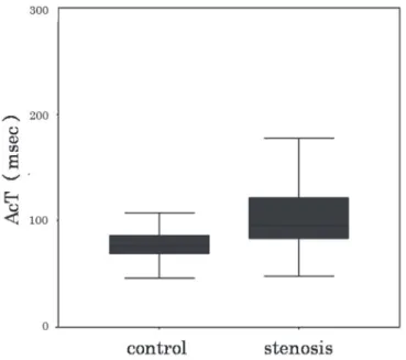
対照群における平均 AcT は 79.0±15.5 msec であり,平 均値 +2 標準偏差である 110 msec を cut off 値と設定し た。
狭窄群(94 血管)における頸部血管超音波検査で,狭 窄 率 と AcT に は 有 意 な 相 関 が み ら れ た(r=0.64, P<0.01)。また,cut off 値では NASCET 60%の狭窄病変 を予想できる結果であった(Fig. 3)。PSV と AcT の間に も有意な相関がみられた(r=0.62,P<0.01)。Cut off 値で は PSV 170 cm/sec の狭窄病変を予想できる結果であっ た(Fig. 4)。
田村 啓和 ほか 2 名
Figure 3 Relationship between degree of stenosis by the duplex NASCET method and AcT (r=0.64, P<0.01).
AcT has extended by the near occlusion of ICA (arrow head) and the severe kink of ICA (arrow). NASCET: North American Symptomatic Carotid Endarterectomy Trial, AcT: acceleration time, ICA: internal carotid artery
Figure 2 Comparison of acceleration time in control group (<NASCET 10%) and the stenosis group (≥NASCET 10%).
あったが,これらは ICA 分岐部から測定部までの間に存 在する蛇行(Fig. 3,4 arrow)や ICA の near occlusion (Fig. 3,4 arrow head)によるものであった。
AcT 110 msec を cut off 値とした場合では,ROC 曲線 より,NASCET 50 % 以 上 で は 感 度 58.1 %,特 異 度 88.2%。NASCET 60%以上では,感度 94.7%,特異度 82.7%。NASCET 70%以上では感度 100%,特異度 75.9%であった(Fig. 5)。 2.血管撮影との比較 血管撮影は 11 例(11 血管)に施行された。患側血管の 平均 AcT は 138.5±26.3 msec,対側は 91.0±24.7 msec で あり,患側血管では対側と比較して,AcT は有意に延長 していた(P<0.01)。また,血管撮影の狭窄率と AcT の間
にも有意な相関がみられた(r=0.70,P<0.01)(Fig. 6)。
血管壁の石灰化により,超音波検査では狭窄度の評 価ができなかった症例を提示する(Fig. 7)。ICA の AcT を計測したところ 122 msec と延長しており,60%以上の 狭窄が疑われた。同部位の血管撮影では,AcT から推 測されたように,内頸動脈起始部に高度狭窄病変が認め られた。
Figure 4 Relationship between PSV of ICA origin and AcT (r=0.62, P<0.01).
AcT has extended by the near occlusion of ICA (arrow head) and the severe kink of ICA (arrow). PSV: peak systolic velocity, ICA: internal carotid artery, AcT: acceleration time
考 察
頸動脈狭窄では,その狭窄の程度によって CEA や CASが考慮される。狭窄率の評価には血管撮影におけ る NASCET 法が一般的であるが6),血管撮影では約 4% の割合で神経症状を伴う合併症があり7),また,造影剤 による副作用も懸念される。 これに対し,頸部血管超音波検査は簡便で非侵襲的 な検査であることから,日常診療で広く使用されてい る。頸部血管超音波検査における狭窄度の評価には, NASCET法,ECST(European Carotid Surgery Trial)法, 面積狭窄率,PSV などが用いられる。その中でも PSV による評価は再現性も高く8),血管撮影での NASCET 法 による狭窄率との相関が知られており,PSV 150 cm/sec 以上では NASCET 法で 50%以上の狭窄,PSV 200 cm/ sec以上では NASCET 法で 70%以上の狭窄が示唆され る3, 9)。しかし,超音波検査では石灰化を伴う狭窄病変は 音響陰影のため,狭窄部における PSV の測定が困難な 場合がある。 そこでわれわれは,AcT と ICA 起始部狭窄の関連に田村 啓和 ほか 2 名
Figure 6 Relationship between degree of stenosis by the angiographic
NASCET method and AcT (r=0.70, P<0.01).
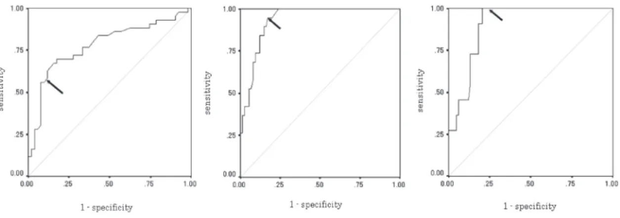
Figure 5 Sensitivity and Specificity of AcT.
A: In the receiver operating characteristic (ROC) curve, when the cutoff value of the AcT was set at 110 ms, the sensitivity was 58.1% and the specificity was 88.2% for an ICA stenosis of more than 50% by the duplex NASCET method.
B: The sensitivity was 94.7% and the specificity was 82.7% for an ICA stenosis of more than 60%. C: The sensitivity was 100% and the specificity was 75.9% for an ICA stenosis of more than 70%.
A B C ついて検討を行った。血管超音波検査において,中枢側 に狭窄病変が存在した場合,その末梢側では血流速波 形の立ち上がりが緩やかとなり,AcT が延長することが 知られている2)。本検討では,AcT と超音波検査での狭 窄率,PSV ともに有意な相関がみられ,AcT 110 msec 以 上で NASCET 60%以上,PSV 170 cm/sec 以上の狭窄が 示唆された。また,ROC 曲線においては,AcT 110 msec を cut off 値とした場合,NASCET 60%以上で感度,特 異度ともに高値を示していた。NASCET 60%は,無症候
がって,AcT は ICA 起始部狭窄病変における,外科的 治療の判断材料の一つになるのではないかと考えられる。 ICA における AcT が延長する要因として,大動脈弁 狭窄(aortic stenosis; AS)が知られている。重症 AS の場 合,総頸動脈(common carotid artery; CCA),内頸動脈, 椎骨動脈(vertebral artery; VA),すべての血管で AcT が
延長するといわれており10),AcT のみで狭窄診断を行う
際には AS や心疾患を除外したほうが良いと思われる。 竹川らによると ICA における AcT を同側 CCA の AcT で除した AcT ratio を用いることで除外が可能であるとし ている。また,AcT ratio と狭窄度を比較し,AcT ratio が 1.5 以上で NASCET 50%以上,2.0 以上で NASCET 70%以上の狭窄を示唆できると報告している11)。本検討 では全例に経胸壁心エコーは施行しておらず,AS の正 確な評価はできていないが,カルテおよび主治医への確 認で明らかな高度 AS 症例は除外した。AS の既往を確 認すれば,AcT のみでも ICA 起始部狭窄病変の評価が 可能であると考えられた。 中枢側に狭窄病変が存在した場合,血流速波形の立 ち上がりが緩やかとなるため,その心収縮期における加 速度(acceleration)についても評価したほうがよいと考え られるが,それについては今後の検討課題としていきた い。また,パルスドプラ測定部位までに屈曲が存在する 例では AcT は延長しており,測定部位についても検討 が必要と考えられる。 今回 cut off 値として設定した値は,当院における基準値 であり,一般的な指標としては,AcT が延長する様々な 要因について検討し,新たな設定が必要と思われる。ま た,本検討は,頸部血管超音波検査における狭窄率との 比較であるため,確定診断には血管撮影も必要である。 しかし,血管撮影による評価との比較において,AcT の 有効性を確認することができ,AcT は頸部血管超音波検 査において,有意狭窄診断の一助となる可能性があると 考えられた。 Figure 7
A: Color Doppler image shows ICA origin. The evaluation of ICA origin is difficult due to the calcification of the vessel wall (arrow).
B: Doppler waveform shows post-stenotic pattern. AcT is 122 ms.
C: Lateral view of a left common carotid angiogram shows 80% ICA stenosis by the NASCET method (arrow).
田村 啓和 ほか 2 名
結 論
AcT は超音波装置上で容易に計測可能なパラメーター の一つである。ICA における AcT の測定は,ICA 起始 部狭窄の有無や狭窄診断に有用であることが示唆され た。 謝 辞 本検討を行うにあたり御協力くださいました,新潟脳外科 病院の新井弘之先生,山崎一徳先生,宮川照夫先生,獨協 医科大学神経内科脳卒中部門の竹川英宏先生に深謝いたし ます。 文 献 1) 脳卒中合同ガイドライン委員会:脳卒中治療ガイドライ ン.協和企画,東京,2009, 227–229 2) 日本脳神経超音波学会・栓子検出と治療学会合同ガイ ドライン作成委員会:頸部血管超音波検査ガイドライ ン.Neurosonology 2006; 19: 40–67 3) 平井都始子,東浦 渉,阪口昇二,他:下肢動脈疾患 の超音波検査─最近の進歩と現状─.脈管学 2004; 44: 727–734
4) Burdick L, Airoldi F, Marana I, et al: Superiority of acceler-ation and acceleracceler-ation time over pulsatility and resistance indices as screening tests for renal artery stenosis. J
Hyper-tens 1996; 14: 1229–1235
5) North American Symptomatic Carotid Endarterectomy Trial Collaborators: Beneficial effect of carotid endarterectomy in symptomatic patients with high-grade carotid stenosis. N Engl J Med 1991; 325: 445–453
6) Cremonesi A, Setacci C, Bignamini A, et al: Carotid artery stenting: first consensus document of the ICCS-SPREAD joint Committee. Stroke 2006; 37: 2400–2409
7) Hankey GJ, Warlow CP, Sellar RJ: Cerebral angiographic risk in mild cerebrovascular disease. Stroke 1990; 21: 209– 222
8) Sabeti S, Schillinger M, Mlekusch W, et al: Quantification of internal carotid artery stenosis with duplex US: comparative analysis of different flow velocity criteria. Radiology 2004;
232: 431–439
9) Koga M, Kimura K, Minematsu K, et al: Diagnosis of inter-nal carotid artery stenosis greater than 70% with power Dop-pler duplex sonography. AJNR 2001; 22: 413–417
10) O’Boyle MK, Vibhakar NI, Chung J, et al: Duplex sonogra-phy of the carotid arteries in patients with isolated aortic stenosis: imaging findings and relation to severity of stenosis. AJR 1996; 166: 197–202
11) 竹川英宏,浅川洋平,李東旭,他:頸動脈狭窄における
収 縮期加速時間の有用性.Neurosonology 2009; 22: 79–82
Usefulness of Acceleration Time for Internal Carotid Artery Origin Stenosis
Hirokazu Tamura,1 Yasuhisa Akaiwa,2 and Kiyoshi Onda31Department of Medical Radiology, Niigata Neurosurgical Hospital, Niigata, Japan 2Department of Neurology, Niigata University Medical and Dental Hospital, Niigata, Japan
3Department of Neurosurgery, Niigata Neurosurgical Hospital, Niigata, Japan
Key words: acceleration time, internal carotid artery, ultrasonography
Calcification of a stenotic internal carotid artery (ICA) hinders accurate evaluation of the stenosis by conventional sonography due to acoustic shadow. We examined the relationship between ICA origin stenosis and acceleration time (AcT). 137 samples (266 vessels) that enforced duplex ultrasonography in our hospital were targeted. The results have shown that there is a significant relationship between stenosis and AcT. An AcT of more than 110 ms suggests that the stenosis is more than 60% by the NASCET method. AcT is thought to be useful for the diagnosis of ICA stenosis. (J Jpn Coll Angiol, 2011,