33
Yonago Acta medica 2011;54:33–36
Squamous Cell Carcinoma Derived from a Skin Flap Used
for Reconstruction of the Hypopharynx
Aigo Yamasaki, Nobuya Monden, Teruaki Takishita, Yasuhiko Yamashita, Tohru Ishikawa, Norihiro Teramoto* and Hiroya Kitano†
Department of Head and Neck Surgery and *Department of Pathology, Shikoku Cancer Center, Matsuyama 791-0280 and †Division of Otolaryngology, Head and Neck Surgery, Department of Medicine of Sensory and Motor Organs, School of Medicine, Tottori University Faculty of Medicine, Yonago 683-8504, Japan
Development of a secondary primary cancer in a skin flap is a rare complication. A 70-year-old man underwent partial laryngopharyngectomy, right neck dissection and re-construction with a radial forearm free flap in 2004. Five years later, an exophytic tumor was found on the hypopharynx reconstructed with the radial forearm free flap. He under-went total laryngopharyngectomy, left neck dissection and reconstruction with a jejunal free flap in June 2009. Pathological findings suggested this was a primary cancer rather than a metastasis. Localization to the epidermis and dermis indicate that the tumor was derived from the surface of the skin flap.
Key words: flap; hypopharynx; secondary primary cancer; squamous cell carcinoma
Various flaps are used to reconstruct the defect af-ter resection of head and neck cancer, and these are associated with complications such as ischemia of a free flap, infection, fistula and stenosis. However, the development of a secondary primary cancer in a flap is rare (Genden et al., 2004). Recurrence of the original primary tumor usually occurs in residual mucosa of the pharynx or oral cavity. In the present report, we described a case of a male patient with a secondary primary squamous cell carcinoma which arose in a skin flap 5 years after the initial treatment of hypopharyngeal cancer. Pathological features and therapeutical procedures of the squamous cell carcinoma were minutely pre-sented.
Patient Report
A 70-year-old man was admitted to Shikoku Can-cer Center in 2004 for treatment of squamous
cell carcinoma of the right pyriform sinus of the hypopharynx (T2N0M0). He underwent partial laryngopharyngectomy, right neck dissection and reconstruction with a radial forearm free flap. We confirmed complete resection of the tumor by path-ological examination. The patient was discharged without postoperative radiation therapy and had regular follow-up thereafter. The patient stopped smoking and drinking after the 1st surgery in 2004. Five years later, in April 2009, he noticed a mass in the anterior neck and visited our hospital in May. Ultrasonography showed that this tumor was located anterior to the thyroid cartilage and was 4 cm in diameter. Cytologic examination of a fine-needle aspiration specimen revealed squamous cell carcinoma. Flexible fiberoptic laryngoscopy re-vealed no tumor in the pharynx or larynx. Gastro-intestinal endoscopy detected an exophytic tumor in the hypopharynx. This tumor was located at Abbreviations: MRI, magnetic resonance imaging; PET/CT, positron emission tomography with computed tomography
34
A. Yamasaki et al.
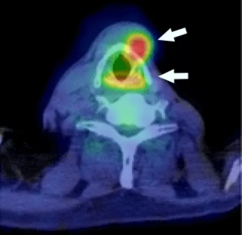
the apex of the right pyriform sinus reconstructed with the radial forearm free flap (Fig. 1). No other tumor was present in the pharyngeal or esophageal mucosa. Endoscopic biopsy of the hypopharyn-geal tumor was performed, leading to a diagnosis of squamous cell carcinoma. Magnetic resonance imaging (MRI) demonstrated only the anterior cervical cancer, and this was located outside the thyroid cartilage, away from the hypopharynx (Fig. 2). Positron emission tomography with computed tomography (PET/CT) detected both the hypo-pharyngeal and anterior cervical tumors (Fig. 3) but showed no distant metastasis. The patient was diagnosed with a secondary primary cancer on the skin flap and cervical recurrence of hypopharyn-geal cancer.
MRI and PET/CT suggested the anterior cervi-cal cancer was adherent to the thyroid cartilage, so we decided that the larynx could not be preserved. We therefore performed total laryngopharyngec-tomy, left neck dissection and reconstruction with a jejunal free flap in June 2009. Postoperation radio-therapy was performed with a total dose of 60 Gy.
Pathological findings
The hypopharyngeal cancer was separate from the anterior cervical cancer and was localized to the right pyriform sinus reconstructed with the 1.0-cm diameter skin flap (Fig. 4). It showed exophytic
Fig. 1. Gastrointestinal endoscopy detected an exophytic
tumor in the hypopharynx. This tumor was located at the apex of the right pyriform sinus that had been reconstruct-ed with a radial forearm free flap (arrow).
Fig. 2. MRI demonstrated only the anterior cervical
cancer, which was external to the thyroid cartilage, away from the hypopharynx (arrow). MRI, magnetic resonance imaging.
Fig. 3. PET/CT detected both hypopharyngeal cancer
and anterior cervical cancer (arrows). PET/CT, positron emission tomography with computed tomography.
Fig. 4. Hypopharyngeal cancer localized to the right
py-riform sinus reconstructed with the 1.0-cm diameter skin flap (arrow).
35
Secondary primary cancer in a skin flap
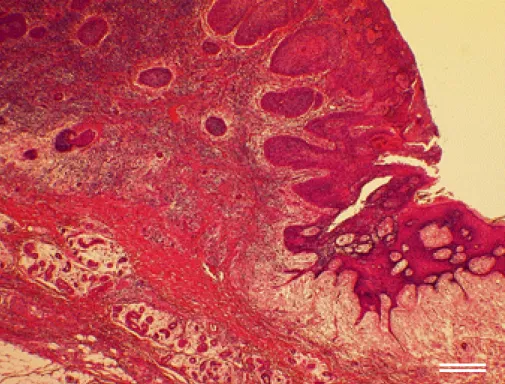
Fig. 6. Most of the cancer was localized to the epidermis and dermis of the skin flap, with little in the subcutaneous
tis-sue. It was diagnosed as well-differentiated squamous cell carcinoma (hematoxylin and eosin). Bar = 340 μm. growth from the skin flap (Fig. 5). Most of the
tumor was contained within the epidermis and der-mis of the skin flap, with little in the subcutaneous tissue. No tumor was present in the pharynx, lar-ynx or cervical esophageal mucosa of the resected specimen. This tumor was diagnosed as well-differentiated squamous cell carcinoma (Fig. 6).
Fig. 5. The hypopharyngeal cancer shows
exophytic growth from the skin flap (hema-toxylin and eosin). Bar = 3 mm.
The anterior cervical cancer was located an-terior to the thyroid cartilage with little invasion, and measured 5.2 3.0 2.4 cm. It extended to the pre-epiglottic space and reached the first tracheal cartilage without invasion of the thyroid gland. Pathological diagnosis was poorly differentiated squamous cell carcinoma. No metastatic lymph nodes were found.
36
A. Yamasaki et al.
Discussion
Pathological findings suggested that the skin flap tumor was a primary cancer rather than a metas-tasis. Localization to the epidermis and dermis showed that this cancer was derived from the sur-face of the skin flap. In contrast, metastatic skin cancer usually occurs in subcutaneous tissue, and leaves the epidermis intact (Reingold, 1966). The development of a secondary primary cancer in a flap is rare. Several factors were reported to cause this complication, including chronic irritation that the skin does not suffer at its normal site, for ex-ample, from smoking, drinking and food (Monnier et al., 2008; Woolgar and Triantafyllou, 2009).
The present patient stopped smoking and drinking after the 1st surgery in 2004, and cancer of the skin flap was detected 5 years later. Hence, physical or chemical stimulation from food, saliva or stomach acid might have been involved in car-cinogenesis. In an autopsy case of squamous cell carcinoma derived from a reconstructed esophagus, papillomatous cancer developed in the epithelium of the skin tube, which demonstrated hyperkerato-sis, papillomatous proliferation and severe anapla-sia (Kanisawa et al., 1959). The authors considered that carcinogenesis had resulted from stimulation by food and drink.
In the present patient, the cancer developed 5 years after the 1st surgery, which seems to be a relatively short period. The latency of 6 to 40 years was reported between reconstructive surgery and development of cancer in the skin flap (Johnson et al., 1983; Deans et al., 1990; Scott and Klaassen, 1992; Sa’do et al., 1994; Iseli et al., 2002). But the period of carcinogenesis is unknown. In treating him, we performed total laryngopharyngectomy and reconstruction with a jejunal free flap because the anterior cervical cancer was adherent to the thyroid cartilage. Cancer of the skin flap tends to be localized to the surface of the flap, and could often be removed by partial resection of the flap (Yoshino et al., 1989). Skin flap-derived cancer is a rare complication with an unknown period of
car-cinogenesis. Careful and long-term observation of patients contributes to improving prognosis of the disease.
References
1 Deans JA, Hill J, Welch AR, Soames JV. Late develop-ment of a squamous carcinoma in a reconstructed phar-ynx. J Laryngol Otol 1990;104:827–828.
2 Genden EM, Rinaldo A, Suarez C, Wei WI, Bradley PJ, Ferlito A. Complications of free flap transfers for head and neck reconstruction following cancer resection. Oral Oncol 2004;40:979–984.
3 Iseli TA, Hall FT, Buchanan MR, Kleid SM. Squamous cell carcinoma arising in the skin of a deltopectoral flap 27 years after pharyngeal reconstruction. Head Neck 2002;24:87–90.
4 Johnson FE, Chang PK, Huvos AG, Strong EW. Neo-plastic changes in transposed deltopectoral skin. Arch Otolaryngol 1983;109:407–409.
5 Kanisawa M, Homma Y, Suzuki K, Katayama T. An autopsy case of squamous cell carcinoma derived from reconstructed esophagus. Acta Pathol Jpn 1959;9(Sup-pl):843–849.
6 Monnier Y, Pasche P, Monnier P, Andrejevic-Blant S. Second primary squamous cell carcinoma arising in cu-taneous flap reconstructions of two head and neck can-cer patients. Eur Arch Otorhinolaryngol 2008;265:831– 835.
7 Reingold IM. Cutaneous metastases from internal car-cinoma. Cancer 1966;19:162–168.
8 Sa’do B, Nakamura N, Higuchi Y, Ozeki S, Harada H, Tashiro H. Squamous cell carcinoma of the oral cav-ity derived from a skin graft: a case report. Head Neck 1994;16:79–82.
9 Sakamoto M, Nibu K, Sugasawa M, Nakatsuka T, Harii K, Ichimura K. A second primary squamous cell car-cinoma arising in a radial forearm flap used for recon-struction of the hypopharynx. ORL J Otorhinolaryngol Relat Spec 1998;60:170–173.
10 Scott MJ, Klaassen MF. Squamous cell carcinoma in a tube pedicle forty years after jaw reconstruction. Br J Plast Surg 1992;45:70–71.
11 Woolgar JA, Triantafyllou A. Histological changes in intra-oral skin flaps. Head Neck Oncol 2009;1:2. 12 Yoshino K, Satou T, Umatani K, Fujii T, Hatta T. A
case of squamous cell carcinoma originated in the trans-ferred skin flap after resection of hypopharynx carci-noma. Otolaryngol-HNS 1989;61:1127–1130.
Received December 13, 2010; accepted January 4, 2011 Corresponding author: Hiroya Kitano, MD