「臍帯血中に検出される
DNA
酸化損傷マーカー8-hydroxy-2'-deoxyguanosine と母子因子との関連性」
弘前大学大学院保健学研究科保健学専攻
提 出 者 氏 名 : 蝦 名 智 子
所
属 : 医療生命科学領域 放射線生命科学分野
指 導 教 員 : 柏 倉 幾 郎
目次
略語一覧
... 2
序 論 ... 3
1.研究の背景 ... 3
2.研究の目的 ... 4
3.研究の対象と方法 ... 4
4.倫理的配慮 ... 9
5.論文の構成 ... 10
Chapter 1 ... 12
1.Introduction ... 13
2.Material and methods ... 14
3.Results ... 15
4.Discussion ... 18
Chapter 2 ... 21
1
.Introduction ... 22
2
.Materials and Methods ... 23
3
.Results ... 24
4.Discussion ... 28
結 論 ... 31
1.本研究の結論 ... 31
2.結語および今後の課題 ... 31
謝 辞 ... 33
引用文献 ... 34
要旨 ... 44
論文一覧表 ... 47
略語一覧
8-OHdG
:8-
ヒドロキシ-2-
デオキシグアノシン(8-hydroxy-2'-deoxyguanosine
)ROS:活性酸素(reactive oxygen species)
CB:臍帯血(placental/umbilical cord blood)
BMI: 肥満度(body mass index)
序 論
1.研究の背景
近年、成人では、がん、動脈硬化、糖尿病、メタボリックシンドローム、高 血圧など様々な疾患の発症や進行に過剰な活性酸素(ROS)による酸化ストレ スが関与することが報告されている1-7)。この
ROS
は、たばこ、アルコール、高 脂質・高糖質摂取、大気汚染物質、紫外線、放射線など生活環境因子やストレ ス、運動負荷によっても生成されることが知られている。妊娠中の女性では、子宮内で急速に発育する胎児胎盤系が大量の酸素を必要とするため
ROS
が過剰 に発生し、酸化ストレスマーカーが増大することが報告されている8-10)。わが国 では、晩婚化に伴う高齢妊娠の増加、食生活の欧米化、女性の喫煙率の増加、環境汚染などに伴い、妊娠中の女性において更に
ROS
が発生しやすい環境に変 化していると考えられる。全妊娠の3~5%に発症し、妊産婦死亡の主要な原因
の一つである妊娠高血圧症候群の病態に酸化ストレスが大きく関与しているこ とが多く報告され8, 11-13)、抗酸化剤を用いた妊娠高血圧症候群発症予防の研究が 行われている14, 15)。また、妊娠糖尿病16, 17) 、早産18)や低酸素性虚血性脳症19-22) などに関与していることが示唆されている。しかしながら、胎児期における酸 化ストレスの評価は十分とは言えず、また胎児期の酸化ストレスと出生後の疾 患との関連についての詳細の多くは不明である。胎児および新生児期は、全身 の器官系の形成や発育・発達において最も重要な時期である。この時期は、Gluckman & Hanson による Developmental Origins of Health and Disease (DOHaD)
23)の
概念に示されるように、環境因子の影響を受けやすく、各器官の発達期の環境 に応じて将来の疾患や健康状態が規定されると考えられている。従って、胎児 期の環境とその後の疾患との関連性を明らかにし、妊娠期から産まれてくる子 どもの疾患を予防する対策を講じることが必要である。そこで、本研究では、臍帯血中に検出される
DNA
酸化損傷マーカー 8-hydroxy-2'-deoxyguanosine(8-OHdG)に着目し、胎児の酸化ストレス状態と妊娠・分娩に関与する各種母子因
子との関連性について検討することとした。2.研究の目的
本研究は、以下の
3
点を目的とした。① 臍帯血中の
8-OHdG
から正期産で出生した児の胎児期における酸化ストレ ス状態を把握する。② 母子因子および分娩の状態が臍帯血中の
8-OHdG
に及ぼす影響を明らかに する。③ 胎盤および胎児の発育と臍帯血中の
8-OHdG
との関連を明らかにする。3.研究の対象と方法
1) 対象
単胎妊娠、経腟分娩、出生時に児の状態が良好であること、研究への協力に 同意が得られていることを対象の条件とした。
2) 方法
① 胎児期の状態を反映し、胎児の末梢血であり非侵襲的に採取することが可能 である臍帯血を用い、
DNA
酸化損傷マーカーである8-hydroxy-2'-deoxyguano
sine
(8-OHdG
)を指標とし胎児の酸化ストレス度を測定した。② 臍帯血中の
8-OHdG
と助産録および分娩経過記録から抽出した妊娠・分娩に 関与する各種母子因子との関連について後方視的に解析した。3)
試料および指標に関する概説以下に胎児の酸化ストレス状態を測定するための試料として用いた臍帯血及 び胎盤の基本的な構成や役割、酸化ストレスの指標として用いた
8-OHdG
の生 成過程や測定方法について述べる。(1)測定に用いた試料について
① 臍帯血(umbilical cord blood)
臍帯血は、胎児と胎盤を循環する胎児血であり、胎盤および胎児と胎盤をつ なぐ臍帯の血管内に含まれる(
Fig.1
)。臍帯内を走行する2
本の臍帯動脈には、二酸化炭素や老廃物を胎児から胎盤を経て母体の血液に送る静脈血が流れる。
一方、
1
本の臍帯静脈には胎盤を経て母体の血液から酸素や栄養分を受けとり、胎児に向かう動脈血が流れる24)。臍帯血は、非侵襲的に採取が可能であること から胎児(新生児)の状態を評価する検査に用いられる。また、臍帯血中には 造血幹細胞が多く含まれていることから白血病・再生不良貧血などの血液疾患 の治療に用いられ、貴重な医療資源でもある。
Figure 1.
胎盤の割面像の模式図25)② 胎盤(
placenta
)胎児と母体をつなぐ臓器であり、胎児由来の羊膜と繁生絨毛膜、母体由来の 基底脱落膜からなる(Fig. 1)。胎盤は、母児間の物質およびガス交換、妊娠維持 や胎児の発育に必要なホルモンを産生しており、胎児の生命維持に不可欠な臓 器である。胎盤の形成は、妊娠
7
週頃から始まり、妊娠4
か月末までに形態的・機能的に完成する。その後、妊娠
10
か月頃まで増大を続け、妊娠末期には直径約
15~20 cm、厚さ約 2.0~2.5 cm、重さ約 500 g(胎児体重の約 6
分の1)にな
る 24, 25)。胎盤形成時、母体および胎児において血管新生が盛んに起こることに
より、子宮への血流量と胎盤血流量が増加する。通常、胎盤は子宮体部に付着 し、胎児が娩出された後に排出される。
③ 胎盤の物質輸送(placental transfer)
解剖学的には、母体血液と胎児血液は直接混じることなく胎盤の絨毛を通し て胎児の発育に必要なガス交換や物質交換を選択的に行うと考えられている。
胎盤での物質輸送様式には、①単純拡散、②促進拡散、③能動輸送、④飲作用
⑤食作用⑥胎盤損傷による通過などがある。胎盤における母体から胎児への酸 素の移行は、単純拡散によって行われている。母体血の酸素分圧は、子宮動脈
で平均
95 mmHg
であるが、胎児血の酸素分圧は臍帯静脈で平均27 mmHg
であり胎児は極めて低酸素の環境で発育している26)。この母児間の酸素分圧の高低 によって、酸素の移行が容易に行われている。また、母体に投与された薬剤の 多くは、単純拡散により胎盤を通過し胎児へ移行している。薬物の単純拡散で は、分子量が小さく、脂溶性が高く、イオン化していないものが通過しやすく27)、 分子量が
1000
を超えるものは胎盤をほとんど通過しない28)。母親に投与される 薬剤の他に、内分泌撹乱物質(ダイオキシンに代表される環境ホルモン)やタ バコに含まれる有害物質も胎盤を通過することが知られている。④ 臍帯血の採取方法
臍帯血の採取は、分娩第
3
期(胎児娩出後から胎盤娩出まで)に採取する方 法と、胎盤娩出後に採取する方法がある。本研究においては、胎児が娩出した 後の分娩第3
期に臍帯血管を穿刺し胎盤が娩出される前に臍帯血を採取した(Fig. 2)。
Figure 2.
臍帯静脈穿刺による採血バック直接採取法29)一部改変(2)酸化ストレスと測定した指標について
① 酸化ストレス(
oxidative stress
)酸化ストレスとは、生体内の活性酸素系の亢進、あるいは消去系(抗酸化シ ステム)の低下により引き起こされる酸化力が抗酸化力を上回った状況“a
disturbance in the pro-oxidant/anti-oxidant system in favour of the former”
と定義さ れている1)。酸化ストレスは、生体内の活性酵素種の量によって規定され、生活 習慣病や老化を促進すると考えられている 30)。また、加齢とともに抗酸化酵素 の量が減少する31) ことも酸化ストレス増大に影響している。生体は酸化ストレ スに対して、まず適応し、それが不可能な場合、組織障害や細胞死を引き起こ す。② 活性酸素種(reactive oxygen species:ROS)
ヒトを含めほとんどの生物は好気性生物であり、呼吸によって酸素を取り込 むことによって生命活動を維持している。体内に取り込まれた酸素の一部は、
エネルギー代謝や感性防御過程において、酸素分子の還元反応により
ROS
を発 生させる。ROS
のうち、反応性の高いヒドロキシルラジカル(・OH
)や一重項 酸素(1O
2)などの分子種は、生体に非特異的な損傷をもたらす毒性の強い物質 である 32)。このROS
がDNA
の酸化的損傷を引き起こし、酸化ストレスに起因 するさまざまな疾患に関与していると理解されている7)。ROS
は、たばこ、アルコール、高脂質・高糖質摂取、大気汚染物質、紫外線、放射線など生活環境因子やストレス、運動負荷によっても生成される。妊娠中 の女性では、子宮内で急速に発育する胎児胎盤系が大量の酸素を必要とするた め
ROS
が過剰に発生し、酸化ストレスマーカーが増大することが報告されている 8-10)。特に、血管新生が盛んに起こり血流の豊富な胎盤が、妊娠中の
ROS
を最も多く発生させる臓器と考えられている。
③
DNA
の酸化的損傷(Oxidative DNA damage)DNA
は、五炭糖とリン酸が交互に繰り返しながらつながってできた2本の鎖が二重らせん構造をなしていることが特徴である。
2
本の鎖の間では、塩基のA
(アデニン)と
T
(チミン)の間で2
本の水素結合、が、C
(シトシン)とG
(グ アニン)の間で3
本の水素結合を形成している(Fig. 3
)。DNA
を直接損傷するROS
は、ヒドロキシルラジカル(・OH)や一重項酸素(1O
2)であり、ROS に よりDNA
損傷の機構や塩基特異性が異なる。ヒドロキシルラジカル(・OH)は、非常に強い酸化還元電位をもっているので、ほとんど全ての塩基を非特異 的に損傷し多様な塩基の分解生成物を産生する。また、糖とも反応して
DNA
鎖 の糖-リン酸結合切断する33)。Figure 3. DNA
の構造模式図④
8-ヒ ド ロ キ シ-2-デ オ キ シ グ ア ノ シ ン (8-hydroxy‐2'-deoxyguanosine:
8-OHdG)
8-OHdG
は、ROSによる生体への影響を反映していると考えられ、Kasaiら34)によって報告されて以来、現在最も広く用いられている酸化ストレスマーカー である。DNA中の
dG(グアニン)は、DNA
の4
種類の塩基のうち最も酸化還 元電位が低いため、ROS
による酸化を受けやすい。ROS
によってdG
(グアニン)が酸化的に損傷されると、dG(グアニン)残基の
C8
位に・OHが付加すること によってC8‐OH‐adduct radical が生成される。その後、 C8‐OH‐adduct radical
の酸化反応により8-OHdG
が生成される(Fig. 4)35)。酸化的損傷を受けたDNA
中の塩基は、修復されないと細胞死や突然変異などを誘導しさまざまな疾病を引き起こす。損傷を受けた塩基は、主に塩基除去修復機構によって修復される。
この修復過程で
8-OHdG
は、細胞外に排出され、生体内で代謝、分解されるこ となく血液を経て速やかに尿中に排泄される。従って、8-OHdG
を測定すること によって、DNA
の酸化的損傷の程度を評価することができる。Shibutani らは
2)、8-OHdG
は化学的に比較的安定な物質であるが、染色体DNA
上に発生した8-OHdG
はDNA
複製時にG⇒T
変異を惹起することから、染色体における8-OHdG
の増加は発がんリスクの上昇に関連すると報告している。Figure 4.
酸化ストレスによるDND
中の8-OHdG
生成⑤
8-OHdG
の測定方法本研究においては、8-OHdG に特異的なモノクロナール抗体を用いた酵素抗 体(Enzyme-Linked Immunosorbent Assay:
ELISA)法
36)により臍帯血中の8-OHdG
を測定した。測定には、尿、血清、末梢血白血球、臓器組織などで測定が可能な高感度
8-OHdG Check ELISA
キットを用いた。また、測定試料に凍結保存された臍帯血血清を用いたため、前処理として限外濾過による高分子成分(蛋白質)
除去を行った後に
8-OhdG
を測定した。4.倫理的配慮
臍帯血採取に関しては、弘前大学医学部倫理委員会から「臍帯血由来造血幹 細胞の分化・増殖ならびに制御因子に関する研究」という研究テーマで承認を
得て実施した。助産録から得られた情報は、ヘルシンキ宣言(ソウル、2008)
及び「疫学研究に関する倫理指針」(厚生労働省、
2008
)に基づき、個人が特定 されないように全てコード化し解析した。対象者へは、提供された臍帯血並び に分離された細胞等は、一切を保存することなく実験終了後医療廃棄物として 処理する旨を文書に明記した。また、提供頂いた臍帯血や得られたデータは研 究目的以外に使用しないこと、提供者個人のプライバシーに関する研究は一切 せず、提供者のプライバシーを保護することについて文書および口頭で説明を 行った。5.論文の構成
本論文は、研究の背景、目的、方法および用語に関する概説を述べた序論に 続き、臍帯血中の
8-OHdG
と母子因子との関連性について検討した2
つの章から 構成される。以下に各章の概要を述べる。第1章(Chapter 1) 臍帯血中の8-OHdGと母子因子との関連性Ⅰ
第 1
章では、臍帯血の8-OHdG
を測定し、母子の妊娠・分娩に関与する各種母 子因子との関連について後方視的に基礎的な解析をした。対象は、単胎妊娠で 経腟分娩により出生した児28
名(男児15
名、女児13
名)とした。臍帯血中の8-OHdG
を測定した結果、臍帯血中の8-OHdG
は0.1~1.39 ng/mL
であり、一般成人と同程度の酸化ストレス度であった。次に、周産期に関連する母子因子と の関連を解析したところ、母体の喫煙群では非喫煙群に比較し臍帯血中の
8-OHdG
値は高く有意差が認められた(0.33 vs. 0.42, P < 0.05)。しかしながら、母体の年齢、在胎週数、分娩所要時間、臍帯動脈血ガス分析値、性別、出生体 重などその他の因子と
8-OHdG
との間に有意差は認められなかった。第2章(Chapter 2) 臍帯血中の
8-OHdG
と母子因子との関連性Ⅱ第
2
章では胎児の発育に関連する胎盤に着目し、正期産児の酸化ストレスと の関連性について検討した。対象は、母体に喫煙歴があるものを除外し経腟分娩により出生した正期産児
60
名(男児35
名、女児25
名)とした。臍帯血中の8-OHdG
は0.11
~1.19 ng/mL
であり、第1章と同様の結果が得られた。対象の出生体重は
2502
~4304 g
であり、胎盤重量は385
~850 g
であった。臍帯血中の8-OHdG
と出生体重に有意差は認められなかったが、胎盤重量及び出生体重1 kg
当たりの胎盤重量との間に正の相関を認めた(r = 0.343, P = 0.007, r = 0.368, P =
0.004)
。次に、成人においては、糖尿病や肥満症の発症・進展に酸化ストレスが関与することが示唆されていることから、母体の体格を、
BMI < 18.5
「やせ」、BMI
18.5–24.9「標準」
、 BMI ≧ 25「過体重/肥満」の3
群に分類し、出生体重および胎盤重量との関連を解析した。母体の
BMI
別に解析をしたところ、「やせ」お よび「標準」群は、臍帯血中の8-OHdG
と胎盤重量に相関は認められなかった が、「過体重/肥満」群では強い正の相関が認められた(r = 0.778, P < 0.05 )。以上の結果は、妊娠中から生まれてくる子どもの将来の疾患を予防すること の可能性を示唆するものである。
Chapter 1
Relationships between 8-hydroxy-deoxyguanosine levels in placental/umbilical cord blood and maternal/neonatal obstetric factors
Abstract
Oxidative stress is associated with the development of various diseases including cancer,
arteriosclerosis, diabetes mellitus, hypertension, and metabolic syndrome. However, little
is known about the involvement of 8-hydroxy-deoxyguanosine (8-OHdG) during the
perinatal period. At present, few studies have investigated the precise correlations
between 8-OHdG levels in cord blood (CB) and the physical conditions of the mother and
neonate. To clarify the involvement of 8-OHdG during the perinatal period, the
relationships between CB 8-OHdG levels and maternal/neonatal characteristics in
vaginal deliveries were determined. The 8-OHdG levels of CB units collected from
singleton gestation vaginal deliveries were analyzed. The relationships between 8-OHdG
levels and perinatal characteristics were analyzed. The 8-OHdG levels in CB ranged from
0.1 to 1.39 ng/mL (median, 0.37 ng/mL). The relationships between 8-OHdG levels and
the perinatal data were analyzed. The 8-OHdG level detected in the nonsmoking group
was significantly lower than that in the smoking group. However, no significant
correlation was observed between 8-OHdG levels and other maternal/neonatal factors,
including umbilical artery acid/base and gas values. Maternal smoking increases the level
of the oxidative DNA damage biomarker 8-OHdG in CB. Because oxidative stress may
influence the long-term health outcomes of infants after birth, understanding maternal
and fetus/neonate stress conditions at delivery may help improve the health of fetuses and
infants.
1. Introduction
Oxidative stress is defined as an imbalance in the pro-oxidant–antioxidant equilibrium in favor of pro-oxidants. It is known that there are relationships between oxidative stress and the development of various diseases such as cancer, arteriosclerosis, diabetes mellitus, hypertension, and metabolic syndrome.
1-6)The concentration of lipid peroxidation in peripheral blood is generally higher in pregnant women than in non-pregnant women because the rapidly growing fetoplacental unit in utero requires a large amount of oxygen.
11, 34, 35)In addition, pregnancy-induced hypertension is associated with maternal oxidative stress.
11, 12, 37, 38)Previous studies suggest that an oxidative DNA damage biomarker, 8-hydroxy-deoxyguanosine (8-OHdG), is elevated in bladder carcinoma, prostate cancer,
39)childhood cancers,
40, 41)diabetes mellitus,
42, 43)coronary heart disease,
10)and myoma uteri,
44)as well as in smokers.
45- 47)Moreover, the levels of 8-OHdG are higher in males than in females.
48, 49)Maternal mental and physical stress during pregnancy not only increase pregnancy complications, such as miscarriage, premature birth, and low birth weight, but also increase the risk of diseases in later life as well as hematopoietic dysfunction in the fetus/neonate.
50, 51)Furthermore, physiologically, pregnancy leads to an increase in free radicals due to the high energy demands of maternal physical functions, and this causes maternal oxidization damage.
The placental/umbilical cord blood (CB) is the peripheral blood of the fetus; in
addition, various hormones and molecules of oxidative stress derived from the maternal
body are generally transported into the CB.
52)Dziaman et al. report that the levels of
8-OHdG in the urine of newborn children are about 2.5 times higher than those of adult
subjects, indicating that 8-OHdG may be a good marker of oxidative stress in
newborns.
53)However, little is known about the involvement of 8-OHdG during the
perinatal period, and there have been few studies investigating the precise correlations
between the levels of 8-OHdG in CB and the physical conditions of the mother and
neonate. Knowing the level of 8-OHdG in CB may facilitate long-term health strategies
after birth. In the present study, to clarify the involvement of 8-OHdG during the perinatal period, the relationships between the levels of CB 8-OHdG and maternal/neonatal characteristics in vaginal deliveries were determined.
2. Material and methods
Subjects and cord blood sample collection
Between November 2010 and April 2011, CB units were collected at a single hospital (Hirosaki National Hospital, Hirosaki, Japan) after obtaining informed consent from all mothers and approval from the Committee of Medical Ethics of Hirosaki National Hospital (Hirosaki, Japan) and the Committee of Medical Ethics of Hirosaki University Graduate School of Medicine (Hirosaki, Japan). The inclusion criteria were singleton gestation vaginal deliveries and birth without resuscitation or immediate rescue procedures. A segment of the umbilical cord was double clamped immediately after neonatal delivery, and the blood was obtained from the umbilical vein before placental delivery (i.e., in utero collection). The CB was collected into a sterile collection bag containing 28 mL citrate phosphate dextrose anticoagulant (CBC-20; Nipro, Osaka, Japan) until the flow ceased. A total of 28 CB units were collected, and serum was separated within 24 h of CB collection. Eppendorf test tubes filled with separated serum were stored at -80°C until analysis for biochemical parameters. Relevant perinatal data (i.e., maternal age, smoking status, gestational age, duration of labor, birth weight, Apgar score, and umbilical artery acid/base status and gas values) were obtained from hospital records.
Quantitative analysis of 8-OHdG
The concentration of 8-OHdG in CB was analyzed using highly sensitive 8-OHdG
ELISA monitoring kits (Jaica, Fukuroi, Japan). Each assay was performed immediately
after thawing of the serum sample. To remove high-molecular-weight proteins, which
interfere with the analysis, each CB serum sample was filtered through an ultrafiltration membrane (molecular weight cut-off, 10000; Amicon). The obtained filtrate was concentrated by a SpeedVac® centrifugal evaporator (Thermo Scientific Savant SPD1010; Thermo Fisher Scientific, Suwanee, GA, USA).
Statistical analysis
Statistical analysis was performed using SPSS software version 16.0 (SPSS Japan, Inc., Tokyo, Japan) and Origin (Origin Lab, Northampton, MA, USA) for Windows.
Descriptive statistics are presented as arithmetic median (range). Data were also analyzed by univariate analysis using the Mann–Whitney U-test, Kruskal–Wallis test, and Spearman rank correlation coefficient depending on the distribution pattern of the data.
The level of significance was set at P < 0.05.
3. Results
Perinatal data of the study population
The perinatal data of the study population are summarized in Table 1. The median
maternal age was 30 years; 57.1% of the mothers were nonsmokers, and 42.9% were
smokers. The median gestational duration was 38.5 weeks; 53.6% of the newborn infants
were male, and 46.4% were female. The median birth weight was 3047 g. The median
Apgar scores at 1 and 5 min were both 9. The median umbilical arterial pH was 7.33, and
base excess was -2.0 mmol/L. The 8-OHdG levels in CB ranged from 0.1 to 1.39 ng/mL
(median, 0.37 ng/mL).
Table 1. Perinatal characteristics of the study population Median (range) Maternal factors
Maternal age (years) 30.0 (16–42)
Gestational age (weeks) 38.5 (32–41)
Maternal smoking status
aNonsmoker 16 (57.1)
Smoker 12 (42.9)
Parity
aPrimipara 20 (71.4)
Multipara 8 (28.6)
Total duration of labor (min) 360.5 (50–2664) First stage of labor (min) 279.5 (38–2497) Second stage of labor (min) 24.0 (1–181)
Oxygen administration
a4 (14.3)
Neonatal factors
Birth weight (g) 3047.0 (2038–3764)
Placental weight (g) 545.0 (400–800)
Neonatal sex
aMale 15 (53.6)
Female 13 (46.4)
Apgar score
1 min 9 (8–9)
5 min 9 (8–10)
Placental/umbilical cord blood
pH
b7.33 (7.21–7.39)
pCO
2(mmHg)
b46.7 (33.3–63.9)
pO
2(mmHg)
b17.0 (8.0–41.0)
HCO
3−(mmol/L)
b24.3 (18.7–28.1)
Base excess (mmol/L)
b-2 (-9 to 1)
8-OHdG (ng/mL)
c0.37 (0.10–1.39)
a
Values represent numbers and percentages;
bumbilical artery;
c
umbilical vein. n=28.
Relationship between 8-OHdG levels and perinatal characteristics
The relationships between 8-OHdG levels and perinatal characteristics were analyzed.
The 8-OHdG level detected in the nonsmoking group was significantly lower than that in the smoking group (0.33 vs. 0.42 ng/mL, P < 0.05; Fig. 5).
Figure 5. The levels of 8-OHdG detected in the CB derived from nonsmokers and smokers. The 8-OHdG level detected in the nonsmoking group (n = 16) was significantly lower than that in the smoking group (n = 12) (P < 0.05).
However, no significant correlation was observed between 8-OHdG levels and other maternal/neonatal factors, including umbilical artery acid/base and gas values. Four cases received oxygen during labor; the 8-OHdG levels in CB ranged from 0.13 to 0.41 ng/mL.
Both above-mentioned groups were further classified as primipara or multipara and male or female. No significant differences were observed between any subgroups (Fig. 6).
0.0 0.5 1.0 1.5 2.0
Smokers
8 -O H d G l ev el ( n g /m L )
Nonsmokers
*
Figure 6. The levels of 8-OHdG detected in the CB derived from the primipara and multipara, and male and female, subgroups. There were no significant differences between any groups (Kruskal–Wallis test).
The relationships between CB 8-OHdG levels and biochemical markers and blood cells detected in the pre-delivery maternal peripheral blood were also analyzed. A significant positive correlation was found between 8-OHdG level and maternal white blood cells (data not shown). However, no significant correlation was observed between 8-OHdG level and maternal CRP, which is an inflammatory marker.
4. Discussion
In the present study, the relationships between CB 8-OHdG levels and perinatal maternal/neonatal characteristics were determined. 8-OHdG is formed when DNA is oxidatively modified by reactive oxygen species (ROS). Thus, 8-OHdG is one of the most sensitive biomarkers of oxidative stress and is, therefore, widely used as a biomarker of oxidative DNA damage.
54)Forlenza et al.
55)report that serum 8-OHdG levels are much lower than urine 8-OHdG levels; serum 8-OHdG levels are about 0.20–
1.26 ng/mL in healthy adults. In addition, Schulpis et al.
56)compared the 8-OHdG levels
0.0 0.5 1.0 1.5 2.0
[B]
Smokers
Smokers Nonsmokers
Multipara PrimiparaMultipara Primipara
8 -OHd G l ev el ( n g /m L )
Nonsmokers [A]
0.0 0.5 1.0 1.5 2.0
Female
Female Male
Male
8 -OHd G l ev el ( n g /m L )
in CB between vaginal and cesarean section deliveries; the mean 8-OHdG level was 0.25–0.27 ng/mL, indicating that there was no significant difference between delivery modes. In the present study, the 8-OHdG levels in CB obtained from singleton gestation vaginal deliveries ranged from 0.10 to 1.39 ng/mL (Table 1), which is the same as that detected in healthy adult serum. In addition, the 8-OHdG levels in smokers were higher than those in nonsmokers (Fig. 1). Tobacco causes the creation of ROS, which lead to DNA damage; this subsequently elevates 8-OHdG levels in urine and serum.
45-47)Previous studies demonstrate that maternal smoking increases the risk of childhood disorders including preterm delivery,
57)intrauterine growth retardation (IUGR), low birth weight,
57,58)cleft lip/cleft palate,
59)congenital heart disease,
60)and attention-deficit hyperactivity disorder.
61)Furthermore, some studies report that 8-OHdG levels are elevated in childhood cancer,
40)diabetes mellitus,
42,43)and coronary heart disease.
62)The results of the present study do not indicate any relationships between 8-OHdG levels and various indicators of fetal developmental outcomes. However, additional approaches regarding the characteristics of oxidative stress during the perinatal period are required to improve the health and developmental outcomes of fetuses and infants.
Maternal leukocyte count is well known to increase during pregnancy. Although the mechanism underlying the increase in leukocytes is unclear, it is reported that the count is affected by cortisol, which increases during pregnancy and as a result of various placenta-derived cytokines.
24)In the present study, a significant positive correlation was observed between CB 8-OHdG level and pre-delivery maternal white blood cell count (data not shown); meanwhile, CB 8-OHdG level was not related to CRP, an inflammatory response marker (data not shown). Because maternal leukocyte count was measured at different times depending on individuals, its precise changes were unclear. However, there may be a relationship between the number of leukocytes and 8-OHdG level as a result of maternal oxidative stress.
In conclusion, the results of the present study indicate that CB 8-OHdG levels in
smokers are significantly higher than those in nonsmokers. However, no relationship was
found between 8-OHdG level and quantity or duration of smoking. Oxidative stress
possibly influences the long-term health outcomes of infants. Although 8-OHdG is a
relatively stable chemical substance, it can lead to G
→T mutations during DNA
replication if generated near chromosomal DNA, increasing the risk of cancer.
2)Future
investigations focusing on leukocytes, 8-OHdG level, and CB inflammatory cytokine
levels are required. Healthcare personnel involved in perinatal medicine should try to
reduce oxidative stress during pregnancy to improve the health of the fetus after birth.
Chapter 2
Association between the 8-OHdG level in placental/umbilical cord blood and maternal/neonatal characteristics at full-term birth
Abstract
The aim of this study was to estimate the relationship between the levels of a DNA damage biomarker, 8-hydroxy-deoxyguanosine (8-OHdG), in placental/umbilical cord blood (CB) and maternal/neonatal characteristics at full-term birth. We used ELISA kits to measure the 8-OHdG levels in CB in mothers with full-term normal vaginal deliveries.
The possible relationships between the 8-OHdG levels and infant birth weight, placental
weight and placental weight per infant birth weight were assessed. The 8-OHdG levels in
the CB ranged from 0.11 to 1.19 ng/mL, with a median of 0.41 ng/mL (mean: 0.43 ± 0.21
ng/mL). Significant positive correlations were observed between the 8-OHdG levels and
placental weight or placental weight per infant birth weight (r = 0.343, P = 0.007, r =
0.368, P = 0.004, respectively). However, no significant correlations were observed
between the 8-OHdG levels and infant birth weight. In addition, when the maternal body
mass index (BMI) values were classified into three groups, significant positive
correlations were observed between the 8-OHdG levels and placental weight per infant
birth weight in the BMI 18.5–25 group and between the 8-OHdG levels and placental
weight in the BMI ≥ 25 group. These findings demonstrate the possibility that the
8-OHdG level in CB is correlated with placental growth and suggest that the maternal
physique affects this correlation.
1. Introduction
Oxidative stress is defined as a disturbance in the prooxidant/antioxidant balance in favor of the former, leading to potential damage.
63)Oxygen is one of the most important elements required to sustain life. However, the oxidizing potential of oxygen can result in toxic biological effects. Some of the oxygen taken up by the body during respiration is used to generate reactive oxygen species (ROS) during energy metabolism or defense against pathogens. ROS may induce oxidative damage to DNA and are associated with various diseases, including cancer, diabetes, metabolic syndrome, hypertension and atherosclerosis.
3-6, 64)Previous studies have suggested that the levels of one of the typical biomarkers of DNA damage, 8-hydroxy-deoxyguanosine (8-OHdG), are increased in patients with bladder carcinoma, prostate cancer,
39)childhood cancers,
40, 41)diabetes mellitus,
42, 43)coronary heart disease
65)and myoma uteri,
41)as well as in smokers.
45-47)Moreover, the 8-OHdG levels are higher in males than females.
48, 49)The concentrations of lipid peroxidation products in the peripheral blood are generally higher in pregnant women than in non-pregnant women because the rapidly growing fetoplacental unit requires a large amount of oxygen in utero.
8-10)In addition, pregnancy-induced hypertension is associated with maternal oxidative stress.
8, 11-13)As the unborn child grows in the uterus, it receives all of its necessary nutrition and
oxygen requirements from the mother through the placenta. Immediately after birth, the
newborn baby must adapt from a hypoxic environment (PO
2of 20–25 torr) to a high
oxygen environment (PO
2of 100 torr) and is readily affected by oxidative stress.
66)Recent reports have suggested associations between fetal oxidative damage and fetal
growth restriction (FGR)
67)and between birth weight and gestational age.
68)However,
there is little information regarding the relationship between the state of oxidative stress
in the newborn and placental weight. We recently reported that the 8-OHdG levels in
placental/umbilical cord blood (CB) collected from a smoking group were significantly
higher than those observed in CB collected from a non-smoking group.
69)In the present study, we evaluated the relationships between the levels of a DNA damage biomarker, 8-hydroxy-deoxyguanosine (8-OHdG), in CB and maternal/neonatal growth characteristics at full-term birth.
2. Materials and Methods
Subjects and CB sample collection
Between August 2010 and February 2012, 60 CB units were collected at a single
hospital (Hirosaki National Hospital, Hirosaki, Japan) after obtaining informed consent
from all mothers and approval from the Committee of Medical Ethics of Hirosaki
National Hospital (Hirosaki, Japan) and the Committee of Medical Ethics of Hirosaki
University Graduate School of Medicine (Hirosaki, Japan). The inclusion criteria were a
singleton birth, full-term gestation, vaginal delivery and birth without resuscitation or the
use of immediate rescue procedures. In the present study, only a non-smoking group of
mothers was assessed in order to exclude the effects of smoking. A segment of the
umbilical cord was double-clamped immediately after neonatal delivery, and blood was
obtained from the umbilical vein before placental delivery (i.e., in utero collection). The
CB was collected into a sterile collection bag containing 28 mL of citrate phosphate
dextrose anticoagulant (CBC-20; Nipro, Osaka, Japan) immediately after delivery. The
serum was separated within 24 hours of CB collection. Eppendorf tubes filled with the
separated serum were stored at −80°C until the analysis of biochemical markers. Relevant
perinatal data, including maternal age, maternal weight, maternal body mass index
(BMI), smoking status, gestational age, duration of labor, birth weight, placental weight,
Apgar score and the umbilical artery acid/base status and gas levels were obtained from
labor and delivery records .
Quantitative analysis of the 8-OHdG levels
The 8-OHdG levels in the CB were measured using highly sensitive 8-OhdG ELISA kits (Jaica, Fukuroi, Japan). Each assay was performed immediately after thawing the serum sample. To remove high-molecular-weight proteins, which interfere with this analysis, each CB serum sample was filtered through an ultrafiltration membrane (molecular weight cutoff: 10,000; Amicon). The filtrate was concentrated using a SpeedVac
®centrifugal evaporator (Thermo Scientific Savant SPD1010; Thermo Fisher Scientific, Suwanee, GA, USA).
Statistical analysis
To determine the involvement of the maternal condition, the placental weight per Kg of infant birth weight was calculated. In addition, in order to identify differences related to maternal physique, the study population consisted of 60 pregnant females who were classified into three groups: underweight (BMI < 18.5), normal weight (BMI 18.5–24.9) and obese (BMI ≥ 25) according to the BMI based on the criteria of the World Health Organization. In the present study, the overweight (BMI 25–29.9) and obese (BMI > 30) groups were combined to form a BMI ≥ 25.0 obese group. The statistical analysis was performed using the SPSS version 21.0 (SPSS Japan, Inc., Tokyo, Japan) and Origin (Origin Lab, Northampton, MA, USA) for Windows software packages. The results are presented as medians (ranges) and percentages. Associations between variables were assessed using Spearman's rank correlation coefficient. Categorical variables were compared using the Chi-square test. A P -value of <0.05 was considered to be significant.
3. Results
Study population characteristics
The characteristics of our study population are summarized in Table 2. The maternal
age ranged from 18 to 43 years, with a median of 31 years; 53.3% of the mothers were
primiparas. The prepregnancy BMI values ranged from 16.5 to 45.8 Kg/m
2, with a
median of 20.9 Kg/m
2. Taking into consideration the wide BMI range, the mothers were
classified into three groups according to the maternal BMI. The proportion of mothers in
the BMI < 18.5, BMI 18.5–24.9 and BMI ≥ 25 groups was 16.7%, 70.7% and 13.3%,
respectively. The birth weights ranged from 2,502 to 4,304 g, with a median value of
3,176 g. A total of 58.3% of the infants were male, and the median gestational age at birth
was 39 weeks. The placental weights ranged from 385 to 850 g, with a median value of
555 g. All newborns were delivered vaginally, 18.3% were treated with maternal oxygen
administration during labor and 6.7% underwent vacuum extraction. The CB 8-OHdG
levels ranged from 0.11 to 1.19 ng/mL, with a median of 0.41 ng/mL (mean: 0.43 ± 0.21
ng/mL). In the study population, there were pregnancy complications, such as
pregnancy-induced hypertension or gestational diabetes mellitus. In addition, none of the
mothers consumed alcohol during pregnancy.
Table 2. Study population characteristics
Median (range) Maternal characteristics
Maternal age (years) 31 (18–43)
Prepregnancy BMI (Kg/m
2)
a20.9 (16.5–45.8)
< 18.5 (%)
c10 (16.7)
18.5 – 24.9 (%)
c42 (70.0)
≥ 25.0 (%)
c8 (13.3)
Gestational weight gain (Kg)
b8.6 ( −4.3–14.8)
Alcohol use
c0 (0)
Parity
Primipara
c32 (53.3)
Multipara
c28 (46.7)
First stage of labor (min) 395 (30–2520) Second stage of labor (min) 22 (1–191)
Oxygen administration
c11 (18.3)
Vacuum extraction 4 (6.7)
Neonatal factors
Gestational age (weeks) 39 (37–41)
Birth weight (g) 3176 (2502–4304)
Placental weight (g) 555 (385–850)
Neonatal sex
Male
c35 (58.3)
Female
c25 (41.7)
Apgar score
1 min 9 (8–9)
5 min 9 (9–10)
CB
Umbilical artery pH 7.31 (7.19–7.45)
8-OHdG (ng/mL) 0.41 (0.11–1.19)
a
BMI based on the self-reported prepregnancy weight.
bGestational weight gain based on
the ratio of self-reported prepregnancy weight to that at delivery.
cThe values are numbers
and percentages. n=60.
Correlations between the 8-OHdG levels and maternal/neonatal characteristics
We assessed the possible correlations between the 8-OHdG levels and maternal/neonatal characteristics. The CB 8-OHdG levels were positively correlated with the placental weights (r = 0.343; Fig. 7A). A more significant positive correlation was observed between the 8-OHdG levels and the placental weights per infant birth weight (r
= 0.368; Fig. 7B). Meanwhile, no significant correlations were found between the 8-OHdG levels and the other maternal/neonatal factors, including the umbilical artery pH values (data not shown). In this study population, 11 mothers were placed on oxygen therapy during labor; their CB 8-OHdG levels ranged from 0.13 to 0.75 ng/mL. However, there were no significant differences between the group treated with oxygen inhalation (0.40 ng/mL (0.11–1.19)) and the group treated without oxygen inhalation (0.44 ng/mL (0.13–0.75)).
Figure 7. Correlations between the CB 8-OHdG level and placental weight (g) (A) and placental weight (g) per infant birth weight (Kg) (B) (n = 60). There were significant positive correlations between these variables.
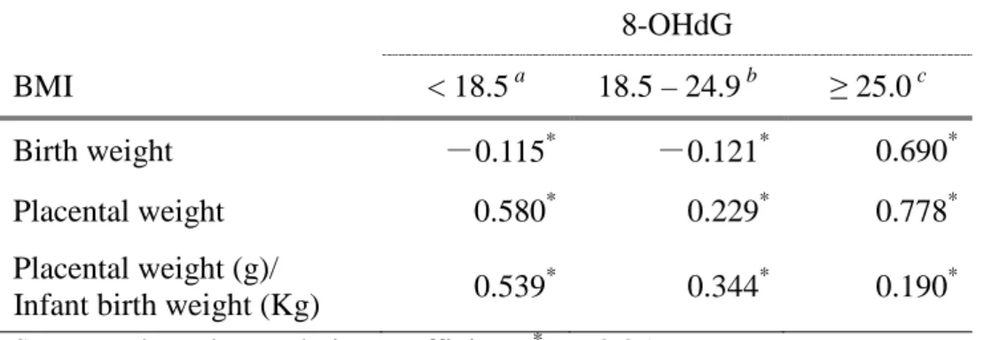
The correlation between the maternal BMI values and 8-OHdG levels was analyzed using Spearman's rank correlation coefficient (Table 3). Consequently, the levels of 8-OHdG in the BMI 18.5–25 group were significantly positively correlated with the placental weights per infant birth weight. Similarly, the 8-OHdG levels were correlated
300 600 900
0.0 0.4 0.8 1.2
100 150 200 250
0.0 0.4 0.8 1.2 [A] [B]
8 -OHd G l ev el s ( n g /m L )
Placental weight (g) r = 0.343
P = 0.007
8 -OHd G l ev el s ( n g /m L )
Placental weight (g)/Infant birth weight (Kg) r = 0.368
P = 0.004
with the placental weights in the BMI ≥ 25 group.
Table 3. Correlations between the 8-OHdG level and infant/placental weight 8-OHdG
BMI < 18.5
a18.5 – 24.9
b≥ 25.0
cBirth weight
-0.115* -0.121*0.690
*Placental weight 0.580
*0.229
*0.778
*Placental weight (g)/
Infant birth weight (Kg) 0.539
*0.344
*0.190
*Spearman’s rank correlation coefficient:
*P < 0.05.
a
n=10,
bn=42,
cn=8.
4. Discussion
All CB samples used in the present study were collected after full-term vaginal deliveries. No pregnancy complications were observed, including fetal growth restriction (FGR). The CB 8-OHdG levels in the new-borns with singleton vaginal deliveries ranged from 0.11 to 1.19 ng/mL (Table 2). Forlenza et al. reported that the serum 8-OHdG levels are much lower than the urine 8-OHdG levels. In their study, the serum 8-OHdG levels were approximately 0.20–1.26 ng/mL in healthy adults.
55)The values obtained in the present study were approximately equal to the levels detected in healthy adults.
Additionally, Schulpis et al. compared the 8-OHdG levels in CB between new-borns with vaginal and cesarean section deliveries
56)and found that the mean 8-OHdG levels were 0.25 ng/mL and 0.27 ng/mL, respectively, which indicates that there are no significant differences between delivery modes. In the present study, no significant associations were observed between these factors. Because no comparisons were made between the 8-OHdG levels in maternal blood and those in CB in the present study, is not possible to draw a definitive conclusion based only on the present data.
The present results showed that the 8-OHdG levels increased in association with the
placental weight in the maternal BMI ≥ 25 group (Table 3, Fig.7). The placenta is the
primary source of maternal oxidative stress during normal pregnancy, and increased lipid peroxidation is a normal phenomenon in pregnancy.
70)In addition, obesity during pregnancy is considered to be a high-risk state because it is associated with many complications.
71)Fujimaki et al. reported that oxidative stress may also contribute to the development of FGR.
67)Meanwhile, the placental tissue is not the only source of ROS, as pregnancy is a pro-inflammatory state associated with an increased number of granulocytes and, consequently, a decreased number of lymphocytes and monocytes.
70)Additionally, the expression of adhesion molecules on the cell surface of circulating leukocytes indicates the activation of leukocytes, resulting in the release of cytokines, which may lead to the generation of superoxide and other ROS as part of the immune response.
72)Considering the above points, the present results suggest the possibility that excessive growth of the placenta associated with maternal obesity increases the oxidative stress of the fetus. However, since the present study was conducted based on hospital records, we were unable to obtain full information regarding the mothers’ diets (nutritional status), including food and beverages, affecting the fetal environment.
Therefore, it was not possible to identify the relationships between maternal lifestyle-related factors and the 8-OHdG levels in CB. More precise approaches are required to clarify the relationships between the 8-OHdG levels and maternal obesity.
More than 95% of the oxygen taken in during breathing is ultimately converted into
water or reduced via hydroxylation reactions by drugs. The remaining fraction of oxygen
is not returned to the environmental air, thus leading to the formation of ROS. Essentially,
an unborn child is exposed to an extremely low oxygen environment. Meanwhile, oxygen
is essential for fetal development, and hypoplasia and placental dysfunction may delay
fetal growth. In cases involving a high ratio of placental weight per infant birth weight,
oxygen is readily supplied from the mother; thus, large amounts of ROS may be
generated. Although further studies should be performed, the correlation shown in Figure
1 suggests the possibility that ROS produced by the placenta contribute to the generation
of 8-OHdG, depending on factors of the maternal physique, such as obesity and BMI.
The relationships among the CB 8-OHdG levels, oxidative stress and unborn child
growth/development have not yet been sufficiently studied. It has been demonstrated that
the intrauterine environment has an effect on the development of future diseases in the
child. Therefore, determining the state of intrauterine oxidative stress and its effects on
the unborn child is extremely important for disease prevention. More precise approaches
are required to elucidate the relationships between the external environment, maternal
lifestyle factors and fetal oxidative stress.
結 論
1.本研究の結論
本研究は、単胎妊娠、正期産で経腟分娩により出生した健康な児を対象に臍
帯血中の
8-OHdG
を測定し、妊娠・分娩に関与する各種母子因子との関連性について検討した。得られた結論を以下に述べる。
1)
臍帯血中の8-OHdG
は0.1~1.39 ng/mL
であり、子宮内の胎児も一般成人と 同程度の酸化ストレス状態あることが示された。母体の喫煙群では、非喫煙 群に比較し臍帯血中の8-OHdG
値は高く有意差が認められ(0.33 vs. 0.42, P <0.05)
、母体の喫煙は胎児の酸化ストレスを増大させる因子となる可能性が示唆された。
2)
臍帯血中の8-OHdG
は0.11~1.19 ng/mL
であり、第1章と同様の結果が得ら れた。胎盤重量及び出生体重1kg
当たりの胎盤重量との間に正の相関を認め られ(r = 0.343, P = 0.007, r = 0.368, P = 0.004)、子宮内で順調に発育した正 期産児では、胎盤の発育は胎児の酸化ストレスを増大させる可能性が示され た。母体「過体重/肥満」群では、臍帯血中の8-OHdG
と胎盤重量に強い正 の相関が認められ(r = 0.778, P < 0.05
)、肥満妊婦では、胎盤の増大は胎児 の発育を促進する一方、酸化ストレスを増大させる可能性があることが示唆 された。3)
臍帯血中の8-OHdG
は、母体の年齢、在胎週数、児の性別、分娩所要時間や 分娩時の児の状態などに関する要因との関連性は認められなかった。2.今後の課題
胎児期における酸化ストレスの評価は十分とは言えない現状であることから、
本研究で得られた臍帯血中の
8-OHdG
値は、胎児の酸化ストレスを評価する上 で基礎的データと成り得る。しかし、正期産で子宮内の発育が良好かつ出生時 の状態が良好な児を対象としたこと、助産録および分娩経過記録にある母体の 基本情報を基に後方視的に解析した為、児の健康状態や疾病との関連性、母体の具体的な生活因子との関連性は明らかにされていない。
本研究の対象には、分娩中に母体へ酸素投与が行われた事例が含まれていた。
基礎的な解析において、酸素投与と臍帯血中の
8-OHdG
とには関連性は認めら れなかったが、酸素投与時間や酸素投与時の母体の酸素飽和度など不明であっ たため十分な解析には至らなかった。胎児が発育する子宮内は、子宮外に比べ 低酸素状態にあり、分娩によって胎児は高酸素環境へ曝される事になり、酸化 ストレスを受けやすい状態にあることが考えられる。また、早産児に対する高 濃度の酸素療法が、その後の後遺症と関連することが知られていることからも、分娩時の酸素投与が児に及ぼす影響について明らかにする必要がある。
わが国においては、高齢妊娠の増加に伴い、肥満妊婦・妊娠高血圧症候群・
妊娠糖尿病または糖尿病合併妊娠、早産、分娩中の胎児機能不全の増加が予測 される。また、食生活の欧米化、女性の喫煙率の増加、環境汚染などにより母 体および胎児の酸化ストレスの増大が懸念される。胎児期の酸化ストレス関連 因子の解明は、生まれてくる子どもの将来の疾患を予防するために有益な情報 になることが期待される。
謝 辞
本博士論文は、筆者が弘前大学大学院保健学研究科博士後期課程において行っ た研究をまとめたものです。本研究を遂行するにあたり終始暖かい激励とご指導、ご 鞭撻を賜りました弘前大学大学院保健学研究科 医療生命科学領域 放射線生命科 学分野 柏倉幾郎教授に深く感謝申し上げます。
本研究において、臍帯血中の
8-OHdG
測定にご協力下さいました弘前大学大学院 保健学研究科・門前暁助教、吉野浩教助教、放射線医学総合研究所 羽澤勝治研究 員、弘前大学大学院保健学研究科・博士後期課程 山口平様に心より感謝申し上げま す。また、臍帯血採取にご協力いただきました、独立行政法人国立病院機構 弘前病 院 産婦人科 尾崎浩士先生およびスタッフの皆様に心より感謝申し上げます。最後に、本研究の実施にあたり、貴重な臍帯血をご提供下さいましたお母様方に、
深く感謝申し上げます。
引用文献