胸部大動脈手術における間歇的逆行性脳灌流法の
有用性についての実験的検討
明 石 興 彦,大 澤 宏,吉 井 新 平,
多 田 祐 輔,松 本 雅 彦
山梨大学医学部第 2 外科 要 旨:【目的】現在大動脈弓部手術における補助手段として,超低体温循環停止法,順行性選択 的脳灌流法,持続的逆行性脳灌流法などが主に用いられているが,未だ確立された方法はない。わ れわれは間歇的逆行性脳灌流法を導入し,良好な臨床成績を得ている。今回,本法の有用性を実験 的に検討した。 【方法】家兎 24 羽を循環停止群,持続的逆行性脳灌流群,間歇的逆行性脳灌流群,対照群の 4 群に 分類し,近赤外線分光法による脳組織酸素化率(SdO2)の持続測定,および実験後の脳組織内水 分含有率により比較検討した。 【結果】SdO2比率(循環停止直前値からの比率)は,間歇的逆行性脳灌流群が持続的逆行性脳灌流 群より低下しにくい傾向を示した。復温時の SdO2比率の最高値も,間歇的逆行性脳灌流群が持続 的逆行性脳灌流群より有意に高値を示した(p < 0.01)。脳組織内水分含有率は,持続的逆行性脳 灌流群に比べ間歇的逆行性脳灌流群が低値を示した。 【結語】間歇的逆行性脳灌流法は,持続的逆行性脳灌流法よりも脳浮腫を軽度に抑え,逆行性灌流 時及び復温時における酸素供給が優れており,有効な脳保護法であることが示唆された。 キーワード 逆行性脳灌流法,超低体温循環停止法,胸部大動脈瘤,近赤外線分光法,脳浮腫 緒 言 現在,胸部大動脈手術に際しての補助手段と して,超低体温併用逆行性脳灌流法は広く臨床 応用され良好な成績が報告されている。この場 合一般的には持続的逆行性脳灌流法が用いられ ている。当教室では 1999 年より,脳浮腫軽減 を目的に,逆行性脳灌流と脱血を繰り返し行う, 間歇的逆行性脳灌流法に変更し,良好な成績を 得ている。 今回われわれは,間歇的逆行性脳灌流法の有 用性を調べるため,空間分解法を応用した近赤 外線分光法を用いて脳組織酸素化率を,さらに 術後脳組織含有水分量を測定することにより実 験的に比較検討した。 材料および方法 実験には,JW 種家兎 24 羽を使用し,超低体 温循環停止群(Hypothermic circulatory arrest: HCA 群 , n = 6), 持 続 的 逆 行 性 脳 灌 流 群 ( Continuous retrograde cerebral perfusion:CRCP 群,n = 6)
,間歇的逆行性脳灌流群(In-termittent retrograde cerebral perfusion: IRCP 群,n = 6),対照群(Control 群,n = 6)の 4 〒 409-3898 山梨県中巨摩郡玉穂町下河東 1110
受付: 2005 年 4 月 27 日 受理: 2005 年 4 月 27 日
群に分類した。 1.麻酔・体外循環 吸入麻酔(GOH)にて麻酔導入後,気管内 挿管した。耳動静脈に乳酸加リンゲル液にて末 梢ルート及び動脈圧ラインを確保し,動脈圧を 持続モニターした。頭部の皮下組織を剥離しポ ケットを作成し,近赤外線プローブを埋め込み 持 続 的 に モ ニ タ ー し た 。 フ ェ ン タ ニ ル 30µg/kg,臭化ベクロニウム 0.12 mg/kg を静 注し,二酸化炭素分圧が 30 ∼ 45 mmHg とな るように調節呼吸とした。その後の麻酔及び循 環管理は,フェンタニル 9µg/kg/hr,ミダゾ ラ ム 1 . 5 m g / k g / h r , 臭 化 ベ ク ロ ニ ウ ム 0.14 mg/kg/hr,ドパミンを持続静注し,平均 血圧を 45 ∼ 80 mmHg に保持した。 胸骨正中切開し,ヘパリン(1,000 IU/kg) 静注後,上行大動脈送血(16G),右房脱血 (12Fr)による体外循環を開始した。体外循環 回路は,ポンプは Sarns 社製のローラーポンプ, 人 工 肺 は TERUMO 社 製 CAPIOX SX を 用 い た。回路充填液は Control 群より採取した同種 他家全血及び乳酸加リンゲル液で行い,マニト ール液(1,000 mg/kg),重炭酸ナトリウム液 (1 mol/l)10 ml を添加し,ヘマトクリット 15 ∼ 20 %の希釈型体外循環とした。送血量は 100 ml/kg/min とし,α-stat 管理1)にて動脈血 酸素分圧が 150 mmHg 以上,二酸化炭素分圧 が 30 ∼ 45 mmHg となるように酸素流量を調 節した。また,pH を 7.3 ∼ 7.5 に保つよう重炭 酸ナトリウム液を適宜投与した。中心冷却は送 血温と直腸温の差を 10 °C 未満に保持し,20 °C で循環停止とした。逆行性脳灌流の送血温は 15 ∼ 18 °C とした。 2.脳保護法 ・ HCA 群 直腸温 20 °C にて,45 分間の超低体温循環停 止とした。 ・ CRCP 群 循環停止後,脱血管を遮断し,両側顎静脈よ り酸素加血を 7 ml/kg/min で逆行性に 90 分間 灌流した。その間,大動脈の送血ラインは大気 開放とし脳灌流血を回収した。灌流終了後 30 分間バイパス加温した。 ・ IRCP 群 循環停止後,まず脱血を行いリザーバーに 40 ∼ 50 ml/kg 貯血した。脱血管を遮断し,酸 素加血を 7 ml/kg/min で両側顎静脈より逆行 性に灌流した。回路血がなくなると右房及び両 側顎静脈より脱血を行った。これを 90 分間繰 り返した。この間,大動脈の送血ラインは大気 開放とし脳灌流血を回収した。灌流終了後 30 分間バイパス加温した。 ・ Control 群 全身麻酔下に他家血輸血のため右心房より脱 血後,塩化カリウムにて心停止とし,直後に脳 組織を採取した。
3.脳組織酸素化率(SdO2: the oxygen satu-ration obtained with the method of DIFFU-SION EQUATION)の測定
近赤外線分光測定法(NIRS: Near Infrared Spectroscopy,島津製作所製 OM-220)の浅部 用プローブを頭頂部の皮下に埋め込み,光拡散 方程式に基づいた空間分解法(SRS: spatially resolved spectroscopy)により酸素化ヘモグロ ビ ン ( oxy Hb), 脱 酸 素 化 ヘ モ グ ロ ビ ン
(deoxy Hb)の定量化指数,および SdO2: oxy
Hb/(oxy Hb+deoxy Hb)を連続モニターした。 比較検討には,主に SdO2比率(循環停止直前 値に対する比率)を用いた。 4.脳組織水分含有率 採取した脳組織(半球)の重量を測定後,オ ートクレーブにて 80 °C で 72 時間乾燥させ乾 燥重量を測定した。脳組織水分含有率は, (湿重量−乾燥重量)÷湿重量× 100(%) の式を用いて算出した。 5.統計学的検討 すべての結果は中央値(四分位値間)で示し た。2 群間の比較には中央値検定を,多群間の 比較検定には Kruskal-Wallis 検定を行い,群間 に差がみられた場合は,Tukey 検定で多重比較 を行った。危険率 0.05 未満で有意とした。
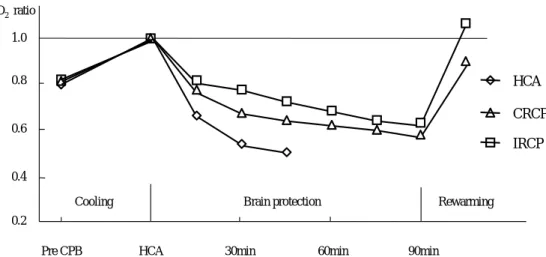
結 果 1.HCA 群・ CRCP 群・ IRCP 群の体重,循 環 停 止 時 の ヘ マ ト ク リ ッ ト 値 ( CRCP 群 ・ IRCP 群では逆行性脳灌流開始時と終了時の平 均により算出),体外循環開始から直腸温 20 °C までの冷却時間,並びに循環停止時の SdO2値 には有意差を認めなかった(Table 1)。 2.SdO2比率 体外循環開始前の SdO2比率は,HCA 群 0.79 (0.70 ∼ 0.88),CRCP 群 0.81(0.70 ∼ 0.91), IRCP 群 0.82(0.77 ∼ 0.93)で差はなく,3 群 とも体外循環による中心冷却に伴って上昇し た。循環停止とした後は,IRCP 群では逆行性 脳灌流を行っている間は上昇し,脱血時には低 下したが,3 群とも時間の経過とともにほぼ一 定の割合で低下した。復温時には,体外循環再 開で一旦上昇したが,体温が上昇するにつれ低 下傾向を示した(Fig. 1)。 循環停止 45 分後の SdO2比率は,HCA 群
Table 1. Characteristics of the experimental groups
HCA CRCP IRCP
Body weight (kg) 3.6(3.5 ∼ 4.0) 3.5(3.3 ∼ 3.6) 3.4(3.4 ∼ 3.6)
Hematocrit (%) 16.5(15.0 ∼ 18.0) 15.8(15.4 ∼ 17.5) 15.5(14.4 ∼ 16.3)
Cooling period (min) 34(23 ∼ 39) 34(29 ∼ 38) 33(30 ∼ 37)
The SdO2pre HCA (%) 72.6(63.8 ∼ 74.3) 62.9(59.0 ∼ 67.5) 64.0(58.1 ∼ 69.7) There were no significant differences between each groups. Values are shown as the median with interquartile ranges.
SdO2; the oxygen saturation obtained with the method of DIFFUSION EQUATION, HCA; hy-pothermic circulatory arrest, CRCP; continuous retrograde cerebral perfusion, IRCP; intermit-tent retrograde cerebral perfusion.
Change of SdO2 ratio
0.2 0.4 0.6 0.8 1.0
Pre CPB HCA 30min 60min 90min
HCA CRCP IRCP Rewarming Brain protection Cooling SdO2ratio
Fig. 1. The median SdO2ratio before the start of CPB was 0.79, 0.81, and 0.82 in the HCA, CRCP, and IRCP groups, respectively. There were no significant differences among the three groups. The SdO2ratio decreased during brain protection time and increased during rewarming time. CPB; cardiopulmonary bypass.
0.51(0.49-0.60),CRCP 群 0.64(0.51-0.86), IRCP 群 0.73(0.63-0.82)で,HCA 群と IRCP 群間には有意差を認めたが,CRCP 群と IRCP 群間には有意差を認めなかった(Fig. 2)。 復温時の最高値は,CRCP 群 0.86(0.67-1.05), IRCP 群 1.03(1.00-1.07)で,両群間に有意差 を認めた。特に IRCP 群では全例 1 以上に回復 した(Fig. 2)。 3.脳組織水分含有率 Control 群 77.4(76.0 ∼ 78.1)%,CRCP 群 79.8(78.9 ∼ 86.8)%,IRCP 群 78.2(77.4 ∼ 79.9)%であった。CRCP 群,Control 群間に は有意差を認めたが,IRCP 群,Control 群間 には有意差を認めなかった(Fig. 3)。 考 察 上大静脈から逆行性に送血することによる脳 保護法は,Mills ら2)の体外循環中の大量空気 塞栓への対処法に始まり,Lemole ら3)の間歇 的逆行性脳灌流法,三木,上田ら4-6)の超低体
温下間歇的逆行性脳灌流併用 open aortic tech-nique,持続的逆行性脳灌流法へと発展してお り,現在,弓部大動脈手術に際しての脳保護補 助手段として,順行性脳分離体外循環法と並び 広く臨床応用され,超低体温循環停止法の脳保 護有効時間を延長し,手術成績の向上,手術術 式の拡大に寄与してきた7-10)。しかし,一方で は持続的逆行性脳灌流法(CRCP)は,脳浮腫 に起因するせん妄をはじめとする中枢神経障害 を高率に伴うことが指摘され11),問題となっ ている。 静脈灌流圧と頭蓋内圧との間には正の相関が あり12),静脈から灌流を行う CRCP では,必 然的に頭蓋内圧が上昇する。また,静脈圧の上 昇は,血液脳関門の透過性亢進をきたし脳浮腫 を呈するとされ13),脳浮腫による毛細血管レ ベルでの血管床の圧迫がおこる。これらは脳灌 流を妨げ,時間とともに脳血流が減少する主た る要因になると考えられる14)。実際にマイク
SdO2ratio (45min)
0 0.2 0.4 0.6 0.8 1.0 IRCP CRCP HCA P < 0.05 0.73 0.64 0.51
Maximal SdO2ratio during the rewarming
0 0.2 0.4 0.6 0.8 1.0 1.2 1.4 IRCP CRCP P < 0.05 1.03 0.86
Fig. 2. Forty-five minutes after brain protection, the median SdO2ratio was 0.51, 0.64, and 0.73 in the HCA, CRCP, and IRCP groups, respectively. There was a significant difference between the HCA and IRCP groups, but not between the CRCP and IRCP groups.
The median maximal SdO2ratio during rewarming was 0.86 and 1.03 in the CRCP and IRCP groups, respectively. There was a significant difference between the two groups. In all animals of the IRCP group, the maximal SdO2recovered.
ロスフェアーを用いた CRCP 時の脳血流分布の 測定では,時間経過とともに脳血流が低下し, CRCP 開始 5 分後では生理的脳血流量の 20 %, 30 分後では 5 %程度に低下したと報告されて いる15)。また,定常流よりも拍動流のほうが CRCP の際の脳浮腫を軽減したことも報告され ている16)。これらを鑑みると,間歇的逆行性 脳灌流法(IRCP)は CRCP よりも灌流血静脈 圧の減圧により脳浮腫の進行を抑制し,さらに 持続定常流による灌流状態に介入することで, より有効な脳灌流を得られると推測できる。実 際に,われわれの臨床応用での成績は良好であ り,また豚を用いた慢性実験のモデルでは17), 逆行性脳灌流時の血液の出納量や術後の行動解 析により,IRCP が CRCP よりも脳浮腫を軽減 することが報告されている。今回の研究でも, 本法は CRCP よりも術後の脳浮腫を抑制し,脳 組織の酸素供給も良好で,その優位性が証明さ れた。さらに,本法の逆行性灌流維持に必要な 輸液・輸血量は,CRCP に比べ少量であった。 復温早期の SdO2が循環停止直前値以上を示 したのは,逆行性脳灌流の送血温が循環停止直 前の送血温より若干低かったためであろうと推 測される。またこのことは IRCP の与える悪影 響が軽微であったことを示している。復温早期 の SdO2は循環停止直前値とほぼ同じ値であ り,その高い再現性は,SdO2の指標としての 有用性も示唆した。近赤外線分光法の原理は, 700 ∼ 1,200 nm の波長の近赤外線が可視光と 比べ散乱しにくく,また頭部では,ヘモグロビ ン,チトクロームオキシダーゼ等の限られた色 素によってのみ減衰することを利用し,それら の吸光度を測定することで脳組織内の濃度を算 出するものである。OM-220 は,一方から 2 波 Fig. 3. The median water content of the cerebral tissues was
77.4%, 79.8%, and 78.2% in the control, CRCP, and IRCP groups, respectively. There was a significant dif-ference between the CRCP and control groups, but not between the control and IRCP groups.
Water content of cerebral tissue
80
90
70
IRCP
CRCP
Control
P < 0.01
(%)
N.S.
78.2
79.8
77.4
長の近赤外線を照射し,距離の違う二つの点で 光の強度を測定し,光拡散方程式を用いた連立
方程式を解くことにより18,19),酸素化ヘモグロ
ビ ン ( oxy Hb), 脱 酸 素 化 ヘ モ グ ロ ビ ン
(deoxy Hb)の定量化指数,および SdO2: oxy
Hb/(oxy Hb+deoxy Hb)を持続的に表示する。 しかしながら,この理論では生体の散乱係数を 一定として計算しているため,直接異なった個 体同士を比較するのは妥当でなく,あくまで同 一個体での相対値として扱うのが適当である。 このため,本研究では循環停止直前の SdO2値 を基準値とし,基準値に対する比率にて検討し た。近赤外線分光法による SdO2の測定は,頭 蓋内に浸襲を加えないため,脳代謝,脳浮腫及 び脳灌流などに影響を与えず,また,今回用い たプローブの計測する 1 ∼ 1.5 cm の深さの脳 酸素組織飽和度は,兎の脳全体の脳組織飽和度 を反映すると考えられ,非常に有用であった。 なお,今回の研究で灌流モデルに用いた家兎 は,内頸静脈系が未発達であり,さらに外頸静 脈系には静脈弁が複数存在するため,人と同様 の上大静脈送血では逆行性脳灌流のモデルとし ては不適当である。本研究では,過去の犬の研 究で静脈弁の影響を最小限にすることが可能で あると報告されている12)両側顎静脈からの送 血とし,逆行性灌流時には,その中枢側及び両 側外頸静脈を遮断した。また下大静脈の遮断は 行わず,奇静脈の結紮も行わなかった。 結 論 ① 間歇的逆行性脳灌流群は超低体温循環停 止群に比べ循環停止時の脳組織酸素化率 が有意に高く保たれた。 ② 間歇的逆行性脳灌流群と持続的逆行性脳 灌流群との脳組織酸素化率の比較では, 逆行性脳灌流時には有意差を認めなかっ たが,復温時には間歇的逆行性脳灌流群 が有意に高値を示した。 ③ 脳組織水分含有率は,間歇的逆行性脳灌 流群と対照群の間に有意差を認めなかっ たが,持続的逆行性脳灌流群は対照群よ り有意に高値を示した。 以上より,間歇的逆行性脳灌流法は持続的逆 行性脳灌流法よりも脳組織への酸素供給及び脳 浮腫の面から有利であり,有効な脳保護法であ ることが示唆された。 本研究は山梨大学動物実験委員会の承認を得 て行われた。 引用文献
1) Swan H. The importance of acid-base manage-ment for cardiac and cerebral preservation dur-ing open heart operations. Surg Gynecol Obstet.
158: 391–414, 1984.
2) Mills NL, Ochsner JL. Massive air embolism dur-ing cardipulmonary bypass. Causes, prevention, and management. J Thorac Cardiovasc Surg. 80: 708–717, 1980.
3) Lemole GM, Strong MD, Spagna PM, Karmilow-icz NP. Improved results for dissecting aneurysms. Intraluminal sutureless prosthesis. J Thorac Cardiovasc Surg. 83: 249–255, 1982. 4) 三木成仁,上田祐一,楠原健嗣ほか.間歇的逆
行性脳灌流下 Open aortic technique による大動 脈弁閉鎖不全を伴った DeBakey Ⅲ型逆行性大動 脈解離に多する血行再建術.日心外会誌 17: 131–133,1987.
5) Ueda Y, Miki S, Kusuhara K, et al. Surgical treat-ment of aneurysm or dissection involving the as-cending aorta and aortic arch, utilizing circulato-ry arrest and retrograde cerebral perfusion. J Cardiovasc Surg 31: 553–558, 1990.
6) Ueda Y, Miki S, Kusuhara K, et al. Deep hypother-mic systehypother-mic circulatory arrest and continuous retrograde cerebral perfusion for surgery of aor-tic arch aneurysm. Eur J Cardiothorac Surg. 6: 36–41, 1992.
7) Reich DL, Uysal S, Ergin MA, Griepp RB. Retro-grade cerebral perfusion as a method of neuro-protection during thoracic aortic surgery. Ann Thorac Surg. 72: 1774–1782, 2001.
8) Coselli JS. Retrograde cerebral perfusion is an ef-fective means of neural support during deep hy-pothermic circulatory arrest. Ann Thorac Surg.
64: 908–912, 1997.
9) Okita Y, Ando M, Minatoya K, et al. Predictive fac-tors for mortality and cerebral complications in arteriosclerotic aneurysm of the aortic arch. Ann
Thorac Surg. 67: 72–78, 1999.
10) Usui A, Yasuura K, Watanabe T, Maseki T. Com-parative clinical study between retrograde cere-bral perfusion and selective cerecere-bral perfusion in surgery for acute type A aortic dissection. Eur J Cardiothorac Surg. 15: 571–578, 1999. 11) Okita Y, Minatoya K, Tagusari O, et al.
Prospec-tive comparaProspec-tive study of brain protection in total aortic arch replacement: deep hypothermic circulatory arrest with retrograde cerebral perfu-sion or selective antegrade cerebral perfuperfu-sion. Ann Thorac Surg. 72: 72–79, 2001.
12) Nojima T, Nakajima Y, Mori A, et al. Experimen-tal study of optimal perfusion pressure during retrograde cerebral perfusion. Nippon Kyobu Geka Gakkai Zasshi. 42: 1307–1314, 1994. 13) Tsuru Y. An experimental study on the
occur-rence of brain edema after retrograde cerebral perfusion. Jpn J Thorac Cardiovasc Surg. 46: 992–999, 1998.
14) Sakahashi H, Hashimoto A, Aomi S, et al. Tran-scranial Doppler measurement of middle cere-bral artery blood flow during continuous
retro-grade cerebral perfusion. Nippon Kyobu Geka Gakkai Zasshi. 42: 1851–1857, 1994.
15) Yamaki F. Gradual change in regional blood flow during retrograde cerebral perfusion. Tokyo Woman’s Medical J. 70: 377–385, 2000.
16) Nojima T, Mori A, Watarida S, Onoe M. Cerebral metabolism and effects of pulsatile flow during retrograde cerebral perfusion. J Cardiovasc Surg. 34: 483–492, 1993.
17) Anttila V, Pokela M, Kiviluoma K, et al. Intermit-tent retrograde cerebral perfusion during pro-longed period of hypothermic circulatory arrest: a study in a chronic porcine model. Scand Car-diovasc J. 34: 116–123, 2000.
18) Matcher J, Kirkpatrick P, Nahid K, Cope K, et al. Absolute quantification methods in tissue near infrared spectroscopy. Proc. SPIE. 2389: 486–495, 1995.
19) Quaresima V, Sacco S, Totaro R, Ferrari M. Non-invasive measurement of cerebral hemoglobin oxygen saturation using two near infrared spec-troscopy approaches. J Biomed Opt. 5: 201–205, 2000.
Intermittent Retrograde Cerebral Perfusion is an Effective Means of Cerebral Protection during Deep Hypothermic Circulatory Arrest
Okihiko AKASHI, Hiroshi OSAWA, Shinpei YOSHII, Yusuke TADA and Masahiko MATSUMOTO Departments of Second Surgery, Faculty of Medicine, University of Yamanashi
Abstract: Although continuous retrograde cerebral perfusion (CRCP) has been widely used, postoperative transient brain dysfunction sometimes occurs due to continuously elevated intracranial pressure. We have introduced intermit-tent retrograde cerebral perfusion (IRCP) and experimentally evaluated its cerebroprotective effect.
[Methods] Twenty-four rabbits were divided into control (n = 6), hypothermic circulatory arrest (HCA, n = 6), CRCP (n = 6), and IRCP (n = 6) groups. Circulatory arrest (at 20 °C) was done for 45 min in the HCA group, and for 90 min with CRCP or IRCP followed by 30 min rewarming in the CRCP and IRCP groups, respectively. IRCP was re-peated at 50 ml/kg with blood drainage into a reservoir and returned via the maxillary vein. The cerebral tissue oxy-gen saturation (SdO2) was monitored using near-infrared spectroscopy and the tissue water content was measured after the experiment.
[Results] During circulatory arrest, the median SdO2ratio (SdO2/pre-HCA SdO2) was 0.51, 0.64, and 0.73 in the HCA, CRCP, and IRCP groups, respectively. The SdO2ratio at rewarming was significantly higher in the IRCP (1.03) than in the CRCP (0.86) group. The cerebral tissue water content was 77.4%, 79.8%, and 78.2% in the control, CRCP, and IRCP groups, respectively.
[Conclusions] IRCP provides higher oxygenation for cerebral tissue, reduces cerebral edema, and is superior to CRCP for cerebral protection.
Key words: retrograde cerebral perfusion, hypothermic circulatory arrest, thoracic aortic aneurysm, Near Infrared Spectroscopy, cerebral edema