Change in Phototactic Behaviour with Growth of
Milkfish, Chanos chanos (FORSSKAL)
著者
KAWAMURA Gunzo, SHINODA Yasushi
journal or
publication title
南総研紀要
volume
1
number
1
page range
75-87
URL
http://hdl.handle.net/10232/15595
Change in Phototactic Behaviour with
Growth of Milkfish, Chanos chanos
(FORSSKAL)
Gunzo KAWAMURA* and Yasushi SHINODA*
Abstract
Phototactic behaviour of milkfish larvae and juveniles was observed in the sea and laboratory, and their retinae were observed histologically. Larvae collection by using a lamp at night and tank experiments revealed that the larvae have strong positive phototaxis. Throughout their growth, there were three shifts in phototactic behaviour in a tank. The photopositive behaviour of the larvae became weak by degrees through a metamorphic stage, and juveniles showed photonegative behaviour. On the 39th day from capture, their behaviour became photopositive again. After they were transfered into a larger tkank, on the 139th day, any significant photopotitive or photonegative behaviour could not be observed. The larvae on capture, have duplex retinae. The rods were found to be a few in number, and a clear retinomotor response was observed on the day of capture. The first change in behaviour from photopositive to photone gative seemed to relate with the development of the rods.
INTRODUCTION
It seems that the behaviour of fish larve in relation to light originally was observed
with special reference to depth distribution or vertical migration of the larvae. Based
on the larger catch of night hauls by silk tow net rather than that of day hauls, it wasassumed that the larvae avoidod the surface waters when illfimination was high. It now
seems that diurnal migration of many larval fishes is not as extensive as previously surmised, and it has been shown that much of the difference between day and night catches is due to the ability of many species to avoid the slow-speed sampler in daylight (WOODHEAD, 1966). Based on the catch of a high-speed sampler, it was suggested that young herring avoided intensive surface light (BRIDGER, 1958). On the other hand, it had been suggested that the depth of vertical distribution of the larvae of herring and
Sardinops caerulea varied with larval size (BRIDGER, 1958). KELLY and BARKER (1961)
similarly showed the vertical distribution of the larvae of Sebastes marinus. Although it might have been expected that light would play a dominant role in governing the depth distribution of different species or stages, the critical studies on fish larval distri
bution are still few.
* nmwm • affljf i ')m-k^m^msi^^m.
76 KAWAMURA and SHINODA : Change in Phototactic Behaviour of Milkfish
From the experiments with larval fish, it was revealed that their reactions to light changes with time (HARDER and HEMPEL, 1954). The change in reaction to light probably relate to ontogenical development of the visual system. In this paper, we report the change in phototactic behaviour with growth of the milkfish, Chanos chanos (FORSSKAL), and relationship between the change and the histological development of
the retina.
MATERIALS AND METHODS
The experiments were done in the field and laboratory during 1979~1980. FIELD EXPERIMENT Larvae collection was carried out along the shore by using a fish lamp. The collection ground was located over a shallow, gently sloping, sandy shore in Kumano Bay, Tanega Island in Kagoshima Prefecture, which had been reported earlier by SENTA et al (1980) as a ground wherein numbers of milkfish larvae can seasonally be captured. A kerosene lamp (350cp) was hung at about 50cm above the water surface from a pole fixed in waist-deep water, and its position was shifted to shore or off shore depending on the tide. After dusk, the larvae were attracted by light and were collected with a bagnet of 1mm mesh size netting with a frame, 62cm deep by 136cm long, attached to the net-mouth. This gear was hand-operated by two wading men for four minutes in the illuminated area (test site) and in a dark area 50-75m away from the lamp (control site). The towing speed was about 70cm/s. The milkfish larvae thus captured in the bagnet were selected from the other larvae and were counted on the beach. The number of the larvae captured was compared statistically between the
test and control site.
LABORATORY EXPERIMENT 250 milkfish larvae captured near shore in day light on July 30, 1979 were transported to the laboratory of the Faculty- of Fisheries, with 50 % sea water. After starving for two days from capture they were fed on cooked egg yolk, carp pellets, and algae which grew naturally in the tanks. One hundred larvae were kept and reared in an experimental tank (170cm long, 34cm wide, 20cm water deep) for the phototaxis experiment. The two long sides of the tank were transparent; the other walls and bottom were opaque and grey in color, of which the inside surface was coarsely ground to prevent a mirror light reflection.
As the animals were found to respond more strongly to a vertical light beam than to a horizontal one, a miner's type flashlamp with a 1.5W bulb was fixed vertically at a point 15cm from a side wall and 20cm above the water surface. In the evening the
room was darkened and the animals were acclimated to the darkness for more than
two hours prior to switching on the lamp. At 5, 10, 15 min after the onset of illumination, the number of animals in the circle of 30cm diameter beneath the lamp was counted. At 20min after the onset, the lamp was moved horizontally toward the opposite wall and was returned to the initial point at the speed of 1.4m/min, and the response of the animals to the moving light beam was observed and the number of
animals which followed the lamp was counted.
A quantitative indication of the phototactic behaviour was given as the amplitude of response to the light (ARL) which is defined thus :
where, N5, N,„, and N15 are the number of animals which stayed in the circle beneath
the lamp at 5, 10, and 15min after the on set of illumination, Score is the response of
the animals to the horizontally moving lignt and fixed light which was scored thus:
Response
Animals were strongly attracted to the fixed light and
followed well to the moving light 20
Significant but weak positive response to the fixed light
was observed and the durability of the following 10 reaction to the moving light was weak
No remarkable positive or negative response to the fixed and moving light was observed
Animals did not respond to fixed light but showed remarkable negative response to the moving light Animals responded negatively to both fixed and moving
light
Therefore the value of ARL can very between 100 and -20. On December 16, on
the 139th day from capture, all animals in the experimental tank were transferred
into a larger experimental tank (4m long, 44cm wide, 20cm water deep) made of opaque
vinyl chloride plate, and observations were made until January 11, 1980.
When animals died in the experimental tank, they were replaced by animals from
the rearing tanks. The water temperature in the experimental tank was kept constant,
but ranged from 28.0 to 30.7°C
On the day of capture and every two or four days after capture, light adapted and
dark adapted animals were preserved for histological observation of the retinae. The
light adapted animals were randomly sampled from the rearing tanks in daylight. The
dark adapted samples were the specimens kept in a dark tank for more than two hours
in the evening, and killed and fixed in BOUIN'S solution. Generally, the eyes were
sectioned at 6 microns with the lens intact and stained by the H. E. or Azan methodfor light microscopy.
It was very difficult to resolve the rods by light microscopy if they existed. There
fore, the appearance of the rods was observed by calculating the ratio of countable
visual cells (which will be cone) to the visual cell nuclei in the outer nuclear layer.
The retinae of adult fish, which were obtained at the Aquaculture Department of Southeast Asian Fisheries Development Center in the Philippines, were fixed in Orth- 1 0
- 2 0
solution for 24h, washed in running tap water for 24h, and were preserved in 70 %lor z,tn, wasueu in running tap waiei iui i-rn, anc* m-n. pi^a^^y^^ m , J '
ethanol.
RESULTS
LARVAE COLLECTION The total number of all fish larvae captured by 21
operations was 2256 individuals in the test site and 3117 in the control site. The total
78 KAWAMURA and SHINODA : Change in Phototactic Behaviour of Milkfish
the control. The statistical test of the difference of the catch between the test and control, shown in Table 1, reveals that more larvae were captured in the test site than
in the control (average of the difference, 2.75 ; confidence limit of the avarage at 99.9/
level, 0.84 —4.65). Although the catch was poor, it is evident that the larvae can beTable 1. Result of larvae co Uection. Number of catch
Date
Test site Control Difference
July 5-6 4 1 3 3 0 3 6 - 7 1 0 1 1 0 1 5 0 5 7 - 8 0 I - 1 4 0 4 0 0 0 2 2 0 4 0 4 1 0 1 1 1 0 8 - 9 2 0 2 2 0 2 4 0 4 2 1 1 5 1 4 30 8 0 8 6 0 6 31 2 0 2 5 0 5 Total 62 7 55 Average 2.75 S. E. ±0.50 Confidence• limit of the average iaf the differences at 99.9/ level : 0.84--4.65
attracted by an artificial light at night. They have positive phototaxis.
GROWTH IN TANK When the larvae were introduced into the tank on the
first day of August, they swam continuously, forming one or two loose schools very close
to the bottom, and showed no preference for any particular area in the tank. In the evening, they dispersed horizontally and vertically under the illumination of the room
lamp. Next day, they showed strong feeding activity on cooked egg yollc and seemed
very healthy in the thank.
The larvae on capture had densely pigmented eyes, transparent bodies, and no pelvic
fins. On the seventh day from capture, all specimens had pelvic fins. On the 18th day,
the finfold completely disappeared in all specimens. Following KAWAMURA and HARA
(1980a), the authors here named this period, from the appearance of the pelvic fins until
the complete disappearance of the finfold, the "metamorphic stage". In the Philippines,
the metamorphic stage starts on the 5th day from capture and lasts until the 15th day
The metamorphosis which took place in our laboratory delayed by the two-day starvation
period following capture. After the metamorphic stage they showed strong synchronized
movements. Although their feeding was very active, they grew with extremely lower
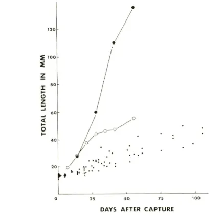
growth rates than the published data (SCHUSTER, 1952 ; RABANAL et al, 1953)(Fig. 1).
According to HUNTER (1977) and HUNTER and KlMBRELL (1980), the growth of
5 100 S o z < o • ♦ 0 25 50 75 100
DAYS AFTER CAPTURE
Fig. 1. Growth of the milkfish reared in different conditions.
Large closed circle ;
average growth at a rate of one fish/m2 (from SHUSTER, 1952), open circle ; at
a rate of 10-100 fish/m2 (from RABANEL et al, 1953) ; small closed circle ; present data at a rate of 173 fish/m2.northern anchovy and Pacific mackerel is accelerated by high temperaures, while meta
morphosis is delayed at lower temperatures.
This seems general in other temperate
fishes as well. Because there is no available data on the growth of milkfish under natural conditions, we could not compare the growth of reared milkfish with wild ones. Hence
we here used the day of capture and metamorphosis as a reference stage to show age of
the animals used.
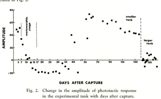
PHOTOTACTIC BEHAVIOUR IN TANK On the fourth day from capture,
when the lamp was turned on, the larvae rushed into the illuminated area and stayed
there for a long period. They so seldom or slowly moved in the light spot that it was
very easy to count the numbers of the animals attracted in the area. When the lamp
was slowly moved, the animals in the bright area obediently moved with the moving
light spot, and the ARL became around 50. This strong photopositive behaviour could
be observed for three consecutive days.
80 KAWAMURA and SHINODA ! Change in Phototactic Behaviour of Milkfish
by day and disappeared on the 11th day. On the 14th day, whch the lamp was turned on, the animals immediately swam away from the bright area. When the lamp was moved, the animals swam forward keeping a certain distance from the moving light spot. Finally, a part of them were driven by the moving light to the opposite wall. The strong phototaxis of the larvae complety turned to negative through the metamorphic stage, and on the 18th day, the ARL showed minimum value. A similar photonegative behaviour
lasted until the 37th day.
On the 39th day, they showed photopositive behaviour again. They darted into the bright zone and moved with the moving light spot taking very high ARL values. This strong photopositive behaviour lasted until the 136th day, although they responded negatively to the light on the 43rd day.
On the 139th day, all animals in the experimental tank were transfered into a larger experimental tank. In the larger tank, they seldom formed a school. At night, individual animals scattered and were resting motionless on the bottom and the strong photopositive behaviour shown in the smaller tank completely disappeared. Immedi ately after the onset of illumination, the animals showed negative responses. A few minutes after that, some of them came into the bright zone, stayed there for a moment, then swam sway again. They repeated such behaviour. No remarkable response to the moving light could be observed. In the larger thank, the ARL largely fluctuated
between 11.0 and -10.0.
The over-all results of the phototactic behaviour observed in the tanks are illus
trated in Fig. 2. a 3 Q. < a. E o ' . ' smaller tank
• • - .
_i_i_ _i_ 5 7 10 • IS 17 20 130 • • •DAYS AFTER CAPTURE
Fig. 2. Change in the amplitude of phototactic response in the experimental tank with days after capture.
larger tank
-A. so
'it
HISTOLOGY OF THE RETINA The larvae on capture had dense ganglion
cells and cells in the inner nuclear layer, but the number of nuclei of the visual cells was very small. For the adults, on the other hand, the former two cells distribute in very low density, while the density of the nuclei of visual cells was very high (Fig. 5). It seems, therefore, that the cells in the inner nuclear layer and the ganglion cells are already formed in the larval stage and disperse tangentially with retinal growth.
dark adapted retina, the masking pigment retreated to the periphery of the retina, the
cone myoids extended and the rod myoids contracted (Fig. 6). The pigment index (ALI,
1959) for the dark adapted retinae gradually decreased with growth of the layer of
pigment epithelium and visual cell layer, but for the light adapted retinae, after the
seventh day, it was almost constant (Fig. 3).All retinal elements, except for the cells in the outer nuclear layer, did not seem to
undergo large changes in development for two months after capture. The density of
the cells in the outer nuclear layer underwent a large change within 12 days. The ratio
between cone elipsoid and the nuclei ranged from 1.4 to 2.1 in the larvae on capture,1.6 to 2.3 on the seventh day, 2.3 to 2.9 on the 10th day, and 2.6 to 3.3 on the 12th
<
*
2&-S 10 15 20
DAYS AFTER CAPTURE
Fig. 3. Ratio between the cone elipsoids and the unclei with days after capture. Arrows show metamorphic stage.
z
| o.
30 o o o 0 c
» .
10 20 30 40 50 60
DAYS AFTER CAPTURE
Fig. 4. Pigment index with days after capture. Open circle ; light adapted, closed circle ; dark adapted.
day. After the 12th day, the ratio did not largely increase until the 63th day (Figs. 4,
6-9).82 KAWAMURA and SHINODA : Change in Phototactic Behaviour of Milkfish
DISCUSSION
The larvae collection by use of fish lamp and the tank experiments revealed that the larvae which appear near the shore exhibit a storong positive phototaxis. This is supported by the fact that, in the Philippines, the larvae collectors use kerosene lamps at night for more efficient larvae collection. KUMAGAI et al. (in prees) carried out a tank experiment using the milkfish larvae and observed strong photopositive behaviour of the
larvae.
According to BLAXTER (1975), the larvae of herring, pilchard, haddock, sand goby, plaice, and lemon sole have pure-cone retinae, and the rods appear at metamorphosis. The milkfisk larvae, on the other hand, already have rods. The milkfish is not only one species which has rods in larval stage. The Japanese anchovy have developed grouped rods and dense retinal tapetum in their larval stage (KAWAMURA et al. in press).
The ratio of cones to the visual cell nulei underwent large changes through the metamorphic stage. This increase in the" ratio coincides with the first change of pho topositive to photonegative behaviour of the larvae. Therefore, it seems that the rod development relates to the first change in phototactic behaviour. However, the second and the third changes in phototactic behaviour can not be explained by the rod development.
YUSA et al. (1971) described a shift from photopositive to photonegative through metamorphosis in Limanda yokohama. A similar shift from photopositive to photo negative occurs in Acanthurus triostegus sandivicensis when they settle in tide pools (MALIAVE, 1977), and Anguilla vulgaris before they enter rivers (DEELDER, 1958). The change in phototaxis is probably a sort of adaptation to a new habitat. According to SCHUSTER (I960), milkfish larvae enter creeks, backwaters or esturies, and the juveniles remain there for an uninterrupted period of four years before trying to return to the sea ; but the period seems to vary depending on the capacity of the backwaters.
MARLIAVE (1977) noted that yolk-sac larvae of Anoplarchus purpurescens will become negatively phototactic if starved, but remained photopositive if fed from the time of hatching ; and warned that observation of behaviour of larvae in the laboratory must be interpreted cautiously if those larvae do not feed or grow. Even though the growth rate of the milkfish in the laboratory was very low, they actively fed on the foods supplied and appeared to be very healthy. Thus we cannot believe that the second
change from photonegative to photopositive will be attributed to very slow growth of
the juveniles in the tank. Their photopositive behaviour was very strong. It might be resolved by ecological observations whether the change was a natural behavioural change or an induced one caused by the rearing condition.BURI (1981) observed wild juvenile milkfish in a mangrove swamp in the Philippines,
and reported that during night schooling habit was more or less abandoned and indivi ual fish were found scattered and resting motionless over the substrate. These fish did not react to light or shadow movements. Ae he did not explain the size or age of the juveniles, we can not compare the behaviour observed in the tank directly with the wild ones observed in the mangrove swamp. Although the animals in the larger tank
were a little more active, their behaviour resemble that of the wild ones. Therefore
the behaviour obseved in the larger tank seems reliable, but we can not reject the
Only from the tank experiments, it would be very difficult to reveal the phototactic
behaviour of fishes. We should always refer the observations of wild fish in the field.We desire more ecological observations in the field. Moreover, the behaviour through
vision may relate not only to development of the eyes, but also to development of the
optic tectum. The optomotor reaction undergoes a change through the metamorphic
stage. The optomotor reaetion is also somewhat weak in the larvae, but becomes strong
and almost perfect in the juveniles (KAWAMURA and HARA, 198a). This change cannot
be attributed only to the development of the retina. The relationship,
if it exists,
between the development of the behaviour by vision and the optic tectum is a point
of interest which arises from the developmental study.
REFERENCES
An, M. A., 1959, The ocular structure, retinomotor and photobehavioural responses
of juvenile Pacific salmon. Can. J. Zoo., v. 37, p. 965 —996.BLAXTER, J. H., 1975, The eyes of larval fish, in "Vision in fishes" (ed. by All, M. A.),
Plenum Press, New York, pp. 427~443.BRIDGER, J. P., 1958, On efficiency tests made with a modified Gulf III high-speed tow
net. J. cons. perm. int. Explor. Mer, v. 23, p. 357-365.
BURI, P., 1981, Ecology on the feeding of milkfish fry and juveniles, Chanos chanos
(FORSSKAL), in the Philippines. This bull, v. 1, p. 25-42
DEELDER, C L., 1958, On the behaviour of elvers (Anguilla vulgaris TURT.) migrating
from the sea into fresh water. J. cons. perm. int. Explor. Mer, v. 24, p. 135-146.HARDER, W and HEMPEL, G., 1954, Studien zur Tagesperiodik der Aktivitat von
Fischen-I. Versuche an Plattfischen. Kurze Mitt. Inst. Fischdiol Univ. Hamb.,
no. 5, p. 22 —31.
HUNTER, J. R., 1977, Behavior and survival of northern anchovy Engraulis mordax larvae.
Calif. Coop. Oceanic Fish. Invest. Rept, no. 19, p. 138-146.
and KlMBRELL, C A., 1980, Early life history of Pacific mackerel, Scomber
japonicus. Fish. Bull, v. 78, no. 1, p. 62 —76.KAWAMURA, G. and HARA, S., 1980a, The optomotor reaction of milkfish larvae and
juveniles. Bull Japan. Soc. Sci. Fish., v. 46, p. 929-932.and
, 1980b, On the visual feeding of milkfish larvae and juveniles
in captivity, ibid., v. 46. p.1297- 1300.
KELLY, G. F. and BARKER, A. M., 1961, Vertical distribution of young redfish in the
Gulf of Maine. R-v. Reum. Cons. perm. int. Explor. Mer, v. 150, p. 220-233.MARLIAVE, J. B., 1977, Development of behaviour in marine fish.
Memorial Univ.
Mar. Sci Res. Lab. Tech. Rep., p. 240-267.
RABANAL, H. R., ESQUERRA, R. S. and NEPOMUCENO, M. N., 1953, Studies on the
rate of growth of milkfish, Chanos chanos FORSKAL, under cultivation. Proc.
Indo-Pacif. Fish. Coun., 4th meeting, no. 2, p. 171-180.SCHUSTER, W H., 1952, An annotated bibliography on the culture of milkfish Chanos
chanos FORSKAL. Occ. Pap. Indo-Pacif. Fish. Coun., v. 52/3, 24p., I960, Synopsis of biological data of milkfish Chanos chanos FORSKAL, 1775.
FAO Fisheries. Biology Synopsis, no. 4, 57p.
84 KAWAMURA and SHINODA : Change in Phototactic Behaviour of Milkfish
of milkfish, Chanos chanos (FORSSKAL) fry in southern Japan.
Bull Fac. Fish.
Nagasaki Univ., v. 48, p. 19-26.WOODHEAD, P. M. J., 1966, The behaviour of fish in relation to light in the sea.
Oceanogr. Mar. BioL Ann. Rev., v. 4, p. 337-403.YUSA, T, FORRESTER, C. R. and IlOKA, C, 1971, Eggs and larvae of Limmanda yokohama
(GUNTHER). Fish. Res. Bd. Can. Tech. Rep., no. 236, 21p.
Plate 1
Fig. 5. Gross sections showing the retinal elements of an adult milkfish. C ; cone, G ;
ganglion cell, N ; visual cell nuclear. 5A ; ventral retinal region, 5B ; dorsal
retinal region. Scale ; 50 microns.
Plate 2
Fig. 6. Retinae of larvae on capture. 6A ; light adapted, 6B ; dark adapted. C ;cone,
B ; bipolar and amacrine cells, C ; ganglion cell, N ; visual cell nuclear.Fig. 7. Retinae of larvae on the seventh day from capture. 7A ; light adapted, 7B ;
dark adapted. Scale ; 50 microns.
Plate 3
Fig. 8. Retinae of juveniles on the 15th day from capture. 8A ; light adapted, 8B;
dark adapted.
Fig. 9. Light adapted retina of a juvenile on the 63th day from capture. 9A ; dorsal
retinal region, 9B ; ventinal region. Scale ; 50 microns.Plate 1
iSii
fM^ •':•:•
m
HHUJ'•:••••••••
:.
5BKAWAMURA and SHINODA: Change in Phototactic Behaviour of Milkfish Plate 2 vtsiA 6A 6B M ^V.-^.-V^;.^.. *•*»*/.-. 7B
Plate 3
.v^^.t/;^;.:..*-;;-'/. jj. •
ilitlll
i^saM^MMjsfs -safe' „ ' * • ' .
: