INTRODUCTION
The incidence of colorectal cancer in Japan has increased over the past four decades, and it was sug-gested that this increase is linked to increased con-sumption of Western foods characterized by high-fat and less fiber diets. Such dietary habits are known
to be correlated with high fecal concentrations of bile acids. In agreement with this, epidemiological studies showed that fecal concentrations of bile acids were high in patients with colon cancer as well as in people from countries with a high incidence of colon can-cer (1, 2). Thus, all these findings suggest that a strong relation between fecal bile acids and human colon carcinogenesis. Bile acids have also been studied extensively in animal models of colon cancer (3-5). In our previous studies, the effects of human bile acids on azoxymethane (AOM)-induced aberrant crypt foci (ACF), which is a good surrogate marker for colon carcinogenesis, were investigated in the rat
ORIGINAL
Effect of ursodeoxycholic acid on azoxymethane-induced
aberrant crypt foci formation in rat colon :
in vitro
po-tential role of intracellular Ca
2+Mohammed Abdul Momen
1, Yasumasa Monden
1, Hitoshi Houchi
2and
Atsushi Umemoto
11
Second Department of Surgery, and2
Division of Pharmacy, University Hospital, The University of Tokushima School of Medicine, Tokushima, Japan
Abstract: The studies were conducted to examine the precise nature of the suppressive effect of ursodeoxycholic acid (UDCA) on colonic aberrant crypt foci (ACF) formation. Fischer 344 rats were treated with a single dose of azoxymethane (AOM) (20 mg/kg, s.c.) and fed basal diet (MF) supplemented with UDCA (0.4%) during an initiation or a post-initiation stage. ACF were enumerated at the 2nd, 5th and 8th weeks after AOM administration (15-18 rats/group). The number of ACF in the UDCA treated group was decreased significantly in the initiation and post-initiation stages at the 2nd (P<0.01, P<0.0001) and 8th weeks (P<0.001, P<0.0001), respectively, compared with untreated controls. In the time-course experiments, the effect of continuous feeding of UDCA (0.4%) on ACF formation was evaluated. ACF number was decreased significantly (P<0.005) until the 16th week. UDCA showed a significant dose-dependent suppression of ACF number from a range of 0.1-0.4% UDCA. To approach the subcellular mecha-nisms of the effect of bile acids, the intracellular free Ca2+concentration ([Ca2+]
i) of bile
acid-treated rat colonic cancer cells (ACL-15) was examined. DCA and CDCA, which are pro-motive on ACF formation, induced a rapid increase in [Ca2+]
i, while UDCA and CA, which
are suppressive or non-effective on ACF formation, did not. These findings suggest that the promotive effect of bile acids may involve intracellular Ca2+signaling.
J. Med. Invest. 49 : 67-73, 2002
Keywords : ursodeoxycholic acid, aberrant crypt foci, colon cancer, bile acid, intracellular Ca2+
Received for publication January 5, 2002 ; accepted January 31, 2002.
Address correspondence and reprint requests to Atsushi Umemoto, Second Department of Surgery, The University of Tokushima School of Medicine, Kuramoto-cho, Tokushima 770 - 8503, Japan and Fax : + 81- 88 - 633 -7144.
The Journal of Medical Investigation Vol. 49 2002
67
colon (6, 7). In these studies using dietary intake and intracolonic administration of bile acids, ursodeoxycholic acid (UDCA) showed a significant suppressive effect on ACF formation, whereas other bile acids, i.e. cholic acid (CA), deoxycholic acid (DCA), chenodeoxycholic acid (CDCA) and lithocholic acid (LCA), showed either promotive effects or no effect on the colonic mucosa. The preventive effect of UDCA on colonic tumor formation was also demonstrated in rats (5). Although controversies continue with respect to find-ing a correlation between the ACF parameters and tumor outcome, as a precursor lesion of the colon cancer (8, 9), it has been established that ACF is a good biomarker to assess the effects of colon carcino-gens as well as chemopreventive agents (10, 11). UDCA may be one of the hopeful chemopreventive agents for high-risk groups for colon cancer. In the present study, the effects of UDCA in different stages of colon carcinogenesis by AOM treatment were ex-amined in Fischer 344 (F344) rat colon and the time-course and dose-response of the effects of UDCA on ACF formation were also examined.
Although, the bile acids are greatly involved in the development of colon tumors and ACF, the exact mechanism of the bile acids action remains unclear. Increasing evidence suggests that colonic malignant transformation involves various mutations activating proto-oncogenes such as K-ras (12), inactivating tumor suppressor genes such as p53 and APC (13), and inactivating genes involved in DNA mismatch repair. On the other hand, epigenetic changes in sig-nal transduction elements, such as protein kinase C (PKC) may also be involved in multistage process-es of colon carcinogenprocess-esis (14, 15). Bile acids, which have various effects on colonic ACF formation, are also known activators of PKC that have been impli-cated in the development of colonic carcinoma (16). Since PKC is activated by intracellular calcium ions, it is possible to have an association between the ef-fect of bile acids and their intracellular calcium in-duction. It was also reported that calcium ions (Ca2+
) are a versatile mediator of many cellular biological processes, including cell proliferation (17). The free Ca2+
acts as a proliferative signal for many normal cells (18). Cytoplasmic calcium is also a pluripotent regulator of cell function including the cell cycle and apoptosis (19, 20). Thus, the agents that affect the intracellular calcium level, could be considered as important substances for cell functions, and we hy-pothesized that bile acid activity on cellular prolifera-tion leading to ACF formaprolifera-tion may be in part through the alteration of the intracellular calcium level.
There-fore, we aimed to assess the intracellular Ca2+
induc-tion in rat a colon cancer cell line (ACL -15) by bile acids, which are either promotive or suppressive/ ineffective in colonic ACF formation in rats.
MATERIALS AND METHODS
ChemicalsCA, CDCA and DCA were purchased from Sigma Chemical Co. (St. Louis, MO). UDCA was from Mitsubishi-Tokyo Pharmaceutical Co. Ltd. (Tokyo, Japan). All the bile acids were sodium salt forms and these were at least 97-99% pure according to the manufacturer’s indications. AOM was purchased from Sigma Chemicals Co. (St. Louis, MO). Fura-2-AMTM
was obtained from Dojindo Laboratories (Kumamoto, Japan). Micro-cover glass (13 mm in diameter) was obtained from Matsunami Glass Ind. Ltd. (Osaka, Japan).
Animals
Six-week-old, male, F344 rats were purchased from SLC Co. (Hamamatsu, Japan) and housed in polycarbonate cages with wood chips in a specific pathogen-free laboratory of our animal facilities, three rats/cage. They were kept under constant conditions of tem-perature (22±2℃) and humidity (55±5%) with a 13 hr light/11 hr dark cycle. All the rats were provided with laboratory chow basal diet (MF) (Oriental Yeast Co. Ltd., Tokyo, Japan) unless specified and the beds were changed twice a week. The arrival weight and food consumption of the animals were recorded.
Study design
Study-1(Initiation and post-initiation)
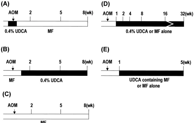
Male F344 rats (6 weeks old) were treated with a single dose of AOM (20 mg/kg, 10 mg/ml in sterile saline) subcutaneously. Rats were allocated to one of 3 experimental groups, the control and two UDCA groups (initiation and post-initiation, 15-18 rats/group). In the initiation group, the rats were fed MF (Orien-tal Yeast, Co., Ltd., Tokyo, Japan) supplemented with the UDCA (0.4%) ad libitum from four days before to three days after the AOM injection (for 1 week), and thereafter the rats were fed MF alone (Fig. 1 A). In the post-initiation group, the MF was changed to a UDCA-supplemented diet (0.4%) one week after AOM injection (Fig. 1B). The control group was fed the MF during the entire experimental period (Fig. 1C). Rats (n=5-7) were sacrificed at the 2nd, 5th and 8th weeks.
M. A. Momen et al. Effect of bile acids on colon carcinogenesis
68 M. A. Momen et al. Effect of bile acids on colon carcinogenesis
The isolated colons were collected and washed with 0.9% NaCl water solution. The lumen of the colon was filled with 10% phosphate-buffered formalin (pH 7.4) for 3 min. These were incised in a longitudinal direc-tion and fixed flat between two pieces of filter paper in the buffered formalin. The colons were stained with 0.2% methylene blue (Sigma Chemical Co., St. Louis, MO) and the number of ACF along the colon was counted by their position in every 2 cm under light microscope (21) from the anus to proximal colon.
Study-2 (Time-course)
In this study, the rats (6 weeks old) were treated with AOM as Study-1. One week later, the rats were divided into two groups and were fed ad libitum the MF diet supplemented with UDCA (0.4%) or MF alone, until the end of the experiment (Fig. 1D). Rats (n=4 -5) were sacrificed at the 2nd, 4th, 8th, 16th and 32nd weeks to count the colonic ACF.
Study-3 (Dose-response)
After treatment with AOM as a single dose and MF feeding for 1 week, the rats were divided into five groups (6 rats/group). From the beginning of the second week rats were fed ad libitum the MF sup-plemented with different concentrations of UDCA (0.05%, 0.1%, 0.2% and 0.4%) or MF alone (Fig. 1E).
At the end of 5th week the rats were sacrificed to evaluate the colonic ACF.
Statistical analyses in all studies were carried out using analysis of variance (ANOVA), and P<0.05 was considered significant.
Cell culture
The rat colon cancer cell line (ACL -15 cells) was obtained from Riken Cell Bank (Tsukuba, Japan). ACL-15 cells were grown in a 5% CO2-95% air
humidi-fied atmosphere at 37℃ and maintained for 3-5 days as monolayer cultures in Dulbecco’s modified Eagle’s minimum essential medium (DMEM ; Nissui, Tokyo, Japan) supplemented with 10% fetal bovine serum (FBS), 2 mM glutamine, penicillin (100 units/ml), streptomycin (100µg/ml) and gentamycin (40 µg/ml). The cells were plated on 13 mm-diameter round cover glasses in 35-mm culture dishes at a density of 1× 10⁶
cells/dish.
Measurement of intracellular free [Ca2+
]i
The cells were incubated at 37℃ for 30 min in 1 ml of calcium containing phosphate-buffered saline PBS(+)/
4µM fura-2/acetoxy methyl ester, a fluorescent Ca2+
indicator. Then, the cells on cover glasses were trans-ferred to a small incubation bath (approx. 0.5 ml) that was continuously rinsed with PBS(+) on the plat-form of a microscope. Together with the onset of
Fig. 1 Study protocols. (A) Study-1 (initiation), (B) Study-1 (post-initiation), (C) Study-1 (control). Rats (5 -7 rats/group in each time point) were sacrificed at the 2nd, 5th and 8th weeks. (D) Study-2 (time-course, 4 -5 rats/group at each time point were sacrificed). (E) Study-3 (dose-response, 6 rats/dose-group). AOM ; azoxymethane, UDCA ; ursodeoxycholic acid, MF ; basal diet.
69
The Journal of Medical Investigation Vol. 49 2002 69
the intracellular free Ca2+
concentration ([Ca2+
]i) mea-surement, PBS(+) rinsing the cells was immediately replaced by 1 mM bile acid-containing PBS(+). Fluo-rescence of the cells was measured using a fluores-cence spectromicroscope (excitation, 340/380 nm ; emission, 510 nm). About ten cells that showed ap-parently normal morphological features, that were isolated from surrounding cells were selected for measurement in each bile acid case. The [Ca2+
]iwas calculated using the previously reported equation (22).
RESULTS
Effect of UDCA on ACF formation
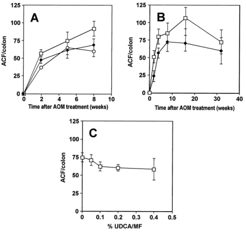
In Study-1, the UDCA (0.4%) supplemented diet decreased the ACF number both during the initia-tion and post-initiainitia-tion stages (Fig. 2A). After two weeks of UDCA treatment, the ACF numbers in the initiation and post-initiation groups decreased signifi-cantly to 83.8% and 64.7% of the control group (av-erage : 56.8 ACF/colon), respectively (P <0.01, P < 0.0001). At the 5th week of enumeration, the num-bers in the initiation and post-initiation groups de-creased to 82.3% and 87.0% of the control (average :
74.4 ACF/colon), respectively, but these suppressions were not significant. At the 8th week, the ACF num-bers in the initiation and post-initiation groups de-creased significantly to 74.6% and 65.0% of the con-trols (average : 92.0 ACF/colon), respectively (P < 0.001, P <0.0001). At the end of 8th week, the total numbers of aberrant crypts (AC) in the initiation and post-initiation groups showed decreasing tendencies by 77.8% and 66.2% of the control (average : 283.0 AC/colon), respectively, but they were not signifi-cantly different from controls.
In the time-course study (Study-2), the numbers of ACF in the UDCA supplemented diet group were 46.0%, 70.7%, 85.1%, 66.4% and 82.5% of controls in the 2nd, 4th, 8th, 16th and 32nd weeks, respective-ly (Fig. 2B). The numbers of ACF decreased signifi-cantly in the UDCA supplemented diet group at the 2nd (P<0.001), 4th (P<0.005) and 16th weeks (P<0.005), but at the 32nd week, the ACF number was not sig-nificantly decreased. The maximum suppressive ef-fect of UDCA was observed at the 2nd week (46.0% of controls), whereas it was minimum (82% of con-trols) at the 32nd week, the end of the experiment. In the dose-response study (Study-3), dietary ad-ministration of UDCA decreased the number of ACF in a dose-dependent manner. The suppressive effect of UDCA on ACF formation was significant in UDCA
Fig. 2. Effect of ursodeoxycholic acid (UDCA) on azoxymethane (AOM)-induced aberrant crypt foci (ACF) formation in the rat colon.
(A) Study-1 (□, control ; ◆, initiation ; ○, post-initiation). UDCA (0.4%) decreased the number of ACF both in the initiation and post-initiation stages of colon carcinogenesis. (B) Study-2 (time-course) (□, control ; ◆, UDCA). (C) Study-3 (dose-response)
M. A. Momen et al. Effect of bile acids on colon carcinogenesis
70 M. A. Momen et al. Effect of bile acids on colon carcinogenesis
of 0.1% (P <0.05), 0.2% (P <0.01) and 0.4% (P <0.005). UDCA of 0.05% did not decrease the numbers of ACF significantly (Fig. 2C).
Effect of bile acids on [Ca2+
]iof ACL-15 cells Measurement of intracellular Ca2+
by fluorescence microscopy revealed that CDCA and DCA induced a rapid increase in [Ca2+
]ieven when extracellular Ca2+
was abolished (Fig. 3C-D). CDCA and DCA (1 mM) increased [Ca2+
]ito a peak of approximate-ly 120 nM and 100 nM, respectiveapproximate-ly, followed by its gradually decrease to a steady level. In contrast, UDCA and CA did not induce an increase in [Ca2+
]i(Fig. 3A-B).
DISCUSSION
In our previous studies, we found that UDCA sup-pressed the carcinogen-induced colonic ACF forma-tion in rats (6, 7). In the present study, we also ob-served an ACF-suppressive effect of UDCA. To observe the ACF suppressive effect of UDCA in detail, we investigated the effect of UDCA on ACF using differ-ent protocols. In study-1A of the presdiffer-ent study, it was shown that UDCA could decrease the AOM-induced ACF number during the initiation as well as during the post-initiation, suggesting that UDCA affects both stages of colon carcinogenesis. In the time-course
study that examined the long-term effect of UDCA, we found that UDCA continued to significantly sup-press the ACF number compared with controls until the 16th week of the experiment (70.8 vs. 106.6), at the 32nd week the ACF number of UDCA and MF fed groups (60.0 vs. 72.7 ACF/colon) did not differ so much as the early period of the experiment. In the present dose-response study of UDCA we ob-served that the MF diet supplemented with 0.05% and 0.1% of UDCA suppressed the ACF number gradu-ally, but unexpectedly this suppressive effect of UDCA did not increase so markedly at doses higher than 0.1%. The exact cause(s) of these variable effects of UDCA remains to be clarified. However, it was re-ported that orally supplemented bile acids are frequent-ly converted to other bile acids by the enterohepatic circulation as well as by enzymatic action of gut flora (7, 23, 24). In our previous study with dietary admin-istration of UDCA and other bile acids, the fecal bile acids analysis revealed that similar to other bile acids, UDCA was also converted to muricholic acid (50.8%), LCA (10.2%), hydodeoxycholic acid (8.7%), DCA (4.1%), CDCA (0.4%) and others (7). These converted acids are known as ACF promoting bile acids and this con-version was suggested to be changed in terms of qual-ity and quantqual-ity in long-term administration. This may be one of the causes for UDCA not to continue its ACF suppressive effect beyond a certain dose and time. Moreover, rat colonic mucosal cells may change
Fig. 3. Effect of bile acids on [Ca2+]iin ACL-15 cells. (A) UDCA, (B) CA, (C) CDCA, (D) DCA. Time 0 indicates when the bile acid (1 mM) contacted the cells. Each value indicates the “mean±SD” of about ten cells measured.
71
The Journal of Medical Investigation Vol. 49 2002 71
their response to UDCA after long-term administra-tion.
The mechanism(s) involved in tumor promotion by bile acids remains unclear. Similarly, the mecha-nisms involved in the anticarcinogenic effects of UDCA on experimental colonic cancer also remain to be clarified. Alterations in protein kinase C (PKC) are involved in colonic malignant transformation in humans (14) and in animals (15). PKC constitutes a gene family of serine/threonine protein kinases that play central roles in transmembrane signaling events, and are involved in diverse biological processes, including cellular proliferation and differentiation (25). Bile acids, which are known as activators of PKC (16), can also stimulate the membrane phospholipase C that converts the phosphatidylinositol biphosphate to inositol triphosphate (IP3) and diacylglycerol (DAG).
IP3acts on endoplasmic reticulum and this released
calcium ions into the cytoplasm. These calcium ions in addition to the DAG, ultimately activate the PKC for further activities. Therefore, bile acids can acti-vate the PKC via the induction of intracellular cal-cium ions. In the present study, CDCA and DCA which were previously reported to increase the num-ber of ACF in the rat colon (6, 7), induced a rapid increase in the [Ca2+
]iin ACL-15 cells. Thus, the ACF promotive effect of CDCA and DCA in animal model studies agrees with the present in vitro finding of their calcium ion induction. From this result we can say that the effect of CDCA and DCA may be, in part, related to the increase in [Ca2+
]i. However, CA, which showed no effect (7) or suppressive effect (26) on ACF formation, and UDCA, which suppressed the ACF number (6, 7), did not show any effect on [Ca2+
]i. Although the ineffective nature of the CA on colonic ACF formation is consistent with its effect of unchanged [Ca2+
]i, the suppressive effect of CA and UDCA on ACF formation is not correlated with their [Ca2+
]i induction. Other mechanism(s) in this suppression process may be involved and remain to be clarified. In conclusion, regardless of the exact mechanism(s) of bile acids to produce colonic ACF, the present find-ings demonstrated that the ACF promotive activity of bile acids in rat colonic mucosa may be through the involvement of PKC that is activated partly by intracellular calcium ions. Therefore, it is suggested that there is a possible relation between the intracellular calcium ion induction by bile acids and the effects of bile acids on ACF formation.
ACKNOWLEDGEMENTS
This study was supported by a Grant-in-Aid for Cancer Research from the Ministry of Health and Welfare, Japan. A doctoral fellowship to Md. Abdul Momen from the Ministry of Education, Science, Sports and Culture of Japan is gratefully acknowl-edged.
REFERENCES
1. Reddy BS, Wynder EL : Metabolic epidemiology of colon cancer. Fecal bile acids and neutral stero-ls in colon cancer patients and patients with adenomatous polyps. Cancer 39 : 2533-2539, 1977 2. Spigelman AD, Owen RW, Hill MJ, Phillips RK : Biliary bile acid profiles in familial adenomatous polyposis. Br J Surg 78 : 321-325, 1991
3. Narisawa T, Magadia NE, Weisburger JH, Wynder EL : Promoting effect of bile acids on colon carcinogenesis after intrarectal instillation of N-methyl-N’-nitro-N-nitrosoguanidine in rats. J Natl Cancer Inst 53 : 1093-1097, 1974
4. Narahara H, Tatsuta M, Iishi H, Baba M, Uedo N, Sakai N, Yano H, Ishiguro S : K-ras point muta-tion is associated with enhancement by deoxycholic acid of colon carcinogenesis induced by azoxymethane, but not with its attenuation by all-trans-retinoic acid. Int J Cancer 88 : 157-161, 2000
5. Earnest DL, Holubec H, Wali RK, Jolley CS, Bissonette M, Bhattacharyya AK, Roy H, Khare S, Brasitus TA : Chemoprevention of azoxymethane-induced colonic carcinogenesis by supplemental dietary ursodeoxycholic acid. Cancer Res 54 : 5071-5074, 1994
6. Seraj MJ, Umemoto A, Kajikawa A, Mimura S, Kinouchi T, Ohnishi Y, Monden Y : Effects of dietary bile acids on formation of azoxymethane-induced aberrant crypt foci in F344 rats. Cancer Lett 115 : 97-103, 1997
7. Lin C, Umemoto A, Jabed MS, Mimura S, Monden Y : Ef-fect of bile acids on formation of azoxymethane-induced aberrant crypt foci in colostomized F344 rat co-lon. Cancer Lett 169 : 121-126, 2001
8. Hardman WE, Cameron IL, Heitman DW, Contreras E : Demonstration of the need for end point vali-dation of putative biomarkers : failure of aberrant crypt foci to predict colon cancer incidence. Can-cer Res 51 : 6388-6392, 1991
9. Thorup I, Meyer O, Kristiansen E : Influence of a dietary fiber on development of dimethylhydrazine-induced
M. A. Momen et al. Effect of bile acids on colon carcinogenesis
72 M. A. Momen et al. Effect of bile acids on colon carcinogenesis
aberrant crypt foci and colon tumor incidence in Wistar rats. Nutr Cancer 21 : 177-182, 1994 10. Pereira MA, Barnes LH, Rassman VL, Kelloff
GV, Steele VE : Use of azoxymethane-induced foci of aberrant crypts in rat colon to identify poten-tial cancer chemopreventive agents. Carcinogenesis 15 : 1049-1054, 1994
11. Wargovich MJ, Chen C-D, Jimenez A, Steele VE, Velasco M, Stephens LC, Price R, Gray K, Kelloff GJ : Aberrant crypts as a biomarker for colon can-cer : evaluation of potential chemopreventive agents in the rat. Cancer Epidemiol Biomark Prev 5 : 355-360, 1996
12. Pretlow TP, Brasitus TA, Fulton NC, Cheyer C, Kaplan EL : K-ras mutations in putative preneoplastic le-sions in human colon. J Natl Cancer Inst 85 : 2004-2007, 1993
13. Vogelstein B, Kinzler KW : The multistep nature of cancer. Trends Genet 9 (4) : 138-141, 1993 14. Kopp R, Noelke B, Sauter G, Schildberg FW,
Paumgartner G, Pfeiffer A : Altered protein kinase C activity in biopsies of human colonic adeno-mas and carcinoadeno-mas. Cancer Res 51 : 205-210, 1991
15. Craven PA, DeRubertis FR : Alterations in pro-tein kinase C in 1,2-dimethylhydrazine induced colonic carcinogenesis. Cancer Res 52 : 2216 -2221, 1992
16. Pongracz J, Clark P, Neoptolemos JP, Lord JM : Expression of protein kinase C isoenzymes in colorectal cancer tissue and their differential activation by different bile acids. Int J Cancer 61 : 35-39, 1995
17. Metcalfe JC, Moore JP, Smith GA, Hesketh TR : Calcium and cell proliferation. Br Med Bull 42 :
405-412, 1986
18. Hickie RA, Wei JW, Blyth LM, Wong DY, Klaassen DJ : Cations and calmodulin in normal and neo-plastic cell growth regulation. Can J Biochem Cell Biol 61 : 934 -941, 1983
19. Santella L, Kyozuka K, De Riso L, Carafoli E : Calcium, protease action, and the regulation of the cell cycle. Cell Calcium 23 : 123-130, 1998 20. Nicotera P, Orrenius S : The role of calcium in
apoptosis. Cell Calcium 23 : 173-180, 1998 21. Bird RP : Observation and quantification of
aber-rant crypts in the murine colon treated with a colon carcinogen:preliminary findings. Cancer Lett 37 : 147-151, 1987
22. Grynkiewicz G, Poenie M, Tsien RY : A new gen-eration of Ca2+
indicators with greatly improved fluorescence properties. J Biol Chem 260 : 3440-3450, 1985
23. Hill MJ : The enterohepatic circulation. In : Bustos-Fernandez L eds. Colon, Structure and Function, Plenum, New York, 1983, pp. 233-252 24. Sarwal AN, Cohen BI, Raicht RF, Takahashi M, Fazzini E : Effects of dietary administration of chenodeoxycholic acid on N-methyl-N-nitrosourea-induced colon cancer in rats. Biochim Biophys Acta 574 : 423-432, 1979
25. Nishizuka Y : Intracellular signaling by hydrolysis of phospholipids and activation of protein kinase C. Science (Washington DC) 258 : 607-614, 1992
26. Magnuson BA, Bird RP : Reduction of aberrant crypt foci induced in rat colon with azoxymethane or methylnitrosourea by feeding cholic acid. Can-cer Lett 68 : 15-23, 1993
73
The Journal of Medical Investigation Vol. 49 2002 73
![Fig. 3. Effect of bile acids on [Ca 2+ ] i in ACL-15 cells. (A) UDCA, (B) CA, (C) CDCA, (D) DCA.](https://thumb-ap.123doks.com/thumbv2/123deta/6796924.1165202/5.892.95.541.761.1182/fig-effect-bile-acids-acl-cells-udca-cdca.webp)
