Fukushima Medical University
福島県立医科大学 学術機関リポジトリ
This document is downloaded at: 2021-11-08T00:36:43Z
Title
The telomerase plays a pivotal role in collateral development under ischemia by suppressing aging-induced production of oxidative stress, expression of p53 and pro-apoptotic proteins(
本文 ) Author(s) 國分, 知樹
Citation
Issue Date 2019-03-22
URL http://ir.fmu.ac.jp/dspace/handle/123456789/973
Rights © International Heart Journal Association. For secondary use, please request the International Heart Journal Association for permission.
DOI
Text Version ETD
1
The telomerase plays a pivotal role in collateral development under ischemia by suppressing aging- induced production of oxidative stress, expression of p53
and pro-apoptotic proteins.
Tomoki kokubun, MD
Department of Cardiovascular Medicine, Fukushima Medical University
2
Summary
Aging is not only a major risk factor for impaired collateral growth under ischemia but also shortens the telomere length, which are regulated by telomerase. We examined the role of telomerase activity during impaired collateral growth during aging in ischemic skeletal muscle. Unilateral hindlimb ischemia was generated in old, young, and old mice chronically administered a telomerase activator. In old mice, blood flow recovery and development of capillary density in ischemic hindlimbs were reduced compared to those in young mice, and these changes were restored to equal levels by administration of TA- 65, a telomerase activator. During the early phase of ischemic muscle changes in old mice, expression of telomerase reverse transcriptase and telomerase activity were both low compared to those in young mice and old mice treated with TA-65. Levels of reactive oxygen species (ROS), DNA double-strand breaks, and expression of p53, p16, and Bax/Bcl-2 were all elevated in ischemic muscles of old mice compared to those in the muscles of young mice and old mice treated with TA-65 treatment; these factors were maintained at low levels equivalent to those seen in young mice during the experiment.
Expression of HIF1-α/VEGF and PGC1α were decreased in old mice compared to those in young mice and old mice treated with TA-65. Collateral growth under ischemic conditions is impaired in aged animals due to low telomerase activity, increased ROS, resultant DNA damage, and expression of tumor suppressor and pro-apoptotic proteins.
These data suggest that telomerase activation enhances collateral growth and rescues ischemic tissue in old individuals.
Key words: aging, telomerase activity, ischemic artery disease
3
senescence is a major risk factor for cardiovascular disease including peripheral artery disease, and impaired angiogenesis occurs with aging. 1,2) A previous report demonstrated that recovery of blood flow after hindlimb ischemia is impaired in older mammals compared with young mammals.3) However, the mechanisms underlying this phenomenon remain unclear.
Telomeres are chromatin structures composed of repetitive noncoding DNA sequences (TTAGGG in humans) at the ends of chromosomes that protect this region from degradation and recombination.4) Telomerase is a ribonucleoprotein composed of a catalytic subunit, telomerase reverse transcriptase (TERT) and a telomerase RNA template component (TERC) that prevents telomere shortening by adding telomeric DNA repeats to the ends of chromosomes.4) Telomere length is maintained by telomerase complex in normal stem cells; however, it gradually shortens with age in most somatic cells where telomerase is either absent or expressed at low levels. Telomere shortening can have a critical impact on stem cell proliferation, consequently inducing vascular cell senescence.5,6) On the other hand, apart from telomere elongation, accumulating evidence indicates that telomerase plays a role in telomere-independent mechanisms involved in vascular aging including in cellular proliferation, gene expression regulation and mitochondrial function.7,8) However, the role of telomerase during impaired collateral flow recovery in aging after hindlimb ischemia remains unclear. Therefore, we performed the present study to examine whether telomerase activation improves blood flow after acute hindlimb ischemia in aged mice.
In this study, limb perfusion recovery in old mice treated with TA-65 was observed and compared to young, 8-week-old (8W) mice and old, 80-week-old (80W) mice. TA-65 is a single small organic molecule that is obtained from an extract of the roots of the plant
4
Astragalus membranaceus, which is commonly used in traditional Chinese medicine and has been identified as an effective telomerase activator.9-11) In previous study, it has been reported that TA-65 treatment of mice resulted in not only a significant 10-fold increase in TERT mRNA and protein level in a liver and other tissue but also increase in mRNA levels of Jun B and c-Myc, two transcription factors regulated by the Mitogen-activated protein kinase (MAPK) pathway, which is a known potential mediator of TA-65 action.
Therefore, TA65 dependent-telomerase activation occurs through transcription factors regulated by the MAPK pathway, which may directly or indirectly regulate the mTERT promotor.8, 9 We demonstrate a pivotal role for telomerase activation in angiogenesis after acute hindlimb ischemia in old mice that occurs by suppressing age-related ROS and DNA damage and subsequently suppressing the expression of p53, p16, and apoptotic proteins.
Methods Animals
This study conformed to the Guide for the Care and Use of Laboratory Animals published by the U.S. National Institutes of Health (NIH Publication, 8th edition, 2011). Our research protocol was approved by the Fukushima Medical University Animal Research Committee [permit number 29074 (24 July 2017)], and all animal experiments were conducted in accordance with the guidelines of the Fukushima Medical University Animal Research Committee. All efforts were made to minimize animal suffering.
Young and old wild-type C57BL/6 male mice [young: 8 weeks (W); body weight (B.W.), 18.4±0.2 g; old: 80W; B.W., 29.3±0.5 g] were housed and bred in a room maintained at
5
22±3°C with a relative humidity of 50±10% and a 12-h light-dark cycle. Mice were given water ad libitum.
Animal experiments
Young mice were given saline, and old mice were administered either saline or the telomerase activator TA-65 (T.A. Sciences, Inc. NY, USA).
In reference to doses used in past papers, TA-65 was dissolved in saline and administered at a dose of 25 mg/kg/day by oral gavage.9,12) Unilateral hindlimb ischemia was induced by the method described below at day 21 after saline or TA-65 administration. Saline and TA-65 administration were continued after hindlimb ischemia was induced, and mice were euthanized with isoflurane aspiration and a lethal dose of pentobarbital (60 mg/kg body weight i.p.) on postoperative day (POD) 3 or POD 21 followed by examinations described in context. We selected POD 3 and 21 based on the findings of our previous reports.13)
Hindlimb ischemia
The left femoral artery was exposed under tribromoethanol anesthesia (200 mg/kg/body weight, i.p.) and was ligated at two positions, just below the inguinal ligament and proximal to the saphenous-popliteal bifurcation using 6-0 sutures. Sham operations occurred without femoral artery ligation but with a skin incision made in the right hindlimb.
Measurement of hindlimb perfusion
Scanning laser Doppler perfusion imaging (Moor Instruments, Wilmington, DE, USA)
6
was used to measure hindlimb perfusion under 1.125% isoflurane/O2 anesthesia. The ratio of ischemic/nonischemic laser Doppler blood flow was measured before and on postoperative days 0, 1, 7, 14, and 21.
Measurement of reactive oxygen species (ROS)
An OxiSelectTM In Vitro ROS/RNS Assay Kit (Green Fluorescence) was used to measure levels of ROS according to the protocol provided by the manufacturer (Cell Biolabs, Inc.
San Diego, CA, USA). Twenty milligrams of harvested soleus muscle tissues were cut to pieces 5 mm away from the Achilles tendon and homogenized in 1 ml of ice-cold PBS.
Homogenized samples were centrifuged at 10,000 g for 5 min to remove insoluble particles. The supernatant of each sample was used. We added 50 μl of each sample in duplicate and hydrogen peroxide (H2O2) standards to a black 96-well plate with 50 μl of the provided catalyst solution and incubated the mixture for 5 min at room temperature to accelerate the oxidative reaction. Next, we added 100 μl of dichlorodihydrofluorescein (DCFH) solution included in the kit to each well, covered the plates to protect them from light and incubated the samples at room temperature for 30 min. Finally, fluorescence was read with a fluorescence plate reader (SpectraMaxⓇ i3, Molecular Devices) at 480 nm excitation/530 nm emission. A H2O2 standard curve was used to quantify total free radical content in skeletal muscle.
Measurement of telomerase activity
The telomerase repeated amplification protocol (TRAP) assay is a popular method to determine telomerase activity in mammalian cells and tissue samples.14) Telomerase activity was measured using the Telo TAGGG Telomerase PCR ELISAPLUS Assay Kit
7
(Cat. No. 12013789001, Roche Diagnostics GmbH, Mannheim, Germany), a photometric enzyme immunoassay for quantitative determination of telomerase activity that utilizes TRAP. Tissues extracts were prepared from frozen tissue pellets stored at -80°C by adding 200 μl of lysis reagent according to the manufacturer’s instructions. The lysis mixture was homogenized and incubated on ice for 30 min. The lysate was then centrifuged at 16,000 x g for 20 min at 4°C, and the supernatant was collected. We measured protein concentrations by standard methods and then performed the TRAP reaction as described below.
First, negative controls for each sample were prepared by heating at 95°C for 10 min.
Then, we prepared 30 μl of master mix (25 μl of reaction mixture, 5 μl of the provided internal standard) per sample. We constructed 50 μl reaction mixtures for each sample (30 μl of master mix, X μl of sample per 10 μg of total protein and added nuclease free water for a total volume of 50 μl), negative controls (30 μl of master mix, X μl of heat-treated sample per 10 μg of total protein and added nuclease free water for a total volume of 50 μl) and control templates (30 μl of master mix, with 1 μl of control template added to a separate tube, 1 μl of lysis reagent added to another tube and 19 μl of nuclease-free water added to each tube), all of which were added to a new 0.2-ml tube suitable for PCR. Then, microtubes were transferred to PCR thermal cycler (GeneAmpⓇ PCR system 9700, Applied Biosystems) for combined primer elongation/amplification reaction using the following protocol [1 cycle for 20 min at 25°C (primer elongation); 1 cycle for 5 min at 94°C (telomerase inactivation); 30 cycles for 30 sec at 94°C, 30 sec at 50°C, 90 sec at 72°C and 1 cycle for 10 min at 72°C (amplification, PCR)]. After 2.5 μl of amplification products were mixed with 10 μl of denaturation reagent, we added 100 μl of hybridization buffer according to the supplier’s instructions. Then, we transferred the 100-μl samples
8
to a precoated MP module included in the kit and incubated for 2 hours at room temperature with agitation at 300 rpm. Then, the MP modules were washed three times, and 100 μl of anti-DIG-HRP was added to each well and incubated for 30 min with agitation at 300 rpm. After washing the plates three times, 100 μl of prewarmed TMB substrate solution was added to the wells, and the wells were incubated at room temperature for 20 min with agitation for color development. Then, 100 μl of stop reagent was added to each well to stop color development. Finally, we measured sample absorbance at 450 nm using a microplate reader (with a reference wavelength of approximately 690 nm).
Relative telomerase activity (RTA) was calculated using the following formula:
(AS-AS0)/AS,IS
RTA = × 100%
(ATS8-ATS8,0)/ATS8,IS
AS: absorbance of the sample
AS,0: absorbance of heat- or RNase-treated samples
AS,IS: absorbance of the internal standard (IS) of the sample ATS8: absorbance of the control template (TS8)
ATS8,0: absorbance of the lysis buffer
ATS8,IS: absorbance of the internal standard (IS) of the Control template (TS8)
Western Blotting
Harvested soleus muscles frozen at -80°C were pulverized in liquid nitrogen and
9
suspended in lysis buffer with a protease inhibitor. After determining protein concentrations using a standard protocol (Bradford protein assay, Bio-Rad), equal amounts of protein (20 μg/sample) were analyzed by 7.5%, 10% or 15% sodium dodecyl sulfate polyacrylamide gel electrophoresis (SDS-PAGE) gels depending on the molecular weight of the targeted proteins. Gels were transferred onto nitrocellulose membranes that were blocked with tris-buffered saline Tween 20 (TBST) containing 5% bovine serum albumin (5% BSA) and immunoblotted using the following primary antibodies at 4°C with gentle agitation overnight: anti-p53 (PAb122) (Enzo Life Sciences, Inc., Farmingdale, NY, USA, 1:1000), anti-p21 (sc-6246) (Santa Cruz Biotechnology, Inc.
1:1000), anti-p15/16 (sc-28260) (Santa Cruz Biotechnology, Inc. 1:1000), anti-histone H2A.X (phosphorS139) (ab81299) (Abcam plc, Cambridge, UK, 1:1000), anti-Bax (#2772) (Cell signaling Technology, Inc. 1:1000), anti-Bcl-2 (#2876) (Cell signaling Technology, Inc. 1:1000), anti-TERT (NB100-317) (Novus Biologicals, LLC., Littleton, CO, USA, 1:1000), anti-HIF-1α (#14179) (Cell signaling Technology, Inc. 1:1000), anti- VEGF (sc-507) (Santa Cruz Biotechnology, Inc. 1:1000), anti-PGC1 alpha (ab54481) (Abcam plc, 1:1000) and anti-β-Actin (sc-47778) (Santa Cruz Biotechnology, Inc.
1:10,000). Membranes were then washed in TBST and incubated with appropriate anti- mouse or anti-rabbit horseradish peroxidase-conjugated secondary antibodies (Santa Cruz Biotechnology, Inc. 1:10,000). Antibody -bound protein was visualized using enhanced chemiluminescence. Relative intensities of protein bands were quantified using NIH ImageJ, version 1.48 (Scion Image, NIH, Bethesda, MD, USA). Data were normalized to β-Actin and are expressed as fold change to the nonischemic skeletal muscle of young mice in the same period for each analysis.
10
Immunohistochemistry
Capillary formation in ischemic soleus muscles was analyzed by immunostaining for CD31 and quantified as capillaries per muscle fiber. Immunohistochemistry was performed using paraffin-embedded tissue (IHC-P).
First, we deparaffinized and rehydrated slides by immersion in the following solutions:
xylene, four washes, 2 min each→100% ethanol, two washes, 2 min each→95% ethanol, 2 min each→90% ethanol, 2 min each. Slides were washed with PBS three times for 5 min each. For antigen retrieval, we brought slides to a boil in Histo VT One (10X, pH 7.0, Nacalaitesque, INC. Kyoto, JPN) buffer and maintained at subboiling temperature for 10 min. We allowed slides to cool on the bench top for approximately 30 min. We incubated slides in 0.3% hydrogen peroxide in methanol for 20 min to quench endogenous peroxidase activity. We then washed the slides in PBS three times for 5 min each. We added primary antibody (rabbit anti-mouse CD31 antibody, Santa Cruz Biotechnology) diluted in 2% BSA-PBS and 0.05% sodium azide (1:50~1:100) and incubated them overnight at 4°C in a humidified chamber. Slides were washed with PBS three times for 5 min each the following day. We then added secondary antibody (HRP-labeled Gout Anti-rabbit IgG, HistofineⓇ Simple StainTM Mouse MAX PO(R), Nichirei Biosciences, Tokyo, JPN) and incubated the slides at room temperature for 30 min. Slides were subsequently washed with PBS three times for 5 min each.
3,3’-diaminobenzidine tetrahydrochloride (DAB; Wako Pure Chemicals Industries, Ltd.
Tokyo, JPN) solution was prepared by mixing 100 ml of 0.05 M Tris buffer, 20 mg of DAB and 17 μl of 30% H2O2 (just before use). Slides were immersed in DAB solution for 5 min to 10 min until chromogenic reaction turned the epitope sites brown. After washing slides with water, we stained the nuclei with hematoxylin and eosin (H.E.).
11
Slides were dehydrated by moving slides through the following solutions for 1 min per section: 70% ethanol → 95% ethanol → 100% ethanol, two washes → xylene, four washes. Finally, we added mounting media to the slides and topped them with coverslips.
Capillary counts were performed under light microscopy (200x). The number of capillaries was measured in five randomly chosen fields from three different sections in each tissue block.
Statistical Analysis
All values are expressed as the means ± S.E.M. One-way repeated measures ANOVA with Tukey’s post hoc test used to compare hindlimb blood flow data. For all other data sets, one-way factorial ANOVA with Tukey’s post hoc test were performed to compare multiple groups. P<0.05 was considered to be statistically significant. All statistical analyses were performed using SPSS software (ver. 24.0, IBM, Armonk, NY, USA).
Results Limb blood flow recovery
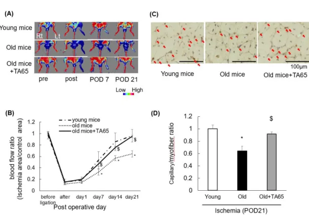
Blood flow recovery was impaired in ischemic hindlimbs of old mice compared to those of young mice (Figure 1A and B). TA-65 treatment in old mice improved blood flow recovery. Histologically, TA-65 treatment also increased capillary density as measured by CD31 staining of muscle tissue in old mice, which reflected improved blood flow recovery (Figure 1C and D). These data support the notion that collateral flow recovery under ischemic conditions is impaired in aging and that telomerase activation by TA-65 treatment could improve age-related impairment of angiogenesis.
12
Levels of ROS in skeletal muscle tissue
Oxidative stress increasing with age, 15) and high levels of ROS are cytotoxic and impair angiogenesis.16) We previously reported that age-related increases in ROS levels after hindlimb ischemia impair limb blood flow recovery.17) We also demonstrated that mitochondrial ROS scavenging in old mice contributes to collateral flow recovery after hindlimb ischemia.13) Therefore, We initially measured levels of ROS in nonischemic and ischemic soleus muscles at POD 3. No difference was found among nonischemic animals (Figure 2A). Levels of ROS in ischemic limbs of old mice were elevated compared to those in nonischemic regions and were prominent compared to those in the limbs of young mice. Furthermore, ROS levels were attenuated by TA-65 treatment in old mice. These findings suggest that the levels of ROS increase in aging tissue under ischemia and are attenuated by TA-65 treatment.
DNA damage in skeletal muscle tissue
It has been reported that ROS, telomere dysfunction, UV exposure, etc., cause DNA damage responses (DDRs), in particular double-strand breaks (DSBs); DSBs are the most serious type of DNA damages and induce apoptosis and cellular senescence, resulting in vascular aging.18) DSBs trigger activation of many factors including phosphorylation of histone variant H2AX (γH2AX), which plays an important role in the DDR and is required for assembly of DNA repair proteins at sites containing damaged chromatin and for activation of checkpoint proteins that cause cell cycle arrest.19) Therefore, γH2AX is a useful tool for detecting DNA damage. To evaluate DNA damage in response to hindlimb ischemia, we measured γH2AX expression in soleus muscle by Western blotting.
On POD 3 and POD 21, γH2AX expression increased in ischemic limbs compared to
13
nonischemic limbs (Figure 2B). In ischemic muscles, the expression levels of γH2AX were high in old mice compared to young mice and were attenuated in old mice treated with TA-65, resulting in levels equal to those in young animals. These results suggest that DNA damage in skeletal muscle induced by ischemia increases with aging and is suppressed by TA-65 treatment.
TERT expression and telomerase activity
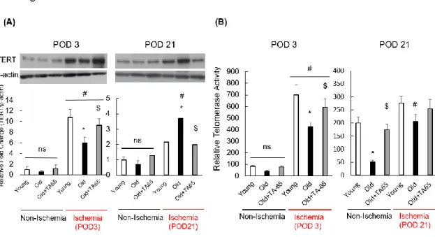
The enzymatic activity of telomerase has been proposed to be primarily controlled by transcriptional regulation of TERT. TERT gene transfer into endothelial progenitor cells (EPCs) contributed to improvement in angiogenesis in a murine model of hindlimb ischemia.20) Thus, impaired TERT expression and telomerase activity may contribute to impaired angiogenesis in the elderly. We measured TERT expression and telomerase activity to investigate the effects on aging in response to ischemia and to confirm telomerase activation by TA-65. On POD 3, TERT expression and telomerase activity were significantly elevated in ischemic limbs compared to nonischemic limbs (Figure 3A and B). In ischemic muscles, TERT expression and telomerase activity in old mice were reduced compared to those in young mice and were upregulated in old mice by TA-65 treatment. In contrast, on POD 21, TERT expression was higher in old mice than in young mice, and the same tendency was found in TERT levels between young mice and old mice treated with TA-65. TA-65 treatment regulated TERT levels at POD 21 and POD 3. In contrast, the effect of TA-65 on telomerase activity at POD 21 in ischemic limbs in old mice was negligible.
It has been reported that TERT expression is upregulated in response to cellular stress, such as that caused by ischemia.21) On POD 21, blood flow recovery in ischemic
14
hindlimbs of young and TA-65-treated old mice were improved to nearly the same level as in nonischemic mice, as described above. Therefore, we proposed that there was a reduced influence of hypoxia and ischemia in young mice and in old mice treated with TA-65 during the late phase of ischemia. These data suggest that TERT expression and telomerase activity that were upregulated in response to ischemia were attenuated in the elderly, especially during the early phase of hindlimb ischemia, and were recovered after TA-65 treatment.
Expression of p53/p21, p15/16 and apoptotic factors
Telomere shortening or DNA damage induces DDR. When damage becomes irreversible, the DDR pathway induces cellular senescence, cell-cycle arrest and apoptosis. The transcription factor p53, also known as a tumor suppressor gene, is widely recognized as one of the downstream effectors in the DDR pathway that plays an important role through activating pathways via posttranslational modifications, such as acetylation or phosphorylation. Considering our findings that TA-65 decreased ROS levels and DNA damage, we assumed that TA-65 treatment would attenuate p53 expression. Thus, we examined whether TA-65 treatment downregulated p53 or p21 expression in ischemic aged skeletal muscles. In nonischemic groups, p53 expression on POD 3 and POD 21 in old mice tended to be high compared to that in young mice and TA-65 treated mice (Figure 4A). p53 expression was higher in ischemic old mice than in nonischemic mice and was attenuated by treatment with TA-65 (Figure 4B). Expression of p21 showed the same tendency, and a similar trend was recognized in p53/p21 expression on POD 3 and POD 21 (Figure 4C). The p16/RB (retinoblastoma protein) pathway is also another major pathway of senescence. Aging and many inducers of cellular senescence, such as ROS,
15
increase p16 activity.22) We measured p16 expression at POD 3 and POD 21, and found that expression was higher in ischemic old mice than in nonischemic limbs and was decreased in TA-65-treated old mice (Figure 4D). These findings suggest that a decline in ROS and subsequent DNA damage with TA-65 treatment in aged skeletal muscle attenuate p53/p21 and p16 expression in response to ischemia. Apoptosis, a programmed form of cell death, is considered an anti-angiogenetic mechanism.23) Several studies have reported that telomerase activity is essential for endothelial cell differentiation and protection from apoptosis.24) The B-cell leukemia/lymphoma-2 (Bcl-2) family of proteins are the best-characterized protein family involved in regulation of apoptotic cell death and includes anti-apoptotic and pro-apoptotic members. Activated p53 in response to DNA damage mediates mitochondrial control of apoptosis by promoting pro-apoptotic members (Bax) and suppressing anti-apoptotic Bcl-2.25) Therefore, we examined whether telomerase activation prevents apoptosis under conditions of ischemia. The Bax/Bcl-2 ratio was higher in ischemic old mice than in young mice, and TA-65 treatment of old mice decreased the Bax/Bcl-2 ratio to that of young mice at both POD 3 and POD 21 (Figure 4E). These data suggest that telomerase activation by TA-65 is protective against ischemia-induced apoptosis in aged skeletal muscle and contributes to angiogenesis.
HIF-1α-VEGF signaling pathways
Ischemia-induced angiogenesis is impaired in aging and is mediated by hypoxia inducible factor-1 (HIF-1α).26) HIF-1α is a transcription factor that mediates adaptive responses to hypoxia and ischemia. HIF-1α regulates expression of angiogenetic factors, such as vascular endothelial growth factor (VEGF), via the HIF-1α-VEGF signaling pathways.27) p53 is known as a potent negative regulator of HIF-1α and VEGF during hypoxia.28) To
16
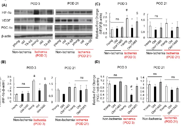
examine the angiogenetic effects of TA-65 treatment, we measured expression of HIF-1α and VEGF by Western blot analysis (Figure 5A). At POD 3, expression of HIF-1α and VEGF were significantly increased in young mice compared to nonischemic mice (Figure 5B and C). This elevation was suppressed in old mice and recovered with TA-65 treatment.
However, on POD 21, expression of HIF-1α was not different among the three groups.
Expression of VEGF in young mice was higher than that in old mice, and there was a tendency for increased expression of VEGF in old mice treated with TA-65 compared to those that were not treated, though this change did not reach statistical significance. These data suggest that telomerase activation by TA-65 is associated with elevated HIF-1α and VEGF expression in aged skeletal muscle at comparatively early phases of ischemia. On POD 21, blood flow recovery in ischemic hindlimbs of young mice and TA-65-treated old mice improved to nearly the same level as in nonischemic mice described above.
Therefore, we propose that hypoxia and ischemia exert less of an influence in young mice and in old mice treated with TA-65 than in untreated old mice during the late phase of ischemia.
Expression of peroxisome proliferator activated receptor γ coactivator 1α (PGC-1α) Recent studies have reported that telomere dysfunction and associated DNA damage responses promote mitochondrial dysfunction.29) p53 induced by telomere dysfunction binds to the PGC-1α promoter and suppresses expression of PGC-1α. Repression of PGC- 1α impairs mitochondrial biogenesis and function, resulting in reduced ATP generation and increased levels of ROS.30) In light of our previous experiments illustrating that telomerase activation by TA-65 attenuates ROS and DNA damage along with decreasing p53 levels in aged ischemic skeletal muscle, we assumed that TA-65 treatment would
17
preserve PGC1α expression under conditions of ischemia. Expression of PGC-1α did not differ in the nonischemic skeletal muscles at POD 3 or POD 21. In ischemic skeletal muscles at POD 3, expression of PGC-1α was significantly lower in old mice than in young mice, and TA-65 treatment of old mice maintained PGC-1α levels at similar levels as those in young mice (Figure 5D). However, there were no differences in PGC-1α expression in ischemic skeletal muscle at POD 21. Considering the reduction in ROS, protection from apoptosis, and preservation of PGC-1α expression by TA-65 treatment in ischemic aged mice, these data suggest that telomerase activation improves mitochondrial dysfunction in aged mice after hindlimb ischemia, especially during early phases of ischemia.
Discussion
Senescence is a dominant risk factor for most forms of cardiovascular disease.31) Telomerase is closely related with cellular senescence and is suspected to be a potential anti-aging therapy.32) Our present study demonstrates the crucial role of telomerase activity for improving blood flow after acute hindlimb ischemia in old mice. Telomerase activation therapy in aged skeletal muscle suppresses age-related ROS and DNA damage in response to ischemia, contributing to angiogenesis through the tumor suppressors p53 and p16 and pro-apoptotic proteins.
The tumor suppressors p53 and p16 are upregulated in response to various cellular stresses, including DNA damage, oxidative stress and hypoxia.19,33-35) Upregulation of p53 induces dramatic and multiple cellular responses including apoptosis and anti- angiogenesis via the HIF-1α/VEGF pathway.36,37) In the present study, as in our previous study13), p53 was upregulated by aging and in response to ischemic DNA damage, which
18
seemed to promote apoptotic processes and inhibit angiogenetic processes. Telomerase activation markedly downregulated p53 expression in response to ischemia. Considering that DDR following DNA damage is the most important pathway for p53 upregulation, reduction of ROS and DNA damage by telomerase activation are the most likely reasons for downregulation of p53. On the other hand, it has been reported that p16 also modulates VEGF expression through HIF-1α.38) Thus, telomerase activation under conditions of ischemia may contribute to angiogenesis not only through p53 downregulation but also through p16 reduction, resulting in collateral flow recovery.
Furthermore, apoptosis is considered an anti-angiogenetic mechanism, and p53 activated due to cellular stress mediates mitochondrial control of apoptosis.23,25) Considering the observed downregulation of p53 and the Bax/Bcl-2 ratio in ischemic muscle, telomerase activation might contribute to inhibiting apoptosis, resulting in angiogenesis and collateral flow recovery. In this study, we examined apoptosis in isolated muscle including vessel, because it is difficult to isolate capillary. Therefore further study is needed like an in vitro study to clarify the pure contribution of TA-65 to angiogenesis and apoptosis.
It has been proposed that the enzymatic activity of telomerase is primarily controlled by transcriptional regulation of TERT, as shown in our results at POD 3. In contrast, there are many reports that telomerase activity is regulated by several factors, such as ROS, exercise capacity and cellular stress related to the magnitude of ischemia,32,39,40) which may affect TERT expression and telomerase activity in chronic ischemic conditions, as we did not observe a positive correlation between telomerase activity and TERT expression in ischemic old mice at POD 21. Therefore, further study is needed to investigate the effect of telomerase on various conditions, including acute and chronic
19
ischemic states. TERT has also been proposed to play a role in regulating oxidative damage-induced apoptosis. Oxidative stress triggers nuclear export of TERT to the mitochondria.41) TERT has been shown to localize to mitochondria and contributes different mitochondrial functions (such as modulating mtDNA integrity, improving respiratory chain function and affecting ROS production).30) Therefore, we believe that the main source of ROS is mitochondria. As previously presented, the chronic scavenging of superoxide that is dropped from mitochondria ETc-complex in aged skeletal muscle like a condition with attenuated telomerase activity, improved collateral growth induced by ischemia through regulation of p53 as a mimicking effect of TA-65 treatment.13) Therefore, mitochondria ETc-complex activity or drop of superoxide from ETc-complex pathway might be modified by telomerase activity and TA-65 decrease generation of superoxide in ETc-complex in mitochondria, though further study is needed to clarify the telomerase effect to mitochondria. Thus, antioxidant effect of telomerase or TA-65 is to reduce the generation of superoxide from ETc-complex in mitochondria. PGC-1α is a master regulator of mitochondrial physiology and metabolism. Furthermore, PGC-1α induces VEGF and contributes to angiogenesis.42) Telomere dysfunction activates p53, which in turn binds and represses PGC-1α promotors, inducing mitochondrial dysfunction that promotes ATP synthesis and increased ROS.29) Considering the observed reduction in ROS, protection from apoptosis, and preservation of PGC-1α expression by telomerase activation during the early phase of ischemia in old mice, increased telomerase activity could improve mitochondrial dysfunction and contribute to angiogenesis through preservation of PGC-1α in aged skeletal muscle after hindlimb ischemia.
There are a few limitations to this study. First, we did not measure telomere length in this study. In recent years, evidence of the additional functions of telomerase other than
20
telomere maintenance have been accumulating.7,8) Human adult somatic cells do not express telomerase activity, whereas most adult mouse somatic cells including skeletal muscle express detectable amounts of telomerase.43) In fact, it has been reported that there is no significant difference in telomere length between young and aged muscle stem cells, known as satellite cells, from uninjured wild-type mice.44) Considering the lack of differences in telomere length in response to aging in the skeletal muscles of mice, the rapid onset of the anti-apoptotic process and angiogenetic differences in old mice treated with TA-65 detectable at day three in vitro seems to depend on telomerase rather than on telomere length. Second, we evaluated only two time points, POD 3 and POD 21 (reference our previous study).13) It is presumed that signal transduction including TERT may dramatically change, especially during the early phase of ischemia, but we did not demonstrate sequential changes. Third, we did not perform measurements in muscles composed of fast-twitch muscle fibers, such as the gastrocnemius. It has been reported that age-related mitochondrial function, one of the important targets of telomerase, varies in different muscle types,45,46) thus the further study is needed to investigate the telomerase performance in other muscle types. Finally, cycloastragenol (CAG), the main component of TA-65, is a molecule isolated from Astragalus. There is a few report that CAG enhanced antioxidant capacity in itself in a D-galactose-induced senescence mouse model and its antioxidant effects may be related to the hydroxyl found in the chemical structure of CAG.47) Since oxidative stress is a main reason for telomere dysfunction and anti-angiogenesis, we could not completely exclude the possibility that the telomere protective and angiogenetic effects of TA-65 may be related to not only telomerase activation but also its direct antioxidative effects.
21
Conclusion
We showed a possible role for telomerase in regulating cellular processes during senescence other than telomere elongation, such as differentiation and angiogenesis, through the tumor suppressors p53 and p16. Our data suggest that telomerase plays a pivotal role in collateral development, and its pharmaceutical activation may be a novel therapeutic option to enhance collateral flow recovery to rescue ischemic tissue in elderly individuals.
Acknowledgments
We thank Tomiko Miura and Chikako Endo for the care of the mice and for laboratory assistance.
Disclosures None.
22
References
1. Lloyd-Jones D, Adams RJ, Brown TM. Heart disease and stroke statistics--2010 update: a report from the American Heart Association. Circulation. 2010; 23; 121 (7):
e46-e215.
2. Soga Y, Iida O, Takahara M, et al, Two-year life expectancy in patients with critical limb ischemia. JACC Cardiovasc. Interv. 2014; 7 (12): 1444-9.
3. Rivard A, Fabre JE, Silver M, et al, Age-dependent impairment of angiogenesis.
Circulation. 1999; 99 (1): 111-20.
4. Greider CW, Blackburn EH. Identification of a specific telomere terminal transferase activity in Tetrahymena extracts. Cell. 1985; 43 (2 Pt 1): 405-13.
5. Rodier F, Campisi J. Four faces of cellular senescence. J Cell Biol. 2011 21; 192(4):
547-56.
6. van Steensel B, Smogorzewska A, de Lange T. TRF2 protects human telomeres from end-to-end fusions. Cell. 1998; 92 (3): 401-13.
7. Hartwig FP, Nedel F, Collares TV, Tarquinio SB, Nör JE, Demarco FF. Telomeres and tissue engineering: the potential roles of TERT in VEGF-mediated angiogenesis. Stem Cell Rev. 2012; 8 (4): 1275-81.
8. Fu W, Begley JG, Killen MW, Mattson MP. Anti-apoptotic role of telomerase in pheochromocytoma cells. J Biol Chem. 1999; 274 (11): 7264-71.
9. Bernardes de Jesus B, Schneeberger K, Vera E, Tejera A, Harley CB, Blasco MA. The telomerase activator TA-65 elongates short telomeres and increases health span of adult/old mice without increasing cancer incidence. Aging Cell. 2011; 10 (4): 604-21.
10. Harley CB, Liu W, Blasco M, et al, A natural product telomerase activator as part of a health maintenance program. Rejuvenation Res. 2011; 14 (1): 45-56. doi:
23
10.1089/rej.2010.1085. Epub 2010.
11. Salvador L, Singaravelu G, Harley CB, Flom P, Suram A, Raffaele JM. A Natural Product Telomerase Activator Lengthens Telomeres in Humans: A Randomized, Double Blind, and Placebo Controlled Study. Rejuvenation Res. 2016; 19 (6): 478- 484.
12. Mouraret N, Houssaïni A, Abid S, et al. Role for telomerase in pulmonary hypertension. Circulation. 2015; 131(8): 742-755.
13. Miura S, Saitoh SI, Kokubun T, et al. Mitochondrial-Targeted Antioxidant Maintains Blood Flow, Mitochondrial Function, and Redox Balance in Old Mice Following Prolonged Limb Ischemia. Int J Mol Sci. 2017; 18 (9): 1897.
14. Kim NW, Piatyszek MA, Prowse KR, et al. Specific association of human telomerase activity with immortal cells and cancer. Science. 1994; 266 (5193): 2011-5.
15. Muller FL, Lustgarten MS, Jang Y, Richardson A, Van Remmen H. Trends in oxidative aging theories. Free Radic Biol Med. 2007; 43 (4): 477-503.
16. Radomska-Leśniewska DM, Hevelke A, Skopiński P, et al. Reactive oxygen species and synthetic antioxidants as angiogenesis modulators: Clinical implications.
Pharmacol Rep. 2016; 68(2): 462-71.
17. Yamauchi H, Miura S, Owada T, et al. Senescence marker protein-30 deficiency impairs angiogenesis under ischemia. Free Radic Biol Med. 2016; 94:66-73.
18. Hoeijmakers JH 2009. DNA damage, aging, and cancer. N Engl J Med 361: 1475–
1485
19. Inoue E, Tano K, Yoshii H, et al. SOD1 Is Essential for the Viability of DT40 Cells and Nuclear SOD1 Functions as a Guardian of Genomic DNA. J Nucleic Acids. 2010;
2010: 795946.
24
20. Murasawa S, Llevadot J, Silver M, Isner JM, Losordo DW, Asahara T. Constitutive human telomerase reverse transcriptase expression enhances regenerative properties of endothelial progenitor cells. Circulation. 2002; 106(9): 1133-9.
21. Zurek M, Altschmied J, Kohlgrüber S, Ale-Agha N, Haendeler J. Role of Telomerase in the Cardiovascular System. Genes (Basel). 2016; 7(6): E29.
22. Dai J, Zhu X, Yoder MC, Wu Y, Colman RW. Cleaved high-molecular-weight kininogen accelerates the onset of endothelial progenitor cell senescence by induction of reactive oxygen species. Arterioscler Thromb Vasc Biol. 2011; 31 (4): 883-9.
23. Dimmeler S, Zeiher AM. Endothelial cell apoptosis in angiogenesis and vessel regression. Circ Res. 2000; 87 (6): 434-9.
24. Zaccagnini G, Gaetano C, Della Pietra L et.al. Telomerase mediates vascular endothelial growth factor-dependent responsiveness in a rat model of hind limb ischemia. J Biol Chem. 2005; 280 (15): 14790-8.
25. Chipuk JE, Kuwana T, Bouchier-Hayes L, et al. Direct activation of Bax by p53 mediates mitochondrial membrane permeabilization and apoptosis. Science. 2004;
303 (5660): 1010-4.
26. Rivard A, Berthou-Soulie L, Principe N, et al. Age-dependent defect in vascular endothelial growth factor expression is associated with reduced hypoxia-inducible factor 1 activity. J Biol Chem. 2000; 275 (38): 29643-7.
27. Bosch-Marce M, Okuyama H, Wesley JB et al. Effects of aging and hypoxia- inducible factor-1 activity on angiogenic cell mobilization and recovery of perfusion after limb ischemia. Circ Res. 2007; 101(12): 1310-8.
28. Ravi R, Mookerjee B, Bhujwalla ZM, et al. Regulation of tumor angiogenesis by p53- induced degradation of hypoxia-inducible factor 1alpha. Genes Dev. 2000; 14 (1): 34-
25
44.
29. Sahin E, Colla S, Liesa M et. al. Telomere dysfunction induces metabolic and mitochondrial compromise. Nature. 2011; 470 (7334): 359-65.
30. Sahin E, DePinho RA. Axis of ageing: telomeres, p53 and mitochondria. Nat Rev Mol Cell Biol. 2012; 13 (6): 397-404.
31. Lähteenvuo J, Rosenzweig A. Effects of aging on angiogenesis. Circ Res. 2012; 110 (9): 1252-64.
32. Jäger K, Walter M.Therapeutic Targeting of Telomerase. Genes (Basel). 2016; 7 (7):
39.
33. Vousden KH, Prives C. Blinded by the Light: The Growing Complexity of p53. Cell.
2009; 137 (3): 413-31.
34. Jacobs JJ, de Lange T. Significant role for p16INK4a in p53-independent telomere- directed senescence. Curr Biol. 2004; 14 (24): 2302-8.
35. Yaswen P, MacKenzie KL, Keith WN et al. Therapeutic targeting of replicative immortality. Semin Cancer Biol. 2015; 35 Suppl: S104-S128.
36. Amaral JD, Xavier JM, Steer CJ, Rodrigues CM. The role of p53 in apoptosis. Discov Med. 2010; 9 (45): 145-52.
37. Farhang Ghahremani M, Goossens S, Nittner D, et al. p53 promotes VEGF expression and angiogenesis in the absence of an intact p21-Rb pathway. Cell Death Differ. 2013;
20 (7): 888-97.
38. Zhang J, Lu A, Li L, Yue J, Lu Y. p16 Modulates VEGF expression via its interaction with HIF-1alpha in breast cancer cells. Cancer Invest. 2010; 28 (6): 588-97.
39. Chung J, Khadka P, Chung IK. Nuclear import of hTERT requires a bipartite nuclear localization signal and Akt-mediated phosphorylation. J Cell Sci. 2012; 125 (Pt 11):
26
2684-97.
40. Babizhayev M.A., Yegorov Y.E. Tissue formation and tissue engineering through host cell recruitment or a potential injectable cell-based bio composite with replicative potential: Molecular mechanisms controlling cellular senescence and the involvement of controlled transient telomerase activation therapies. J. Biomed. Mater. Res. Part A.
2015; 103A: 3993–4023.
41. Cong Y, Shay JW. Actions of human telomerase beyond telomeres. Cell Res. 2008;
18 (7): 725-32.
42. Arany Z, Foo SY, Ma Y, et al. HIF-independent regulation of VEGF and angiogenesis by the transcriptional captivator PGC-1alpha. Nature. 2008; 451 (7181): 1008-12.
43. Kipling D. Telomere structure and telomerase expression during mouse development and tumorigenesis. Eur J Cancer. 1997; 33 (5): 792-800.
44. Tichy ED, Sidibe DK, Tierney MT, et al. Single Stem Cell Imaging and Analysis Reveals Telomere Length Differences in Diseased Human and Mouse Skeletal Muscles. Stem Cell Reports. 2017; 9 (4): 1328-1341.
45. Picard M, Taivassalo T, Ritchie D, et al. Mitochondrial structure and function are disrupted by standard isolation methods. PLoS One. 2011; 6 (3): e18317.
46. Kruse SE, Karunadharma PP, Basisty N, et al. Age modifies respiratory complex protein and I homeostasis in a muscle type-specific manner. Aging Cell. 2016; 15 (1):
89-99.
47. Yongjie Yu, Limin Zhou, Yajun Yang, et al. Cycloastragenol: An exciting novel candidate for age-associated diseases. Exp Ther Med. 2018; 16(3): 2175-2182.
27
Figure 1. Changes in ischemic limb blood flow and capillary density.
Recovery of ischemic limb blood flow measured by Doppler flow imaging was impaired in old mice compared to young mice. TA-65 treatment in old mice improved blood flow recovery. (A) Representative data. (B) Changes in blood flow. Histologically, TA-65 treatment increased capillary density measured by CD31 staining of the muscle with old mice. (C) Representative data. The arrow shows a capillary. (D) Summarized data. POD:
postoperative day. Data are expressed as the means ± S. E. M. n=18, each, *P<0.05 vs.
young mice. $ P<0.05 vs. old mice.
28
Figure 2. ROS generation and DNA damage in ischemic muscle.
Levels of ROS (A) and γH2AX (B) in ischemic limbs in old mice were elevated compared to nonischemic regions and ischemic muscles of young mice; these changes were attenuated in old mice by TA-65 treatment. POD: postoperative day. Data are expressed as the means ± S. E. M. n=18, each, *P<0.05 vs. young mice. $ P<0.05 vs. old mice. # P<0.05 vs. nonischemic muscle.
29
Figure 3. TERT expression and telomerase activity.
On postoperative day (POD) 3, TERT expression (A) and telomerase activity (B) were elevated under conditions of ischemia compared to nonischemic conditions. In ischemic groups on POD 3, TERT expression levels and telomerase activity in old mice were lower than those in young mice and were upregulated in old mice with TA-65 treatment. Data are expressed as the means ± S. E. M. n=18, each, *P<0.05 vs. young mice. $ P<0.05 vs.
old mice. # P<0.05 vs. nonischemic muscle.
30
31
Figure 4. Expression of p53, p21, p16 and apoptotic factors.
Representative Western blots shown in (A). Expression levels of each protein were normalized by -actin. p53 (B) and p21 (C) expression levels were higher in ischemic old mice than in nonischemic groups and were attenuated in old mice by administration of TA-65. On postoperative day (POD) 3 and 21, p16 (D) expression levels and the Bax/Bcl- 2 ratio (E) were higher in ischemic old mice than in nonischemic mice. In ischemic groups, p16 expression levels and the Bax/Bcl-2 ratio in old mice were higher than those in young mice and were attenuated in old mice by administration of TA-65. Data are expressed as the means ± S. E. M. n=18, each, *P<0.05 vs. young mice. $ P<0.05 vs. old mice. # P<0.05 vs. nonischemic muscle.
32
Figure 5. Expression of HIF-1α, VEGF, and PGC-1α.
On postoperative day (POD) 3 after induction of ischemia, expression of HIF-1α and VEGF were increased in young mice compared to nonischemic mice (A, B, C). These elevations were suppressed in old mice and upregulated by TA-65 treatment. However, on POD 21 after induction of ischemia, expression of these factors were not different among the three groups. Furthermore, in ischemic skeletal muscles on POD 3, expression of PGC-1α was reduced in old mice compared to young mice, and TA-65 treatment preserved PGC-1α to a similar level as in young mice (D). However, there were no differences in PGC-1α expression in ischemic skeletal muscle on POD 21. (A) Representative Western blots. (B, C, D) Summarized data. Data are expressed as the means ± S. E. M. n=18, each, *P<0.05 vs. young mice. $ P<0.05 vs. old mice. # P<0.05 vs. nonischemic muscle.
33
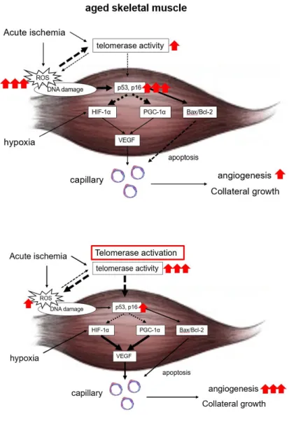
Figure 6. Working hypothesis.
Telomerase is activated by ischemia; however, senescence increases reactive oxygen species (ROS) and attenuates telomerase activity and inhibits collateral growth by increased p53 and p16 activity under conditions of ischemia. Therefore, telomerase activation may be a novel therapeutic option for enhancing collateral flow recovery and rescuing ischemic tissue in elderly individuals.
34
Abbreviations:
TERT telomerase reverse transcriptase TERC telomerase RNA template component ROS reactive oxygen species
POD post-operative days CVD cardiovascular disease PAD peripheral artery disease H2O2 hydrogen peroxide
DCFH dichlorodihydrofluorescin
TRAP Telomerase Repeated Amplification Protocol RTA The relative telomerase activity
DDR DNA damage response DSBs double strand break γH2AX histone variant H2AX RB retinoblastoma protein
HIF-1α Hypoxia inducible factor-1α VEGF vascular endothelial growth factor
PGC1α proliferator activated receptor γ co-activator 1α Bcl2 B-cell leukemia/lymphoma 2
Bax Bcl2-associated X