INTRODUCTION
Clear cell meningioma has recently been identified and included in the World Health Or- ganization (WHO) classification as a peculiar variant that differs from conventional men- ingioma by affecting younger patients, arising more often in spinal or cerebellopontine loca- tions and showing a higher recurrence rate
1,2). However, It is obvious that the rarity of this tu- mor increases the difficulty of correct diagnosis
3).
Lumbar meningioma are less common
4). When they occur, as is the case with thoracic and cervical meningiomas, they are characteris- tically invested with arachnoid and dural mater.
The present case report illustrates a rare case characterized by unusual multiple lumbar lesions, absence of dural attachment, dense ad- herence to the fascicles and a histological and immunohistochemical appearance that led to the diagnosis of clear cell meningioma.
Takanori Murakami, Kenji Imoto, Tsuneo Takebayashi, and Toshihiko Yamashita
Department of Orthopaedic Surgery, Sapporo Medical University South 1, West 16, Chuo-ku, Sapporo, Hokkaido, 060-8543
Japan
ABSTRACT A case of multiple clear cell meningioma of
cauda equina in a 10-year-old girl is reported.
She was referred with 3-month history of pain- ful episodes of backache and a markedly sore right leg and admitted for further examination.
No neurological deficiency was recognized in physical examination. Magnetic resonance imag- ing scan clearly exhibited heterogenous tumors at the level of Th12 and L2 following roent- genogram that did not show any abnormal find- ings. Tumor resection was performed by thoraco-lumbar en block laminectomy with T- Saw. Subsequently, resected laminas were re-
placed to obviate possible spinal kyphosis. The patient's recovery was uneventful. Clear cell meningioma was discerned by pathological ex- amination, whose findings including granules consistent with glycogen and ultrastructure of meningioma.
Younger patients who undergo multiple laminectomy have been reported to incur kyphotic change at involving the spine. In order to obviate such deformities, we think that the posterior element of the spine should be recon- structed.
Key words :
Clear cell meningioma, Spinal tumor, Spinal reconstruction
Address correspondence to Takanori Murakami, Department of Orthopaedic Surgery, Sapporo Medical University
South 1, West 16, Chuo-ku, Sapporo, Hokkaido, 060
!8543 Japan TEL: +81
!11
!611
!2111 (ext. 3333) FAX: +81
!11
!641
!6026
Clear Cell Meningioma of Cauda Equina in a 10-year-old Child
〈Case Report〉
Tumor Res.36,33−38(2001) 33
CASE REPORT
A 10-year-old girl was referred with a 3- month history of painful episodes of backache and a markedly sore right leg. Symptom had progressed and finally led to hospital admission for detailed examinations and possible surgical treatment.
Examination
Physical examination revealed that there
was no neurological deficiency, including muscle weakness, sensory disturbance, bowel-bladder disfunction, morbid reflex or tension sign of nerve root.
Roentgenogram did not show any abnormal findings. None of the following features were ob- served ; abnormal interpedicle distance, pedicle sign on antero-posterior view of lumbar spine nor scalloping of vertebral body or enlargement of the foramen on lateral view (Fig. 1).
In magnetic resonance imaging scan, het- erogenous tumors of about 2.0 cm in diameter were recognized on the Th12 and L2 levels of spine. Tumors with iso signal on T1 weighted imaging and low signal on T2 weighted imaging with well contrast-enhancing were recognized (Fig. 2 ).
Myelogram revealed the shadow of a in- tradural tumor with complete blockage of en- hancement of dural sack at the level between L 2 and 3 (Fig. 3).
Surgical Procedure
Thoraco-lumbar en block laminectomy from T12 to L3 was performed with T-Saw
5)with
view to later lamina reconstruction. Sequentially intradural exposure disclosed a 1.0 × 1.5 cm el- liptical tumor at the level between T12 and L1 and a 2.0 × 3.5 cm elliptical tumor at the level between L2 and L3. The cephalad tumor was strongly adhered to two fascicles and it was im- possible to dissect the tumor from nerve tissue.
On the other hand, the caudal tumor was lightly adhered to nerve and therefore easy to sepa- rate. Both tumors had no dural adherence what- soever. After transecting the fascicles surround- ing the tumors, tumors with involved fascicles were removed by microdissection without viola- tion of the capsule.
Subsequently, resected laminas were re-
Fig. 1 Roentgenogram did not show any abnormal finding. 1A: antero-posteriorview of lumbar spine: 1B: lateral view of lumbar spine.
T.MURAKAMI et al.
34
placed and attached firmly to base bone with nylon suture for the precaution of possible spi- nal kyphosis (Fig. 4).
Postoperative Course
The patient's postoperative recovery was uneventful. Postoperative magnetic resonance imaging study of the lumbar spine at 2 years after surgery demonstrated no residual tumor at the operative site and no recurrence.
Fig. 2 MRI reveals multiple spinal tumors at the level of Th12 and L2. 2A: tu- mor at L2 level with low signal on T2 weighted imaging on axial view. 2 B: well enhanced tumors at the level of Th12 and L2 on saggital view.
Fig. 4 CT scan shows recapped lamina on L2.
Fig. 3 Myelogram exhibited the shadow of a spinal tumor with complete block of en- hancement at L2/3 level.
36(2001) Clear Cell Meningioma of Cauda Equina 35
Pathological Examination
Grossly, the two tumors consisted of ellipti- cal , well-circumscribed firm pale-gray nodules measuring 1.5 × 1.0 cm and 2.0 × 3.5 cm respec- tively.
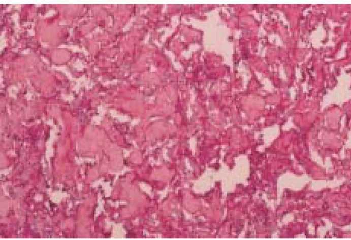
Microscopically they were characterized by sheets and vaguely defined nests of cells. Hya- line fibrosis was present. The cells had abun- dant clear cytoplasm with round uniform bland- appearing nuclei without mitoses. Occasionally the cells formed small whorls, as is characteristi- cally seen in more typical meningiomas (Fig. 5).
Histochemical staining revealed the pres- ence of a small-to-moderate amount of periodic acid Schiff-positive diastase-sensitive granules consistent with glycogen (Fig. 6). Immunohisto-
chemical examinations with vimentin, EMA and S-100 were less positive.
On ultrastructural examination, the tumors were characterized by moderately abundant cy- toplasm literally filled with glycogen particles.
Also interdigitation of cell membranes were rec- ognized. The cells were joined by junctional complexes, most often well-formed desmosomes, characterized as meningioma (Fig. 7).
Based on pathological examinations, the tu- mors were diagnosed as clear cell meningioma.
DISCUSSION
Clear cell meningioma is an uncommon, ag- gressive and morphologically unique variety of meningioma that has been reported to occur in the spine and cerebellopontine angle
6,7,8). It shows no sex predilection and affects young patients more often than conventional varieties of men- ingioma
7). It seems to be a more aggressive tu- mor, with higher incidence of recurrence. Char- acteristically, this tumor is composed of uniform clear cells and has a prominent hyalinized colla- genous stroma
2). Typical light microscopic and ultrastructural features indicative of menin- gothelial differentiation are usually sparse or al- most completely absent in clear cell men- ingioma
9).
Clear cell meningioma is an exceedingly
Fig. 7 Ultrastructure of tumor (× 4000). Remarkable cytoplasmic glycogen depositions including variable intermediate filament, imbrication of cell membranes and well-formed desmosomal junctions were recognized.Fig. 5 Light microscopic appearance of tumor (PAS,
× 200). The cells had abundant clear cytoplasm with round uniform bland-appearing nuclei.
Fig. 6 Histochemical staining revealed the presence of periodic acid Schiff-positive diastase-sensitive granules consistant with glycogen (PAS, × 200).
T.MURAKAMI et al.
36
rare tumor of meninges. Only 14 cases have been described, six of which were spinal in- tradural tumors, five lumbar and one tho- racic
3,10,11). Our case is clearly rare at the point of younger age and multiple involvement of lum- bar spine.
Generally speking, under light microscopic observation, there is an obvious lack of men- ingiomatous features, thus making diagnosis dif- ficult. It is reported that ultrastructurally the tumor have features common to all men- ingiomas, such as junctional complexes, inter- digitation of cells and well-structured desmo- somes, which are very helpful to assess the cor- rect diagnosis
7,12). In our case, the glycogen in cy- tosol with periodic acid Schiff-positive diastase- sensitive granules and characterization of the ul- trastructure in spite of negative reaction in im- munohistochemical examinations were helpful for diagnostic purpose.
Clear cell meningioma can be confused with other clear cell tumors or clear cell-like tumors, for example renal cell carcinoma, chordoma, he- mangiopericytoma, chondroma or untypical os- teosarcoma
1,3). Lee et al. reported the magnetic resonance imaging features of clear cell men- ingioma
13). However, it appears to be difficult to make a correct diagnosis by clinical fingings alone.
The biologic behavior of meningiomas is variable. Clinical prognosis is correlated with histopathologic parameters
3,14,15). Zorludemir et al.
reported that recurrence was noted in 61% of cases, local discontinuous spread in 15%, wide- spread cranial to spinal metastasis in 8% and mortality in 23%
7,16). The present case has not reffered any recurrence in the 2 years since sur- gery. However a much longer follow-up will be necessary with this patient.
Younger patients undergoing multiple laminectomy are reported to incur kyphotic change at involving the spine within 5 years in 3 to 7 % of the cases
17). In order to obviate the possibility of such deformity, we performed en block laminectomy with T-Saw and replaced the resected laminas in order to reconstruct the
posterior element of the spine after resection of tumor. It is now 2 years since this surgery was undertaken and this case has not, so far, exhib- ited any deformity.
In summary, we reported a rare case of spi- nal clear cell meningioma in 10-year-old girl. It was characterized at the point of younger age and multiple involvement of lumbar spine clini- cally. Correct diagnosis was attained with patho- logical examination characterized by rich glyco- gen in cytosol and ultrastructure of men- ingioma. Additionally, we performed en block laminectomy with T-Saw and replaced the re- sected laminas to reconstruct posterior element of the spine to obviate the possible kyphotic de- formity.
REFERENCES
1.Francesc A, Josep L. Clear Cell Meningioma of the Lumbo-sacral Spine With Chordoid Features. Ultrastruct Pathol 1999, 23: 51
!58.
2.Scheutgayer BW. Tumors of meninges: pro- posed modifications of the World Health Or- ganization classification. Acta Neuropathol 1990, 80: 343
!354.
3.Marco P, Stephan P, Tibor M, Jorge CN.
Clear Cell Meningioma: Report of a Spinal Case. Gen Diaan Pathol 1995, 141: 261
!267.
4.McCutcheon IE. The biology of men- ingiomas. J Neurooncol 1996, 29: 207
!216.
5.Tomita K, kawahara N. The treadwire saw:
A new device for cutting bone. J Bone Joint Surg Am1996, 78: 1915
!1917.
6.Shiraishi K. Glycogen-rich meningioma.
Case report and short review. Neurosurg Rev 1991, 14: 61
!64.
7.Zorludemir S, Scheithauer BW, Horose T, Van Houten C, Miller G, Meyer FB. Clear cell meningioma: a clinicopathologic study of a potentially aggressive variant of men- ingioma. Am J Surg Pathol 1995, 19, 493
!505.
8.Jennifer GC, Keith YC, Marc KR, Cheryl AM, Fred JE. Intraparenchymal Clear Cell Meningioma of the Brainstem in a 2-
36(2001) Clear Cell Meningioma of Cauda Equina 37
Year-Old Child. Pediatr Neurosurg 1998, 28:
27
!30.
9.Zuppan CW, Liwnicz BH, Weeks DA. Men- ingioma with chordoid features. Ultrastruct Pathol 1994, 18: 29
!32.
1 0.Robert NNH, Susan CJ. Nondural-based lumbar clear cell meningioma. J Neurosurg 1996, 84: 264
!266.
1 1.Matsui H, Kanamori M, Abe Y, Sakai T, Wakaki K. Multifocal clear cell meningioma in the spine: a case report. Neurosurg Rev 1998, 21: 171
!173.
1 2.George S, John K, Hope MB. Meningeal Tu- mors of Childhood. Cancer 1989, 63: 1205
!1210.
1 3.Lee W, Chang KH, Choe G, Chi JG, Chung CK, Kim IH, Han MH, Park SW, Shin SJ, Koh YH. MR imaging features of clear-cell meningioma with diffuse leptomeningeal seeding. AJNR Am J Neuroradiol 2000, 21:
130
!132.
1 4.Dubois A, Sevely A, Boetto S, Delisle MB, Manelfe C. Clear-cell meningioma of the cauda equina. Neuroradiology 1998, 40: 743
!747.
1 5.Smith DA, Cahill DW. The biology of men- ingiomas. Neurosurg Clin N Am 1994, 5:
201
!215.
1 6.Pimentel J, Fernandes A, Pinto AE, Fon- seca I, Moura NJF, Lobo AJ. Clear cell men- ingioma. Variant and clinical aggressive- ness. Clin Neuropathol 1998, 17: 141
!146.
1 7.Raimondi AJ. Laninotomy and total recon- struction of the posterior spinal arch for spi- nal canal surgery in childhood. J Neruro- surg 1976, 45: 555
!560.
(Accepted for publication, Jan. 21, 2002)
T.MURAKAMI et al.
38