• 検索結果がありません。

Gastrointestinal Endoscopy for Patients with High Levels of Serum CEA and CA19-9

N/A
N/A
Protected

Academic year: 2021

シェア "Gastrointestinal Endoscopy for Patients with High Levels of Serum CEA and CA19-9"

Copied!
9
0
0

読み込み中.... (全文を見る)

全文

(1)

Gastrointestinal Endoscopy for Patients with High Levels of Serum CEA and CA19-9

Norihiro S UZUKI

1)

, Atsushi K ATAGIRI

*1)

, Shinya N AKATANI

1)

, Kazuo K IKUCHI

1)

, Toshihiko G OCHO

1)

, Fumito Y ANAGISAWA

1)

,

Kazuya I NOKI

1)

, Kenichi K ONDA

1)

, Masayuki T OJO

1)

, Yutaro K UBOTA

2)

, Kazuo K ONISHI

1)

, Fuyuhiko Y AMAMURA

2)

,

and Hitoshi Y OSHIDA

1)

Abstract : Serum levels of tumor markers, such as carcinoembryonic antigen (CEA)

and carbohydrate antigen 19-9 (CA19-9) , are often measured to detect potential malignancy. When these levels are high, the presence or absence of malignancy is confirmed via a more detailed examination using gastrointestinal (GI) endoscopy and computed tomography. The rate of confirmation of malignancy upon such a follow-up is unknown. This study aimed to investigate the malignancy detection rate via GI endoscopy for patients with high levels of serum CEA and CA19-9.

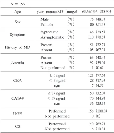
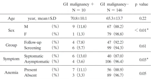
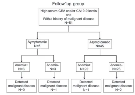
All patients who underwent such GI endoscopy between January 2018 and Febru- ary 2019 at Showa University Hospital were included in this study. The patients were divided into a follow-up group and a screening group, depending on the purpose of measuring their serum CEA/CA19-9 levels. There were 156 patients who underwent GI endoscopy because of high CEA/CA19-9 levels within the study period. Advanced malignant lesions were detected in 10 patients (6.4%) , including seven cases of colorectal cancer and three cases of upper GI malignancies. In the screening group, six cases (5.7%) of GI malignancies were detected, none of which were found in asymptomatic patients without anemia. In the follow-up group, four cases (7.8%) of GI malignancies were detected; three patients were asymptomatic, and one patient had anemia. Our findings suggest that high serum CEA/CA19-9 levels in asymptomatic patients without anemia and without a history of malig- nancy do not indicate the presence of malignancy. However, high serum CEA/

CA19-9 levels may indicate the potential presence of GI malignancies for patients with a history of malignant tumors, even if they are asymptomatic and do not have anemia.

Key words : CEA, CA19-9, endoscopy, malignancy, cancer

Introduction

 Gastrointestinal (GI) cancer is one of the leading causes of morbidity and mortality from malignant disease in Western and Asian countries. Globally, colorectal cancer is the fourth and Original

1)

Department of Medicine, Division of Gastroenterology, Showa University School of Medcine, 1-5-8 Hatanodai, Shinagawa-ku, Tokyo 142-8666, Japan.

2)

Endoscopic Center, Showa University Hospital

To whom corresponding should be addressed.

(2)

gastric cancer the sixth most common malignancy

1)

. In Japan, population-based cancer screening is conducted for gastric, colorectal, lung, breast, and uterine cancer. Because of the high inci- dence of gastric cancer, population-based screening is recommended for individuals older than 50 years, either annually, by performing conventional double-contrast barium radiography, or bienni- ally, by performing upper GI endoscopy

2,3)

. In Japan, a fecal immunochemical test is performed for population-based colorectal cancer screening.

 The most effective modality to detect both gastric and colorectal cancer is an endoscopic examination, and the gold standard for cancer diagnosis is a pathological examination, usually by endoscopic biopsy. Therefore, upper GI endoscopy and colonoscopy are recommended if abnor- malities are found during primary screening. Private medical facilities in Japan offer various kinds of individual screening at the patientʼs own cost. These tests often entail measuring serum levels of tumor markers, such as carcinoembryonic antigen (CEA) and carbohydrate antigen 19-9

(CA19-9) , as only a blood sample is required

4-8)

. CEA levels are useful for surgical treatment planning, posttreatment follow-up, and determining the prognosis of patients diagnosed with colorectal cancer

9)

. Additionally, a meta-analysis reported by Deng et al. suggested that preop- erative elevation of CEA levels is an independent indicator of adverse prognosis

10)

. However, low rates of sensitivity, particularly in the early stages of the disease, prevent the use of any of these serologic markers to diagnose colorectal and gastric cancer

9,11-13)

. Moreover, elevated levels of CEA may also be caused by benign diseases, such as gastritis, peptic gastroduodenal ulcer- ation, diverticulitis, liver disease, chronic obstructive pulmonary disease, diabetes, and any acute or chronic inflammation

14)

. From the viewpoint of lifestyle factors, CEA levels are significantly higher in smokers than in non-smokers

15,16)

.

 When tumor marker levels are high, a more detailed examination is performed with the help of GI endoscopy or computed tomography to confirm the presence or absence of malignancy.

However, the rate of confirmation of malignancy upon such a follow-up is unknown. In this study, we investigated the rate of malignancy, as determined by endoscopic examination, for patients with high tumor marker levels.

Patients and methods

 The present study was conducted according to the guidelines of the Declaration of Helsinki and was approved by the Ethics Committee on Human Studies of Showa University, Tokyo, Japan. This was a retrospective, single-center study of consecutive patients who underwent GI endoscopy between January 2018 and February 2019 at Showa University Hospital.

 Data (age, sex, symptoms at first hospital visit, family history of malignant disease, endoscopic

findings, pathological findings of endoscopic biopsy, blood count, and serum CEA and CA19-

9 levels) of patients who underwent endoscopy because of high serum CEA and CA19-9 levels

were obtained from their electronic medical records. The patients were classified into two groups

according to the purpose of measuring their serum CEA/CA19-9 levels: a follow-up group, with

previously identified malignant tumors, or a screening group, for whom regular cancer screening

was undertaken. The cut-off values of serum CEA and CA19-9 levels in this study were set at

(3)

5 and 34 ng/ml, respectively. Patients with a blood hemoglobin concentration < 14 g/dl for males and < 12 g/dl for females were declared to have anemia, according to the standard reference ranges used at our hospital.

Statistical analysis

 Data are presented as frequencies and percentages. The imputed data were statistically analyzed using JMP Pro 14 for Windows (SAS Institute Inc., Cary, NC, USA) . A descriptive analysis was performed, and differences between the groups were tested using the Chi-square test and the Mann–Whitney U test. A p-value ≤ 0.05 was considered statistically significant.

Results

Patient characteristics

 There were 156 patients who underwent GI endoscopy (upper GI endoscopy and/or colo- noscopy) because of high CEA/CA19-9 levels within the study period. Table 1 describes the patientsʼ baseline characteristics. There were 121 patients with elevated CEA levels (77.6%) and 50 patients with elevated CA19-9 levels (32.1%) . The average hemoglobin concentration was

Table 1. Patient characteristics N = 156

Age year, mean S.D (range) 65.6 13.6 (30-90)

Sex Male      (%)  76 (48.7)

Felmale    (%)  80 (51.3)

Symptom Syptomatic   (%)  46 (29.5)

Asymptomatic (%) 110 (70.5)

History of MD Present    (%)  51 (32.7)

Absent    (%) 105 (67.3)

Anemia

Present     (%)  63 (40.4)

Absent      (%)  92 (59.0)

Not performed (%)  1 (0.6)

CEA

5 ng/ml 121 (77.6)

< 5 ng/ml  28 (17.9)

n,m  7 (4.5)

CA19-9

37 ng/ml  50 (32.0)

< 37 ng/ml  70 (44.9)

n,m  36 (23.1)

UGE Performed  156 (100.0)

Not performed  0 (0)

CS Performed 140 (89.7)

Not performed  16 (10.3)

S.D; standard deviation, MD; malignant disease, n,m; not measured,

UGE; upper GI endoscopy, CS; colonoscopy

(4)

13.6 and 12.5 g/dl for male and female patients, respectively, and 40 male (52.6%) and 23 female

(28.8%) patients were anemic. There were 51 patients (32.7%) in the follow-up group and 105 (67.3%) in the screening group. In total, 139 patients (89.1%) underwent a colonoscopy at Showa University Hospital, one patient (0.64%) underwent colonoscopy at another medical facility, and 16 patients (10.3%) did not undergo colonoscopy, either due to age or because no request was made.

Detected malignant disease

 Malignant lesions were detected in 10 patients (6.4%) . Three of them (1.9%) were diagnosed with malignancy during upper GI endoscopy, and seven (4.5%) were diagnosed with colorectal cancer during colonoscopy. The details of these malignant lesions are presented in Table 2.

 Table 3 summarizes the statistical significance of the cancer detection rate in terms of the

Table 2. Cases with newly detected gastrointestinal malignancies

Case Sex Age Tumor maker Diagnosis Symptom History of MD Anemia CEA CA19-9 Pathology  1 Male 71 CEA Advanced colon cancer + gastric cancer ­  9.1 14 adenocarcinoma  2 Male 68 CEA/CA19-9 Advanced colon cancer + - ­  5.8 491.1 adenocarcinoma( tub2 〜 por)

 3 Male 74 CEA Advanced colon cancer + - ­ 40.8 156.6 adenocarcinoma(tub1)

 4 Male 68 CEA Early colon cancer ­ lung cancer ­  7.1  4.4 adenocarcinoma in adenoma  5 Male 85 CEA/CA19-9 gastric malignant lymphoma + - ­  9.5 3931.8 atypical lymphoid cells  6 Male 69 CEA Advanced EGJ cancer ­ esophageal cancer ­ 33.2   41.8 EGJ carcinoma.sig + tub1  7 Male 77 CEA/CA19-9 Advanced duodenal cancer ­ bile duct cancer + 11.2 1729.7 adenocarcinoma  8 Female 76 CEA Advanced colon cancer ­ - + 25.7 n,m adenocarcinoma(tub2)

 9 Male 74 CEA Advanced colon cancer + - +  5.3 34 adenocarcinoma(tub2)

10 Male 46 CEA Early colon cancer + - +  6.8  18.2 Early colonic adenocarcinoma

( tub1) in adenoma MD; malignant disease, EGJ; esophagogastric junction

Table 3. Significance of cancer detection rate by patient background GI malignancy + GI malignancy­ p value

N = 10 N = 146

Age year, mean S.D 70.8 10.1 65.3 13.7   0.22

Sex M       (%)  9 (11.8)  67 (88.2)

< 0.01

F      (%) 1 (1.3)  79 (98.8)

Group Follow-up   (%) 4 (7.8)  47 (92.2)

  0.61 Screening   (%) 6 (5.7)  99 (94.3)

Symptom Syptomatic  (%)  6 (13.0)  40 (87.0)

   0.03

Asymptomatic (%) 4 (3.6) 106 (96.4)

Anemia Present    (%)  7 (11.1)  56 (88.9)

  0.05

Absent    (%) 3 (3.3)  89 (96.7)

S.D; standard deviation,

; significantly different, M/F; male/female

(5)

patientʼs background. There were statistically significant differences in cancer detection rates between male and female patients (p < 0.01) as well as between symptomatic and asymptomatic patients (p = 0.03) . There was no statistically significant difference in the detection rate of GI malignant tumors between patients with and without anemia (p > 0.05) .

 As summarized in Table 4, there was no correlation between serum CEA levels and the presence of GI malignancy ( p = 0.61) . Similar results were observed for serum CA19-9 levels ( p = 0.16) .

Screening group

 In the screening group, six cases (5.7%) of GI malignancies were detected, of which five were colorectal cancer (four advanced cancer and one mucosal cancer) , and one was gastric malignant lymphoma (see Tables 2 and 3) . Of these six patients, five were symptomatic at the first visit to our hospital, and all patients were found to be anemic via blood tests. In the screening group, there was a statistically significant difference in the detection rate of GI malignancies in patients with or without symptoms and anemia (p = 0.04 and p < 0.01, respectively) . Fur- thermore, no GI malignancies were detected in asymptomatic patients without anemia in the screening group (Fig. 1) . Only 27 patients (25.7%) underwent cancer screening before undergo- ing endoscopy, whereas 78 patients (74.3%) had not previously undergone any cancer screening.

Table 4. Relationship between CEA/CA19-9 and the presence of gastrointestinal malignancy unit Total GI malignancy + GI malignancy­ p value

CEA level  (n = 121) ng/mL, median   7.3   7.2  7.3 0.61

CA19-9 level (n =  50) ng/mL, median 101.0 156.6 34.0 0.16

Fig. 1.   Flow chart of the screening group based on their symptoms and the

presence of anemia. There was no patient with GI malignancy without

symptoms and anemia.

(6)

Moreover, of the six patients with malignant tumors in this group, only one had previously undergone cancer screening (colorectal) , and the remaining five with GI malignancy had never undergone cancer screening.

Follow-up group

 Four cases (7.8%) of GI malignancies were detected in the follow-up group (see Tables 2 and 3) . Of them, one had esophageal gastric junction cancer, one had duodenal cancer, and two had colon cancer (one early and one advanced) . In the follow-up group, six patients (11.8%) were symptomatic when the tumor markers were measured, and only one patient was found to have GI malignant disease, specifically advanced colon cancer. There were 25 (49.0%) patients in the follow-up group who were anemic, and of them, only one was found to have GI malignant disease (advanced duodenal cancer) (Fig. 2) . In the follow-up group, 24 patients (47.1%) had undergone upper GI endoscopy, and 27 patients (52.9%) had undergone colonoscopy. Of the four patients with newly detected GI malignancy in this group, two patients had not undergone GI endoscopy in the previous three years.

Discussion

 In this study, we investigated the incidence of GI malignant disease and its characteristics in patients who had undergone endoscopy due to high serum CEA and CA19-9 levels. Conven- tionally, tumor markers for GI cancer are used as ancillary indicators of therapeutic efficacy and as signs of recurrence of malignant tumors but are less useful as detection tools

9-13)

. However, tumor markers are sometimes used as screening tools for cancer detection because they can be measured with simple blood tests. If the tumor marker level is high, further examination is required to confirm the presence or absence of cancer. In particular, serum CEA and CA19-9

Fig. 2.   Flow chart of the follow-up group based on their symptoms and the

presence of anemia.

(7)

levels are sometimes high in patients with GI cancer. For such patients, it is necessary to per- form highly invasive examinations, such as upper GI endoscopy and colonoscopy. Furthermore, for patients who have had malignant tumors in other organs, the effectiveness of GI endoscopy when high serum CEA and CA19-9 levels are detected during a follow-up hospital visit has not been clarified.

 In the present study, most of the patients had high serum CEA levels. Because of the small number of patients that were negative for CEA, it was difficult to compare these results with those of previous studies regarding the efficacy of using serum CEA levels to detect GI malig- nancies. However, as shown in Table 4, CEA levels were not significantly correlated with the presence of GI malignancies.

 In this study, patients were divided into a follow-up group and a screening group, due to dif- ferences in the reason for measuring CEA and CA19-9 levels and in potential cancer risks. The GI malignancy detection rate was not significantly different between the two groups, suggesting that the serum CEA and CA19-9 levels were not correlated with GI malignancy detection via endoscopy for patients as a whole.

 Serum CEA levels are known to be affected by other factors such as aging and smoking

14-16)

. It is possible that patients in the current study were a mixture of smokers and non-smokers. Patientsʼ smoking habits were not available for this study. However, it is difficult to determine whether elevated CEA levels are due to smoking or malignancy before scanning. Therefore, the presence of a smoking habit seems to be difficult to become the reason for not doing the endoscope.

 This study showed that these patientsʼ symptoms were significantly associated with the detec- tion of GI malignant disease (see Table 3) , anemia tended to be associated with the detection of GI malignancies, although this was not statistically significant. These tendencies were espe- cially significant for patients with high CEA and CA19-9 levels without a history of malignant diseases (screening group) . There was a statistically significant difference in the detection rate of GI malignancies with or without symptoms and anemia in the screening group. Of the six patients with newly detected GI malignancies in the screening group, only one was asymptomatic, and three patients had anemia. The asymptomatic patient who had advanced colorectal cancer had not received population-based cancer screening, and this case could have been detected with- out measuring tumor marker levels if a fecal immunochemical test would have been performed.

 For asymptomatic patients in the study, GI malignancies were more frequently detected in the follow-up group (6.7%) than in the screening group (1.5%) . In the follow-up group, 49.0% and 51.0% of patients had not previously undergone upper GI endoscopies and colonoscopies, respec- tively. This suggested that patients with a history of malignant tumors may need to be screened for GI cancer regularly, even if they are asymptomatic.

 This study suggests that high serum CEA/CA19-9 levels may not be an accurate indication of

GI malignancy for asymptomatic patients without anemia who have never been diagnosed with

cancer. Thus, GI endoscopy for these patients might be unnecessary. By avoiding unnecessary

examinations, medical resources and medical costs can be reduced, and the risk of complications

associated with endoscopy may be avoided. Endoscopy is often considered a major risk for the

(8)

elderly; therefore, it is recommended that such invasive examinations only be performed after confirming the presence of symptoms and anemia.

 Currently, the number of people undergoing population-based cancer screening in Japan is low

17)

. This is reflected in the current study, where a low number of screening group patients had previously undergone population-based cancer screening (25.7%, data not shown) . This may be due to the low tolerability of conventional double-contrast barium radiography, upper GI endoscopy, and the fecal immunochemical test. In recent years, new tumor markers, such as CA11-19

18,19)

, have been identified, and blood-based gene biomarker panels have been developed

20)

. It is essential to develop methods to improve the patient screening rate, thereby increasing the cancer detection rate. However, of the screening tools currently in use, the sim- pler methods are less effective than the more invasive ones. Therefore, new screening methods need to be developed, and the significance of tumor marker tests should be explained to the patient to improve the patient screening rate.

 Second, among patients with high serum CEA/CA19-9 levels, only those who had undergone GI endoscopy were investigated. Therefore, the detection rate of malignant GI tumors for patients with high tumor marker levels in this study cannot be compared with the rates found in previous studies.

 In conclusion, GI endoscopic examination is not recommended for patients with high serum CEA/CA19-9 levels who are asymptomatic, have no anemia, and have no history of malignancy.

Furthermore, for patients with a history of malignant tumors and high serum CEA/CA19-9 levels, GI malignancies may be present, even if the patients are asymptomatic and without anemia.

 There were some limitations to this study. First, this was a single-center, retrospective study with a limited population size. Second, among patients with high serum CEA/CA19-9 levels, only those who have undergone gastrointestinal endoscopy were investigated. Therefore, the detection rate of malignant GI tumors for patients with high tumor marker levels in this study cannot be compared with the rates found in previous studies.

 In conclusion, GI endoscopic examination is not recommended for patients with high serum CEA/CA19-9 levels who are asymptomatic, have no anemia, and have no history of malignancy.

Furthermore, for patients with a history of malignant tumors and high serum CEA/CA19-9 levels, GI malignancies may be present even if the patients are asymptomatic and without anemia.

Acknowledgments

 The authors declare no conflicts of interest. We disclose that a version of this paper was presented at the Japanese Digestive Disease Week (JDDW2019) poster session in Japan on November 23, 2019. This paper has not been published elsewhere.

Conflict of interest

 The authors have declared no conflict of interest.

(9)

References

1) Bray F, Ferlay J, Soerjomataram I, et al. Global cancer statistics 2018: GLOBOCAN estimates of incidence and mortality worldwide for 36 cancers in 185 countries. CA Cancer J Clin. 2018;68:394-424.

2) Hamashima C, Shibuya D, Yamazaki H, et al. The Japanese guidelines for gastric cancer screening. Jpn J Clin Oncol. 2008;38:259-267.

3) Choi IJ. Endoscopic gastric cancer screening and surveillance in high-risk groups. Clin Endosc. 2014;47:497-503.

4) Horie Y, Miura K, Matsui K, et al. Marked elevation of plasma carcinoembryonic antigen and stomach carcinoma.

Cancer. 1996;77:1991-1997.

5) Kodama I, Koufuji K, Kawabata S, et al. The clinical efficacy of CA 72-4 as serum marker for gastric cancer in comparison with CA19-9 and CEA. Int Surg. 1995;80:45-48.

6) Carpelan-Holmstrom M, Louhimo J, Stenman UH, et al. CEA, CA 19-9 and CA 72-4 improve the diagnostic accuracy in gastrointestinal cancers. Anticancer Res. 2002;22:2311-2316.

7) Lai IR, Lee WJ, Huang MT, et al. Comparison of serum CA72-4, CEA, TPA, CA19-9 and CA125 levels in gastric cancer patients and correlation with recurrence. Hepatogastroenterology. 2002;49:1157-1160.

8) Marrelli D, Pinto E, De Stefano A, et al. Clinical utility of CEA, CA 19-9, and CA 72-4 in the follow-up of patients with resectable gastric cancer. Am J Surg. 2001;181:16-19.

9) Locker GY, Hamilton S, Harris J, et al. ASCO 2006 update of recommendations for the use of tumor markers in gastrointestinal cancer. J Clin Oncol. 2006;24:5313-5327.

10) Deng K, Yang L, Hu B, et al. The prognostic significance of pretreatment serum CEA levels in gastric cancer:

a meta-analysis including 14651 patients. PLoS One. 2015;10:e0124151. (accessed 2019 Dec 26) Available from:

https://www.ncbi.nlm.nih.gov/pmc/articles/PMC4400039/pdf/pone.0124151.pdf

11) Macdonald JS. Carcinoembryonic antigen screening: pros and cons. Semin Oncol. 1999;26:556-560.

12) Palmqvist R, Engaras B, Lindmark G, et al. Prediagnostic levels of carcinoembryonic antigen and CA 242 in colorectal cancer: a matched case-control study. Dis Colon Rectum. 2003;46:1538-1544.

13) van der Schouw YT, Verbeek ALM, Wobbes T, et al. Comparison of four serum tumour markers in the diagnosis of colorectal carcinoma. Br J Cancer. 1992;66:148-154.

14) Shapiro M, Scapa E. Elevated carcinoembryonic antigen (CEA) levels in a patient with no malignancy. Hepato- gastroenterology. 2000;47:163-164.

15) Alexander JC, Silverman NA, Chretien PB. Effect of age and cigarette smoking on carcinoembryonic antigen levels. JAMA. 1976;235:1975-1979.

16) Sajid KM, Parveen R, Durr-e-Sabih, et al. Carcinoembryonic antigen (CEA) levels in hookah smokers, cigarette smokers and non-smokers. J Pak Med Assoc. 2007;57:595-599.

17) Cancer Information Service, National Cancer Center, Japan. Cancer Statistics in Japan 2018 (Internet) . (accessed 2019 Dec 23) Available from https://ganjoho.jp/en/professional/statistics/brochure/2018_en.html

18) Overholt BF, Wheeler DJ, Jordan T, et al. CA11-19: a tumor marker for the detection of colorectal cancer. Gastro- intest Endosc. 2016;83:545-551.

19) Kanaan Z, Antaki F. CA11-19: a promising marker for the detection of colorectal cancer. Gastrointest Endosc.

2016;84:753-754.

20) Marshall KW, Mohr S, Khettabi FE, et al. A blood-based biomarker panel for stratifying current risk for colorectal cancer. Int J Cancer. 2010;126:1177-1186.

[Received November 29, 2019 : Accepted January 6, 2020]

Table 1.  Patient characteristics N = 156
Table 3.  Significance of cancer detection rate by patient background GI malignancy + GI malignancy­ p value
Table 4.  Relationship between CEA/CA19-9 and the presence of gastrointestinal malignancy unit Total GI malignancy + GI malignancy­ p value
Fig. 2.    Flow chart of the follow-up group based on their symptoms and the  presence of anemia.

参照

関連したドキュメント

To investigate whether defects in the SPATA17 gene are associated with azoospermia due to meiotic arrest, a mutational analysis was conducted, in which the SPATA17 coding regions

It is shown that plasma endothelial lipase (EL) activity inversely correlated with HDL-C levels, and EL activity in CAD patients was significantly higher than in non CAD

The aim of this pilot study was to measure PBDEs in serum samples from young Japanese males and to examine the relationship between serum PBDE levels and sperm quality.. MATERIALS

If the binding between granulocytic elastase and ulinastatin is stronger than that of granulocytic elastase and a1-PI, and we use an enzyme-linked immunosorbent assay that

The correlation between IL-17 levels and disease duration was not also recognized, although patients with normal serum IL-17 levels showed significantly higher modified Rodnan

The major results of the present study are that patients with SSc have a high frequency of diastolic dysfunction and sympathetic abnormality as detected by myocardial perfusion

The present study demonstrated a correlation between serum fractalkine levels and response to UDCA; although patients with low fractalkine levels (<3 ng/ml) before

We measured blood levels of adiponectin in SeP knockout mice fed a high sucrose, high fat diet to examine whether SeP was related to the development of hypoadiponectinemia induced