Title
Gene therapy of atopic dermatitis and cancer by sustained
expression of interferon-γ in mice( Digest_要約 )
Author(s)
Watcharanurak, Kanitta
Citation
Kyoto University (京都大学)
Issue Date
2013-09-24
URL
http://dx.doi.org/10.14989/doctor.k17862
Right
学位規則第9条第2項により要約公開; 許諾条件により要約
は2013-12-23に公開
Type
Thesis or Dissertation
Textversion
none
Gene therapy of atopic dermatitis and cancer by
sustained expression of interferon-γ in mice
2013
Table of contents
Preface--- 1 Chapter I
Improvement of atopic dermatitis by sustained exogenous expression of IFN-γ in NC/Nga mice via Th1 polarization
Section 1. Sustained expression of IFN-γ ameliorates the development of atopic dermatitis
I-1-1 Introduction--- 3 I-1-2 Materials and Methods--- 4 I-1-3 Results--- 6 I-1-3-a IFN-γ concentration in the serum of NC/Nga mice after injection of IFN-γ-expressing
plasmid DNA--- 6 I-1-3-b IL-12 concentration in the serum of NC/Nga mice after injection of IFN-γ-expressing
plasmid DNA--- 7 I-1-3-c IgE concentration in the serum of NC/Nga mice after injection of IFN-γ-expressing
plasmid DNA--- 8 I-1-3-d mRNA expression of cytokine and chemokine in spleen cells of NC/Nga mice after
injection of IFN-γ-expressing plasmid DNA--- 8 I-1-3-e Skin lesions of NC/Nga mice after injection of IFN-γ-expressing plasmid
DNA--- 9 I-1-3-f Scratching behavior of NC/Nga mice after injection of IFN-γ-expressing plasmid DNA-- 10 I-1-3-g TEWL of NC/Nga mice after injection of IFN-γ-expressing plasmid DNA--- 11 I-1-3-h Histological examination of skin sections of NC/Nga mice after injection of
IFN-γ-expressing plasmid DNA--- 12 I-1-3-i Adverse effects of IFN-γ gene transfer--- 13 I-1-4 Discussion--- 14 Section 2. Sustained IFN-γ expression improves symptoms of acute phase of atopic dermatitis I-2-1 Introduction--- 17 I-2-2 Materials and Methods--- 17 I-2-3 Results--- 19 I-2-3-a Time-course and pharmacokinetic parameters of IFN-γ after hydrodynamic injection of
pCpG-Muγ--- 19 I-2-3-b Effects of IFN-γ gene transfer on the skin lesions of NC/Nga mice--- 20 I-2-3-c Serum concentrations of IgG1 and IgE in NC/Nga mice after IFN-γ gene transfer--- 21
I-2-3-d mRNA expression of Th1, Th2 and Treg cytokines in the spleen of NC/Nga mice after
IFN-γ gene transfer--- 21
I-2-3-e TEWL and scratching behavior of NC/Nga mice after IFN-γ gene transfer--- 22
I-2-3-f Histopathological examination of skin sections of NC/Nga mice--- 23
I-2-3-g Effect of IFN-γ gene transfer on body temperature and liver injury--- 23
I-2-3-h Effect of IFN-γ gene transfer on contact dermatitis--- 23
I-2-4 Discussion--- 24
Chapter II Effects of highly upregulated indoleamine 2,3-dioxygenase 1 on anti-tumor activity of IFN-γ gene transfer in tumor-bearing mice II-1 Introduction--- 26
II-2 Materials and Methods--- 26
II-3 Results and Discussion--- 27
Summary--- 29
Acknowledgments--- 30
List of Publications--- 31
1
Preface
Interferon (IFN)-γ, a cytokine with antiviral, immunomodulatory, and antiproliferative activities, has been extensively studied as a treatment for many diseases, such as viral infections, allergic diseases and cancer. However, a major limitation of IFN-γ-based therapy is the short in vivo half-life of IFN-γ. Therefore, IFN-γ gene transfer has been considered to be a promising method to overcome this limitation because, theoretically, it should be able to extend the duration of action of the short-lived cytokine.
Previous studies in my laboratory showed that the removal of unmethylated cytosine-phosphate-guanine (CpG) dinucleotides from plasmid DNA prolonged the plasmid-mediated transgene expression. A single injection of pCpG-Muγ, a CpG depleted plasmid DNA encoding murine IFN-γ, via tail vein using hydrodynamic injection method resulted in prolonged therapeutic serum concentrations of IFN-γ over 1 month. This sustained transgene expression of IFN-γ could be used as a new therapeutic strategy for chronic diseases, but no such attempts have been made thus far. Therefore, in this thesis, I aimed to investigate the beneficial effects of sustained exogenous expression of IFN-γ in treating atopic dermatitis and cancer in mouse models.
In Chapter I, NC/Nga mice, a model of human atopic dermatitis, at two different stages of the disease were used. In Section 1, NC/Nga mice without dermatitis, which represent ones at a stage before the onset of the disease, were used to study the effects of IFN-γ gene transfer in preventing atopic dermatitis. The mice were injected with pCpG-Muγ or pCMV-Muγ, a conventional short-term IFN-γ-expressing plasmid, into the tail vein using hydrodynamic injection method to, first, examine the importance of the duration of action of IFN-γ in regulating the balance of T helper (Th) cell subsets (Th1 and Th2) in NC/Nga mice. Then, it was examined whether the sustained supply of IFN-γ is necessary for preventing the onset of the disease development. In Section 2, NC/Nga mice with early phase of dermatitis were used to investigate whether the sustained IFN-γ expression is effective in ameliorating or treating the symptoms of the disease.
In Chapter II, the beneficial effect of sustained expression of IFN-γ in treating cancer was investigated. Because it has been reported that IFN-γ induces the expression of indoleamine 2,3-dioxygenase 1 (IDO1), a tryptophan-metabolizing enzyme involved in the induction of tumor immune tolerance. Therefore, the Lewis lung carcinoma (LLC) tumor-bearing wild-type and IDO1 knockout mice were used to investigate whether IFN-γ gene transfer-mediated IDO1 upregulation counteracts the anti-tumor activity of IFN-γ.
2
Chapter I
Improvement of atopic dermatitis by sustained exogenous expression of
IFN-γ in NC/Nga mice via Th1 polarization
3
Section 1
Sustained expression of IFN-γ ameliorates the development of
atopic dermatitis
I-1-1 Introduction
The number of patients with allergies, including those with asthma, pollinosis, and atopic dermatitis, has been increasing in recent decades, especially in developed countries. Atopic dermatitis, one of the most common allergic diseases, is a chronic inflammatory skin disorder accompanied by particular skin lesions. The main characteristics of atopic dermatitis are eczematous skin lesions, epidermal hypertrophy, intense pruritus and infiltration by inflammatory cells. Increased serum immunoglobulin (Ig) E and an excessive production of T helper (Th) 2 cytokines are also frequently observed in patients with atopic dermatitis. It is believed that this disorder is associated with the imbalance of Th lymphocyte subpopulations (Th1 and Th2), which play major roles in the immune response [1]. Under normal conditions, the differentiation of naive T cells to Th1 and Th2 lineages is regulated by cytokines that are secreted from various cells, including themselves, and the Th1/Th2 balance is maintained. However, in atopic dermatitis, the balance shifts to Th2 dominance. This eventually leads to excessive Th2 cytokine production. Th2-like immune responses play an important role in the pathogenic mechanism of atopic disorders, because Th2 cytokines mediate excessive IgE production, a major cause of atopic inflammation [2-5].
Nishiki-nezumi Cinnamon/Nagoya (NC/Nga) mice are a mouse model of human atopic dermatitis. These mice spontaneously develop atopic dermatitis-like skin lesions around the age of 6 to 8 weeks when raised under conventional housing conditions but not under specific pathogen-free conditions.The skin lesions of conventional NC/Nga mice are characterized by erythema, hemorrhage, edema, erosion, scaling and dryness of the skin. Hyperproduction of IgE and Th2-type chemokines have also been reported in these mice. These clinical features are similar to those of human atopic dermatitis [4, 6-8].
IFN-γ, a Th1 cytokine, inhibits the differentiation of naive T cells to Th2 cells, as well as the production of Th2 cytokines from Th2 cells. Thus, IFN-γ has been considered to be capable of correcting the Th1/Th2 imbalance and is effective in the treatment of diseases in which the balance is impaired, such as atopic dermatitis [9]. Despite such positive features, few attempts have been made to use IFN-γ as a pharmaceutical agent for the treatment of atopic dermatitis [10-13]. This is mainly due to the fact that IFN-γ, as well as other IFNs, has a short in vivo half-life, and multiple injections are required to maintain its concentration at levels high enough to prevent dermatitis [14].
Several approaches have been developed to extend the duration of the therapeutic effects of biologically active proteins. Extension of the in vivo half-life of proteins can be achieved by using controlled- or sustained release systems [15-18] or by chemical modification of proteins [19-21]. Pepinsky et al. [22] demonstrated that the high clearance of IFN-β-1a was reduced by PEGylation, and its increased systemic exposure resulted in better antiviral effects. PEGylated IFN-α in combination with an antiviral drug, ribavirin, is now a standard treatment for hepatitis C virus-induced chronic hepatitis. The increased half-life of IFNs obtained by PEGylation has greatly increased their therapeutic efficacy. In addition to these challenges, gene delivery is an option to increase the in vivo half-life of therapeutic proteins, including IFNs. Previous studies in my laboratory demonstrated that the depletion of CpG motifs in plasmid vectors is an effective approach for extending the duration of transgene expression [23, 24]. Additionally, a murine IFN-γ–expressing
4
plasmid DNA, pCpG-Muγ, which contains no CpG motifs except for those in the cDNA region was also developed [25]. A single hydrodynamic injection of pCpG-Muγ resulted in a high and sustained IFN-γ concentration in the serum over 1 month in healthy ICR mice. However, little is known about how such a sustained transgene expression of IFN-γ influences the Th1/Th2 balance under Th2-dominant conditions.
In this section, I injected pCpG-Muγ into a human atopic dermatitis model (NC/Nga mice), to achieve a sustained transgene expression of IFN-γ. A conventional CpG-replete plasmid vector expressing IFN-γ, pCMV-Muγ [24, 26], was also used for comparison to examine the importance of the duration of transgene expression on the immunological changes induced by IFN-γ gene transfer. The expression profile of IFN-γ was first examined in NC/Nga mice, and the effect of the expression on the level of interleukin (IL)-4, -5, -10, -12, -13, -17, and thymus and activation-regulated chemokine (TARC) was evaluated. Then, skin lesions, the intensity of scratching behavior, transepidermal water loss (TEWL), the thickness of the epidermis, and the infiltration of the skin by inflammatory cells were evaluated.
I-1-2 Materials and Methods Animals
Five-week-old male C57BL/6 mice and 6-week-old male NC/Nga mice that were raised under conventional conditions, but had not developed dermatitis, were purchased from Japan SLC (Hamamatsu, Japan) and maintained on a standard food-and-water diet under conventional housing conditions. The protocol for the animal experiments was approved by the Animal Experimentation Committee of the Graduate School of Pharmaceutical Sciences, Kyoto University.
Plasmid DNA
Two types of IFN-γ–expressing plasmid vectors developed in my laboratory were used: pCMV-Muγ, which was constructed by inserting a murine IFN-γ cDNA fragment into the BamHI site of pcDNA3 (Invitrogen, Carlsbad, CA) [26], and pCpG-Muγ, which was constructed by inserting the BglII/NheI IFN-γ cDNA fragment amplified by PCR from the pCMV-Muγ into the BglII/NheI site of pCpG-mcs (InvivoGen, San Diego, CA) [25].
In vivo gene transfer of IFN-γ
Naked plasmid DNA dissolved in isotonic saline solution was injected into the tail vein of mice over 5 sec on day 0 [27, 28]. To adjust the peak level of the IFN-γ concentration after gene transfer, the plasmid dose was fixed at 0.05 and 4.68 pmol/mouse for pCpG-Muγ and pCMV-Muγ, respectively, based on preliminary experiments. pCMV-Muγ was injected twice at an interval of 1 week (days 0 and 7). The body weight and temperature of mice were measured to assess the adverse effects of IFN-γ gene transfer.
Measurement of concentration of IFN-γ, IgE, and IL-4, -12, and -13
Blood samples were obtained from the tail vein at indicated times after gene transfer, kept at 4°C for 2 h to allow clotting, and then centrifuged to obtain serum. Dorsal skin tissue was homogenized in PBS containing protease inhibitors (protease inhibitor mixture; Sigma-Aldrich, Munich, Germany) and then centrifuged for 30 min at 12,000 × g. The concentration of IFN-γ, IgE, and IL-4, -12, and IL-13 in the serum or supernatant of skin homogenate was measured using ELISA kits (Ready-SET-Go! Mouse IFN-γ, IL-4 and IL-13 ELISA, eBioscience, San Diego, CA; OptEIA set Mouse IL-12 and IgE, BD Biosciences, San Jose,
5 CA) according to the manufacturer`s instruction.
mRNA quantification
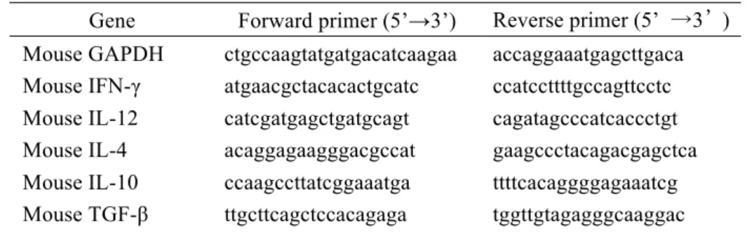
Total RNA was extracted from approximately 100 mg spleen or skin sample using Sepasol RNA I Super (Nacalai Tesque, Kyoto, Japan). The total RNA was cleaned up using an RNeasy mini kit (Qiagen, Hilden, Germany). Reverse transcription was performed using a SuperScript II (Invitrogen) and oligo (dT) primer, according to the manufacturer's protocol. For a quantitative analysis of mRNA expression, real-time PCR was carried out with total cDNA using a LightCycler instrument (Roche Diagnostics, Basel, Switzerland). The oligonucleotide primers used for amplification are listed in Table 1. Amplified products were detected online via intercalation of the fluorescent dye SYBR green (LightCycler-FastStart DNA Master SYBR Green I kit, Roche Diagnostics, Indianapolis, IN). The cycling conditions were as follows: initial enzyme activation at 95°C for 10 min, followed by 45 cycles at 95°C for 10 sec, 60°C for 5 sec, and 72°C for 20 sec. The mRNA expression of genes of interest was normalized using the mRNA level of β-actin.
TABLE 1. Primer sequences for quantitative RT-PCR.
Gene Forward primer (5’→3’) Reverse primer (5’ →3’) Mouse β-actin gcaccacaccttctacaatgag ttggcatagaggtctttacgga Mouse IL-12 catcgatgagctgatgcagt cagatagcccatcaccctgt Mouse IL-4 gcttttcgatgcctggattc gctttccaggaagtctttcagtg Mouse IL-5 agagaagtgtggcgaggagaga cattgcccactctgtactcatca Mouse IL-10 ttgccaagccttatcgga ttctgggccatgcttctct Mouse IL-13 cagctccctggttctctcac ccacactccataccatgctg Mouse IL-17 tccagaaggccctcagacta agcatcttctcgaccctgaa Mouse TARC agtggagtgttccagggatg gtcacaggccgctttatgtt Scoring skin lesions
Skin lesions were scored at indicated times after gene transfer, according to the criteria of Matsuda et al [7]. The scoring was based on the severity of eczema, erosion/excoriation, scaling, erythema/hemorrhage, inflammation of the face, and inflammation of the ear. The total clinical skin severity score was defined as the sum of each of the six signs (none = 0; mild = 1; moderate = 2; and severe = 3). Observation of scratching behavior
On days 7, 14, and 35, scratching behavior was monitored using SCLABA Real (Noveltec, Kobe, Japan), an automated system to analyze the scratching behavior of small animals. Each mouse was put into an acrylic cage, and the behavior of the mice was recorded for 30 min. The number of episodes and the duration of scratching behavior were automatically quantified. In a different set of mice, the scratching behavior was recorded on video for 1 hr on days 7, 14, 35, and 84. The videotape was played back at a later time, and the number of scratching episodes was counted manually. A series of scratching behaviors, starting with the stretching of the hind paws to the head, face, or back and ending with the set-back of the paws, was counted as one bout of scratching [29].
6
TEWL was measured using a VAPO SCAN (AS-VT 100RS, Asahi Biomed, Yokohama, Japan) on the shaved back of mice.
Analysis of skin sections
The dorsal skin of the mice was excised, fixed in 4% paraformaldehyde, and embedded in paraffin. Then, 4 µm-sections were made using a microtome and stained with H&E for histological evaluation or with toluidine blue to detect mast cells. The numbers of lymphocytes, eosinophils, and mast cells on H&E-stained sections (lymphocytes and eosinophils) or toluidine blue-stained sections (mast cells) were manually counted under a microscope and expressed as the number per unit length of skin section.
Effect of IFN-γ gene transfer on 2,4,6-trinitrochlorobenzene-induced dermatitis
To assess whether IFN-γ gene transfer increases the risk for contact dermatitis, C57BL/6 mice were sensitized by painting 25 µl 3% 2,4,6-trinitrochlorobenzene (TNCB) in acetone/olive oil (4:1) on the shaved abdomen (day 0). pCpG-Muγ was injected by the hydrodynamic injection method to TNCB-treated mice 1 day before or 7 day after the TNCB treatment, at a dose of 0.14 µg/mouse. On day 5, the thickness of the ear was measured with a Quick Mini thickness gauge (Mitutoyo, Tokyo, Japan). Then, 20 µl 1.5% TNCB solution was applied to the surface of the ear. The thickness of the ear was measured again 24 h after the second application (challenge), and ear swelling was evaluated by the difference in the thickness before and after the challenge.
Effect of IFN-γ gene transfer on TEWL in tape stripping- and TNCB-induced dermatitis models C57BL/6 mice were shaved on their dorsal skin on day 0 and injected with pCpG-Muγ (0.14 µg/mouse) or saline by the hydrodynamic injection method on the same day. Mice were repeatedly treated with 50 µl 1% TNCB dissolved in acetone/olive oil (4:1) to the shaved skin on days 1, 8, 11, and 13. In a separate experiment, the dorsal skin of C57BL/6 mice was tape stripped on days 1 and 8 and injected with pCpG-Muγ (0.14 µg/mouse) or saline by the hydrodynamic injection method on day 0. TEWL from the stripped or shaved skin was measured as described above.
Statistical analysis
Differences were evaluated by the Student t test, and the level of statistical significance was p < 0.05.
I-1-3 Results
I-1-3-a. IFN-γ concentration in the serum of NC/Nga mice after injection of IFN-γ–expressing plasmid DNA
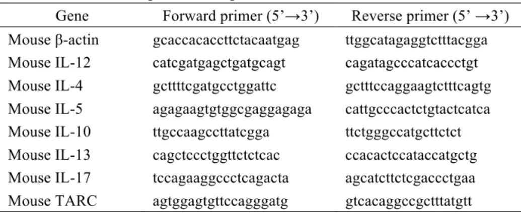
Fig. 1 shows the time courses of the concentration of IFN-γ in the serum after intravenous injection of pCpG-Muγ or pCMV-Muγ. A very high and sustained concentration of IFN-γ was detected in the serum of mice receiving pCpG-Muγ at a dose of 0.05 pmol/mouse. The concentration of IFN-γ was maintained at more than 300 pg/ml over 80 days after a single injection. However, the concentration of IFN-γ had decreased very quickly below the detection limit (25 pg/ml) 3 days after the first injection of pCMV-Muγ (4.68 pmol/mouse). The plasmid was injected again 1 week later, but the peak level was lower than that after the first injection, and the concentration decreased quickly again.
7
I-1-3-b. IL-12 concentration in the serum of NC/Nga mice after injection of IFN-γ–expressing plasmid DNA
It would be expected that a persistent expression of IFN-γ would induce the expression of IL-12, a typical Th1 cytokine that promotes the differentiation of naive T cells into the Th1 phenotype. Therefore, the serum concentration of IL-12 was measured (Fig. 2a). The IL-12 concentration in the untreated group was not constant during the experimental period; it fluctuated, reflecting the acute and chronic phases of the disease [30]. The concentration of IL-12 was significantly increased in the pCpG-Muγ–treated group from days 3 to 42 after injection. The concentration in the pCMV-Muγ–treated group was significantly greater 3 days after the first injection compared with that in the untreated group; thereafter, the profile became superimposed on that of the untreated group. The second injection of pCMV-Muγ on day 7 barely affected the serum concentration of IL-12.
IF N -γ co nc en tr at io n (p g/ m l)
Time after gene transfer (day)
0 20 40 60 80
102
103
104
105 FIGURE 1. Time-course of the
concentration of IFN-γ in mouse serum after injection of IFN-γ-expressing plasmid DNA. NC/Nga
mice were intravenously injected with 0.05 pmol pCpG-Muγ (䖃) or 4.68 pmol pCMV-Muγ (䕕) by the hydrodynamic injection method. The pCMV-Muγ-injected group received a second injection of 4.68 pmol pCMV-Muγ 7 days after the first injection (indicated with a dashed line and an arrow). The results are expressed as the mean 㼼 SD of five mice.
††
Time after gene transfer (day)
0 20 40 60 80 †† ** IL -1 2 co nc en tr at io n (p g/m l) 0 1000 2000 3000 ****† †† ** * * * † * † ** † (a)
FIGURE 2. Time-course of the concentration of IL-12 (a) and IgE (b) in mouse serum after injection of IFN-γ-expressing plasmid DNA. NC/Nga mice were intravenously injected with 0.05 pmol pCpG-Muγ
or 4.68 pmol pCMV-Muγ as described in the legend of Fig. 1. Blood samples from the untreated (䕧), the pCpG-Muγ-injected (䖃) or the pCMV-Muγ-injected (䕕) mice were collected from the tail vein at indicated times after gene transfer. The results are expressed as the mean 㼼 SD of at least three mice. *P<0.05, **P<0.01 compared with the untreated group; †P<0.05, ††P<0.01 compared with the pCMV-Muγ-treated group.
Time after gene transfer (day)
0 20 40 60 80 * Ig E co ncentra tio n (μ g/ m l) 0 50 100 200 †† * † ** * ** † † * (b) 150
8
I-1-3-c. IgE concentration in the serum of NC/Nga mice after injection of IFN-γ–expressing plasmid DNA
Next, the concentration of IgE, a diagnostic marker of atopic dermatitis [31], was measured in the serum, because increased IgE production is a hallmark of Th2 immune responses. Fig. 2b shows the concentration of IgE in the serum of the untreated mice or mice treated with pCpG-Muγ or pCMV-Muγ. The concentration at the onset of treatment was about 10 µg/ml, and it increased with time to more than 100 µg/ml in the untreated group. It also increased in the treated groups, but the increase was significantly inhibited in the pCpG-Muγ–treated group from day 14 after the treatment. In addition, I measured the serum concentrations of IL-4 and -13, which play important roles in the regulation of IgE synthesis [32, 33]. However, the concentrations of IL-4 and -13 in the serum of all groups were below the detection limit (4 pg/ml) of the analysis.
I-1-3-d. mRNA expression of cytokine and chemokine in spleen cells of NC/Nga mice after injection of IFN-γ–expressing plasmid DNA
The effect of IFN-γ gene transfer on the expression of cytokines/chemokines was evaluated by measuring the mRNA expression of IL-12, Th2 cytokines (IL-4, -5, -10, and -13), IL-17, and a Th2 chemokine (TARC) in spleen cells collected from mice 14 days after gene transfer using the primers listed in Table 1. The mRNA expression of these genes in spleen cells was normalized to that of β-actin, and the ratios were compared between the untreated and the pCpG-Muγ–treated mice (Fig. 3). The differences between the groups were very large for IL-5, -10, -12, and -13 and TARC, although they were not statistically significant because of the limited number of samples. The IL-12 mRNA expression was increased by injection of pCpG-Muγ, which was in good agreement with the serum level of IL-12 (Fig. 2a). The mRNA expression of IL-4, -5, -10, -13, and -17 and TARC in the pCpG-Muγ–treated group was lower than that in the untreated group. The mRNA expression in spleen cells 40 days after gene transfer showed no significant differences (data not shown). In addition, the expression of these cytokines in the skin was examined by ELISA and RT-PCR 40 days after gene transfer. However, no significant differences were detected among the groups (data not shown).
9
I-1-3-e. Skin lesions of NC/Nga mice after injection of IFN-γ–expressing plasmid DNA
The results indicated that the Th1/Th2 balance can be shifted to Th1 in the atopic dermatitis model by sustained, but not transient, transgene expression of IFN-γ. The effects of gene transfer on atopic dermatitis were examined in NC/Nga mice. Fig. 4 shows the typical images of the back skin of mice 35 days after the start of treatment. Clinical signs and symptoms were clearly seen on the skin of the untreated mice (Fig. 4a) and the pCMV-Muγ–treated mice (Fig. 4c), indicating that these mice developed a severe dermatitis. The severity of skin damage was scored using a clinical skin score [7]. The untreated and the pCMV-Muγ–treated groups developed dermatitis within a week after the start of the experiment, and the severity of the dermatitis increased with time (Fig. 4d). In contrast, the pCpG-Muγ–treated mice developed much less severe skin inflammation throughout the experimental period (Fig. 4b), and the clinical skin score of the group was significantly lower than that of the untreated or the pCMV-Muγ–treated group (Fig. 4d).
U CpG 1 10 100 1000 U CpG 0.01 0.1 1 U CpG 0.001 0.01 0.1 1 U CpG 0.01 0.1 1 10 100 1000 U CpG 1 10 100 1000 U CpG 0.1 1 10 U CpG 1 10 100 1000
(a) IL-12 (b) IL-4 (c) IL-5 (d) IL-10
(g) TARC (f) IL-17 (e) IL-13 C yt ok in e m R N A /β -a ct in m RNA
FIGURE 3. mRNA expression of cytokines and TARC in spleen cells. Spleens from NC/Nga mice receiving 0.05 pmol pCpG-Muγ were collected 14 days after gene transfer, and the mRNA expression of cytokine and chemokine genes was measured by real-time PCR. The mRNA expression of genes was normalized using the mRNA level of β-actin. In comparison, spleens from untreated mice were treated as above. (a) IL-12, (b) IL-4, (c) IL-5, (d) IL-10, (e) IL-13, (f) IL-17, and (g) TARC. U, untreated mice; CpG, pCpG-Muγ-injected mice. The results are expressed as the mean 㼼 SD of three mice.
10
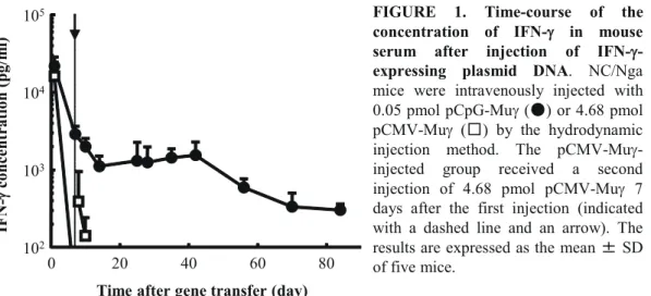
I-1-3-f. Scratching behavior of NC/Nga mice after injection of IFN-γ–expressing plasmid DNA
Fig. 5 shows the number of episodes of scratching and the cumulative time of scratching for a 30-min period. The number and duration of scratching episodes increased with time in the untreated group. The pCpG-Muγ– and pCMV-Muγ–treated groups had significantly fewer episodes and shorter duration of scratching than the untreated group, with significantly better results for pCpG-Muγ–treated mice. Similar results were obtained in a different set of mice whose scratching episodes were counted manually after videotaping (data not shown).
(a) Untreated (b) pCpG-Muγγ (c) pCMV-Muγ FIGURE 4. Typical images of the back skin of NC/Nga mice (a-c) and the time-course of the skin clinical score (d). Photographs were taken at
35 days after the start of treatment: (a) untreated mice, (b) pCpG-Muγ-treated mice and (c) pCMV-Muγ-treated mice. (d) Clinical features of dermatitis were scored at indicated periods of time after the start of treatment. The results are expressed as the mean 㼼 SD of at least
three mice. *P<0.05, **P<0.01
compared with the untreated group; †P<0.05, ††P<0.01 compared with the pCMV-Muγ-treated group.
Time after gene transfer (day)
0 20 40 60 80 Clinica l sk in sco re 0 10 15 20 (d) 5 * * * †† ** †† ** † ** †† ** †† ** ††** †† ** †† ** †† ** † **
11
I-1-3-g. TEWL of NC/Nga mice after injection of IFN-γ–expressing plasmid DNA
Dry skin is a common symptom of atopic dermatitis, which is characterized by extensive water loss through the skin. Thus, the TEWL of the skin was measured on the back (Fig. 6). The TEWL value at day 0 was about 10 g/h/m2 in all groups. In the untreated mice, the value increased to more than 50 g/h/m2 by day 14, and an almost constant value was observed in the period that followed. The elevation of TEWL was significantly inhibited in the pCpG-Muγ–treated group throughout the experimental period. The TEWL of the pCMV-Muγ–treated mice was as low as that of the pCpG-Muγ–treated ones for the first 14 days, but it was significantly higher at days 70 and 84.
**
Time after gene transfer (day)
0 10 20 30 40 Num ber of scra tching 0 50 100 150 (a) ** * ** ** † ** ** † **
Time after gene transfer (day)
0 10 20 30 40 Cum ulative tim e of sc ratching (s ec) 0 50 100 (b) ** * ** ** * ** *
FIGURE 5. Number (a) and cumulative time (b) of scratching episodes. Scratching behavior of NC/Nga mice treated as described in the legend of Fig. 1 were automatically evaluated using SCLABA-Real. (䕧) untreated mice, (䖃) pCpG-Muγ-treated mice and (䕕) pCMV-Muγ-treated mice. The results are expressed as the mean 㼼 SD of at least three mice. *P<0.05, **P<0.01 compared with the untreated group; †P<0.05 compared with the pCMV-Muγ-treated group.
†† **
Time after gene transfer (day)
0 20 40 60 80 TEWL (g /h· m 2) 0 50 100 ** ** ** ** ††**
FIGURE 6. Time-course of the TEWL of the back skin of mice.
TEWL of the skin was measured in the back of untreated mice (䕧), pCpG-Mug-treated mice (䖃) and pCMV-Mug-treated mice (䕕). The results are expressed as the mean 㼼 SD of at least three mice. **P<0.01 compared with the untreated group; ††P<0.01 compared with the pCMV-Muγ-treated group.
12
I-1-3-h. Histological examination of skin sections of NC/Nga mice after injection of IFN-γ–expressing plasmid DNA
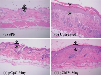
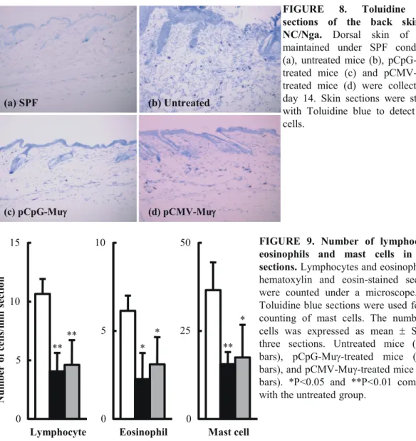
Fig. 7 shows the H&E sections of the back skin of treated and untreated mice at day 14 after the start of treatment. NC/Nga mice maintained under specific pathogen-free (SPF) conditions were used as control mice with healthy skin; the skin sections from the SPF control mice showed no pathological features (Fig. 7a). In marked contrast, there was clear hyperplasia of the epidermis (acanthosis) in the untreated group (Fig. 7b). The sections from the untreated mice also showed an extensive infiltration of lymphocytes and eosinophils. These characteristic features of inflamed skin tissues were not as apparent in the skin sections from the pCpG-Muγ–treated mice (Fig. 7c), which were indistinguishable from the skin sections from the SPF control mice. Compared with the sections from the pCpG-Muγ–treated mice, the ones from pCMV-Muγ–treated mice showed less significant improvement (Fig. 7d). Fig. 8 shows the skin sections in which mast cells were stained with toluidine blue. Again, a significant infiltration of mast cells was observed in the untreated mice (Fig. 8b), but not in the SPF control (Fig. 8a) or the pCpG-Muγ–treated mice (Fig. 8c). The numbers of lymphocytes, eosinophils, and mast cells in skin sections were counted (Fig. 9). The numbers of these cells were significantly less in mice receiving IFN-γ–expressing plasmid DNA compared with untreated mice.
(a) SPF (b) Untreated
(c) pCpG-Muγγ (d) pCMV-Muγ
FIGURE 7. Hematoxylin and eosin sections of the back skin of NC/Nga.
Dorsal skin of mice maintained under SPF conditions (a), untreated mice (b), pCpG-Muγ-treated mice (c) and pCMV-Muγ-treated mice (d) were collected at day 14. Skin sections were stained with hematoxylin and eosin
for histological evaluation and
13 I-1-3-i. Adverse effects of IFN-γ gene transfer
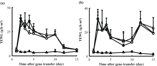
There were no significant differences in the body weight or temperature between the saline-injected mice and the pCpG-Muγ–treated mice. To examine whether IFN-γ gene transfer increases the risk for contact dermatitis, the ear thickness was measured in a mouse model of TNCB-induced contact dermatitis. The challenge with TNCB significantly increased the thickness, but the injection of pCpG-Muγ had no significant effects on the change (data not shown). Fig. 10 shows the time courses of the TEWL from the dorsal skin of tape stripped (Fig. 10a) or TNCB-treated mice (Fig. 10b). Again, no IFN-γ gene transfer-induced increase was observed in any case examined.
FIGURE 8. Toluidine blue sections of the back skin of NC/Nga. Dorsal skin of mice
maintained under SPF conditions (a), untreated mice (b), pCpG-Muγ-treated mice (c) and pCMV-Muγ-treated mice (d) were collected at day 14. Skin sections were stained with Toluidine blue to detect mast cells. (a) SPF (b) Untreated (c) pCpG-Muγγ (d) pCMV-Muγ Eosinophil 0 5 10 Mast cell 0 25 50 Lymphocyte 0 5 10 15 Num ber of cells/m m sectio n ** ** * * ** *
FIGURE 9. Number of lymphocytes, eosinophils and mast cells in skin sections. Lymphocytes and eosinophils in
hematoxylin and eosin-stained sections were counted under a microscope. The Toluidine blue sections were used for the counting of mast cells. The number of cells was expressed as mean ± SD of three sections. Untreated mice (white bars), pCpG-Muγ-treated mice (black bars), and pCMV-Muγ-treated mice (gray bars). *P<0.05 and **P<0.01 compared with the untreated group.
14 I-1-4 Discussion
Because of the multiple functions of cytokines and their complicated network, the effects of externally administered cytokines, including IFN-γ, depend on their pharmacokinetics. Atopic dermatitis, a chronic inflammatory disease with skin inflammation, is characterized by Th2-dominant immunity; therefore, any treatment that normalizes the Th1/Th2 balance can be useful for treatment of the disease. IFN-γ, a typical Th1 cytokine, has been considered to induce a variety of immunological changes, leading to a Th1-dominant state, but its effects on the Th1/Th2 balance in patients with Th2 predominance is not fully understood. The outcome from the previous clinical experiments in which IFN-γ was administered to patients with atopic dermatitis suggested that there would be few significant changes in the immune system of the patients receiving such treatments [10-13]. This can primarily be attributed to the unfavorable pharmacokinetic properties of IFN-γ, which easily passes through the glomerulus of the kidney because it is smaller than the threshold of glomerular filtration, and it disappears from the systemic circulation very quickly [14].
In this section, a sustained level of IFN-γ in the serum was successfully achieved by injecting pCpG-Muγ. An IFN-γ concentration at more than 300 pg/ml, which is greater than the half maximal effective concentration of the protein needed to inhibit virus replication (100 pg/ml), was obtained throughout the experimental period of 84 days. This profile of IFN-γ in the serum was in marked contrast to that obtained by the injection of pCMV-Muγ, which resulted in a very transient IFN-γ concentration in the serum. Such a large difference in the duration of transgene expression is most likely due to the number of CpG motifs in plasmid DNA [24, 34]. A detailed discussion on this sustained expression of IFN-γ from pCpG-Muγ was presented elsewhere [25].
Sustained expression of IFN-γ from pCpG-Muγ induced a variety of immunological changes in NC/Nga mice. In summary, the level of Th1 cytokines IFN-γ and IL-12 (Fig. 2a) was increased and that of IL-4, -5, -10, -13, and -17 and TARC tended to be decreased (Fig. 3); thus, the Th1/Th2 balance shifted toward Th1 dominance. In addition, the level of IgE, a hallmark of allergic reactions, was also significantly decreased. These changes can be considered to be due to the biological activity of IFN-γ, which promotes the
TEWL (g /h· m 2) 0 25 50
Time after gene transfer (day)
0 5 10 15 (a) TEWL (g /h· m 2) 0 20 40
Time after gene transfer (day)
0 5 10 15
(b)
FIGURE 10. Time-course of the TEWL of the back skin of mice in a stripped skin (a) and a TNCB-induced dermatitis model (b). (a) The back skin of C57BL/6 mice was shaved and tape-stripped on days 1 and 8. (䕦) untreated mice, (䕧) stripped mice, (䕿) stripped, saline-treated mice and (䖃) tape-stripped, pCpG-Muγ-treated mice. Results are expressed as mean ± SD of four mice. (b) The back skin of C57BL/6 mice was shaved and applied with TNCB on days 1, 8, 11 and 13. (䕦) untreated mice, (䕧) TNCB-treated mice, (䕿) TNCB-treated, saline-treated mice and (䖃) TNCB-treated, pCpG-Muγ-treated mice. Results are expressed as mean ± SD of four mice.
15
production of IL-12, a typical Th1 cytokine important for differentiation to the Th1 subset. IL-12 is produced from macrophages or NK cells stimulated with IFN-γ. The reduced expression of the Th2 cytokines and TARC also resulted from the expression of IFN-γ, as reported using PBMCs, keratinocytes, or Langerhans cells [35, 36].
A major finding of this section is that the immunological changes induced by IFN-γ gene transfer differ, depending on the vector used (i.e., on the pharmacokinetics of IFN-γ). A variety of changes, including the increase in the IL-12 concentration and the decrease in the IgE level, were observed only in the mice receiving pCpG-Muγ. These results clearly indicate the importance of a sustained concentration of IFN-γ at a level high enough to allow it to modulate the Th1/Th2 balance. In addition, these results may explain the fact that most challenges using IFN-γ protein for the treatment of atopic dermatitis are not very effective, because IFN-γ protein quickly disappears from the systemic circulation [14].
The modulation of Th1/Th2 imbalance by sustained expression of IFN-γ was significantly effective in preventing the onset of symptoms of atopic dermatitis in NC/Nga mice. Rash, scratching, bleeding on the back or ear, acanthosis, and infiltration of lymphocytes, eosinophils, and mast cells were not very apparent in the pCpG-Muγ–treated mice. Inhibition of cellular infiltration into skin would be the consequence of the changes induced by the sustained concentration of IFN-γ; this directly inhibits the infiltration of eosinophils [37, 38], and it could inhibit Th2 cell migration into skin promoted by Th2 chemokines [39-41]. Because Th2 lymphocytes are the cells that release Th2 cytokines [42], the findings in this section strongly suggest that IFN-γ gene transfer results in the inhibition of the differentiation of Th2 lymphocytes. Based on the results obtained, a mechanism of inhibition of atopic dermatitis by sustained expression of IFN-γ is proposed as follows. A sustained concentration of IFN-γ promotes the production of IL-12 from macrophages and NK cells, and these two Th1 cytokines inhibit the production of the Th2 cytokines/chemokines. These changes inhibit the recruitment of immune cells to the skin and prevent the onset of symptoms typical of atopic dermatitis. It is important to prove whether sustained IFN-γ gene transfer is also effective in individuals with dermatitis. Further studies are needed to conclude that IFN-γ gene transfer provides efficacy for patients with atopic dermatitis.
There would be concerns about sustained delivery of IFN-γ, because it is a highly potent Th1 cytokine. However, I observed no significant adverse effects of IFN-γ gene transfer on physiological parameters, such as body weight and body temperature. Also, contact dermatitis, a Th1-associated allergic dermatitis, was not aggravated by the injection of pCpG-Muγ. In addition, the improvement in TEWL observed in NC/Nga mice receiving pCpG-Muγ would not be due to the direct effects of IFN-γ gene transfer, because TEWL was minimally affected by IFN-γ gene transfer in other models (Fig. 10).
The hydrodynamic injection method used for gene transfer in the current study is one of the most efficient methods; using this method, >99% of transgenes were expressed in the liver, followed by kidneys, spleen, lung, and other internal organs [27, 43]. Although there is concern about the toxicity related to the mode of gene delivery [27, 28, 44, 45], a recent report showed that this method of gene delivery can be applied to humans with few toxic effects when plasmid DNA is delivered to a lobe using a balloon catheter [46]. Computer-assisted hydrodynamic gene delivery would also be a less invasive method [47]. Other modes for gene delivery could be used to achieve sustained transgene expression of IFN-γ at a relatively high level, because the dose of pCpG-Muγ used was as low as 0.05 pmol or 0.14 µg/mouse (i.e., approximately 7 µg/kg body weight). In general, greater amounts of plasmid DNA up to 100 µg/mouse (5 mg/kg) have been administered, and an increase in the dose may compensate for the low efficiency of other gene-delivery methods, such as intradermal injection of naked plasmid DNA.
16
In conclusion, it was proved that an extraordinarily sustained IFN-γ expression induces a variety of immunological changes, leading to a Th1-dominant state in the atopic dermatitis model. A short expression of IFN-γ was not effective in inducing such changes, indicating the importance of a sustained concentration of IFN-γ. These results provide a novel strategy for the treatment of atopic dermatitis, in which biologically active IFN-γ protein is supplied to patients from cells transduced with plasmid vector expressing the protein for a long period of time.
17
Section 2
Sustained IFN-γ expression improves symptoms of acute phase of
atopic dermatitis
I-2-1 Introduction
In Section 1, I demonstrated that sustained expression of IFN-γ is effective in preventing the onset of symptoms of atopic dermatitis in NC/Nga mice [48]. To use IFN-γ gene transfer in the treatment of atopic dermatitis patients, it is important to show that such gene transfer is effective not only in preventing the onset of the disease but also in improving the symptoms.
Generally, atopic dermatitis is classified into two phases, acute and chronic. In the acute phase of atopic dermatitis, patients often have raised Th2 cytokines, such as IL-4, IL-5 and IL-13, which induce the class switching of B cells to IgE secretion, but low levels of IFN-γ or IL-12. In contrast, Th1 cytokines, especially IFN-γ, are dominant in chronic phase of atopic dermatitis [49-52].Therefore, the administration of IFN-γ, a typical Th1 cytokine that shifts the differentiation of naive T cells to the Th1 subtype and suppresses the production of Th2 cytokines, appears to be an attractive option for the treatment of acute phase of atopic dermatitis [53, 54].
It has recently been shown that regulatory T cells (Tregs), a subset of T cells, are involved in the modulation of allergic diseases. For example, Tregs and their related cytokines, IL-10 and transforming growth factor (TGF)-β, have been reported to suppress Th2 responses and IgE production [55-57]. Furthermore, IFN-γ as well as the IL-12-specific receptor subunit β2 plays important roles in the production of Tregs [58, 59].
In this section, I aimed to investigate whether IFN-γ gene transfer was able to effectively treat dermatitis induced in NC/Nga mice. To do this, I evaluated the serum levels of IgE and IgG1, the mRNA expression of Th1 and Treg cytokines, TEWL, the number of scratching episodes, the epidermal thickening, and the infiltration of inflammatory cells into the skin. In addition, the effect of sustained IFN-γ gene transfer on contact dermatitis was also examined to assess the safety of the gene transfer process.
I-2-2 Materials and Methods Animals
Seven to eight week-old male NC/Nga mice and seven week-old female C57BL/6 mice were purchased and raised as described in Section 1. The NC/Nga group spontaneously developing dermatitis was used as a model of atopic dermatitis. The protocol for the animal experiments was approved by the Animal Experimentation Committee of the Graduate School of Pharmaceutical Sciences of Kyoto University.
In vivo gene transfer of IFN-γ
The naked plasmid pCpG-Muγ or pCpG-mcs (InvivoGen, Sandiego, CA, USA) was injected into the tail vein of the mice by the hydrodynamic injection method, as described in Section 1, at a dose of 0.05 or 0.2 pmol/mouse on day 0.
18
The concentrations of IFN-γ and IgE in the serum were measured as described in Section 1. The concentration of IgG1 in the serum was measured using ELISA kits (Mouse IgG1 ELISA Quantitation Set, Bethyl Laboratories, Inc., Montgomery, TX, USA) according to the manufacturer’s instructions.
Calculation of the pharmacokinetics parameters of IFN-γ gene transfer
The area under the concentration-time curve (AUC) and the mean residence time (MRT) of IFN-γ after gene transfer were calculated from the concentrations of IFN-γ in the serum of NC/Nga mice using moment analysis [60].
Real time PCR analysis of cytokine expression
RNA was extracted from approximately 100 mg of spleen samples using Sepasol RNA I Super (Nacalai Tesque, Kyoto, Japan). A mixture of recombinant DNase I-RNase-free (TAKARA, Shiga, Japan) and RNase OUT™ recombinant ribonuclease inhibitor (Invitrogen, Carlsbad, CA, USA) was used for DNase treatment. Reverse transcription was performed using a ReverTra Ace® qPCR RT Kit (TOYOBO, Osaka, Japan). The mRNA expression of genes of interest was analyzed using the quantitative real time PCR system as described in Section 1. The sequences of primers used for amplification are listed in table 2. Individual PCR products were analyzed by melting curve analysis and the length of the products was determined by agarose gel electrophoresis. The mRNA expression of each gene was normalized using the mRNA level of GAPDH.
TABLE 2. Primer sequences for quantitative RT-PCR.
Gene Forward primer (5’→3’) Reverse primer (5’ →3 ) Mouse GAPDH ctgccaagtatgatgacatcaagaa accaggaaatgagcttgaca Mouse IFN-γ atgaacgctacacactgcatc ccatccttttgccagttcctc Mouse IL-12 catcgatgagctgatgcagt cagatagcccatcaccctgt Mouse IL-4 acaggagaagggacgccat gaagccctacagacgagctca Mouse IL-10 ccaagccttatcggaaatga ttttcacaggggagaaatcg Mouse TGF-β ttgcttcagctccacagaga tggttgtagagggcaaggac Scoring skin lesions
Skin lesions were scored at indicated times after gene transfer as described in Section 1. Measurement of TEWL
TEWL was measured using an evaporimeter as described in Section 1. Observation of scratching behavior
The scratching behavior was recorded on video for 30 min on days 15 and 21. The number of scratching episodes was counted manually as described in Section 1. On days 30, 33 and 35 after gene transfer, the scratching behavior was monitored using an automated system (SCLABA Real Noveltec Inc., Kobe, Japan) as described in Section 1.
19
The sections of dorsal skin of NC/Nga mice were prepared and stained for histological evaluation and quantification of the numbers of inflammatory cells infiltrating in the skins as described in Section 1. Monitoring of adverse effects of IFN-γ gene transfer
The body temperature of the mice was monitored using a digital rectal thermometer (Physitemp Instruments Inc, Clifton, NJ, USA). To assess liver damage, the serum alanine aminotransferase (ALT) and aspartate aminotransferase (AST) levels of the mice were assayed using commercial test reagents (Transaminase CII-Test Wako, Wako Pure Chemical Industries, Osaka, Japan).
Oxazolone-induced contact hypersensitivity
The plasmid pCpG-Muγ was injected into the tail vain of C57BL/6 mice (female, 7-week-old) by the hydrodynamic injection method at a dose of 0.2 pmol/mouse on day 0 or day 5. Mice were sensitized by the application of 100 µl 2% oxazolone (OX) in ethanol (elicitation phase group) or ethanol alone (induction phase-group) to the shaved abdominal skin on day 1, followed by an application of 10 µl 1% OX to the right ear on day 6. Ethanol was applied to the left ear. For the untreated group, ethanol was applied instead of oxazolone to the abdomen and right ear on either day 1 or 6. At 24 h after the second application, the thickness of both ears was measured using a digital thickness gauge (Quick Mini, Mitutoyo Co., Japan). Ear swelling was calculated from the difference in ear thickness between the hapten- and vehicle-treated ears. Statistical analysis
Statistical significance was evaluated by ANOVA with a post-hoc Tukey-Kramer test for multiple comparisons (clinical skin scores) and Student’s t-test for comparisons between two given groups. The level of statistical significance was set at P<0.05.
I-2-3 Results
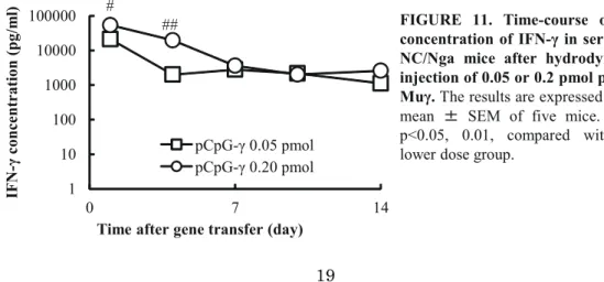
I-2-3-a. Time-course and pharmacokinetic parameters of IFN-γ after hydrodynamic injection of pCpG-Muγ
Figure 11 shows the time-course of the concentrations of IFN-γ in the serum after hydrodynamic injection of pCpG-Muγ. The serum levels of IFN-γ were dependent on the plasmid dose, and the higher dose of 0.2 pmol resulted in significantly higher concentrations for the first 4 days compared with the lower dose. The profiles were analyzed to obtain AUC and MRT values.
1 10 100 1000 10000 100000 0 7 14 pCpG-γ 0.05 pmol pCpG-γ 0.20 pmol
Time after gene transfer (day)
IF N -γ c on ce nt ra tio n (p g/ m l) ## #
FIGURE 11. Time-course of the concentration of IFN-γ in serum of NC/Nga mice after hydrodynamic injection of 0.05 or 0.2 pmol pCpG-Muγ. The results are expressed as the
mean 㼼 SEM of five mice. #, ##
p<0.05, 0.01, compared with the lower dose group.
20
Table 3 summarizes the maximum concentration (Cmax), AUC and MRT after IFN-γ gene transfer. The Cmax and AUC were increased by increasing the plasmid dose, whereas the MRT was hardly affected by the dose.
TABLE 3. Cmax, AUC and MRT of serum IFN-γ after hydrodynamic injection of pCpG-Muγ into mice
Dose (pmol/mouse) Cmax (ng/ml) AUC (ng day/ml) MRT (day)
0.05 21.3 ± 1.5 71.3 ± 7.9 4.67 ± 0.78
0.2 54.0 ± 10a 204 ± 35b 4.21± 0.70
The Cmax values were obtained at 1 day after hydrodynamic injection of pCpG-Muγ, and are expressed as the mean ± standard error of the mean (SEM) of five mice. The AUC and MRT were calculated by moment analysis, and are expressed as the calculated mean ± SEM of five mice. a, b p<0.05, 0.01, compared with the lower dose (0.05 pmol/mouse) group.
I-2-3-b. Effects of IFN-γ gene transfer on the skin lesions of NC/Nga mice
NC/Nga mice at the age of 7 to 8 weeks, which already developed atopic dermatitis-like symptoms, were used to assess the severity of skin lesions. Fig. 12 shows the time-course of the clinical skin score of NC/Nga mice after injection of pCpG-Muγ. The score of mice treated with the lower dose pCpG-Muγ increased with time and was not significantly different from that of the untreated mice for the first two weeks after gene transfer. These results contradicted those of Section 1, in which the lower dose of pCpG-Muγ (0.05 pmol/mouse) was effective in preventing the onset of atopic dermatitis [48]. In contrast, the clinical skin score of mice receiving 0.2 pmol pCpG-Muγ was significantly lower than those of the other two groups over the 5-week observation period. Based on these findings, the dose of the plasmid was set at 0.2 pmol/mouse in the subsequent experiments to study the therapeutic effects of pCpG-Muγ in NC/Nga mice with dermatitis. To confirm the effects of IFN-γ, a control plasmid pCpG-mcs was administered to NC/Nga mice. However, the clinical skin score was hardly affected by this treatment (data not shown).
0 4 8 12 16 20 0 1 2 3 4 5 Cl in ic al sk in sc or e
Time after gene transfer (week)
pCpG-γ 0.05 pmol pCpG-γ 0.20 pmol No treatment * # ***# ** ** ## ##
FIGURE 12. Time-course of the clinical skin score of NC/Nga mice after hydrodynamic injection of pCpG-Muγ. The results are expressed as the mean 㼼 SEM of five (0.05 pmol and 0.2 pmol pCpG-Muγ treated groups) and nine mice
(the untreated group). *, **,
***p<0.05, 0.01, 0.001, compared
with the untreated group; #,##p<0.05,
0.01, compared with the lower dose group.
21
I-2-3-c. Serum concentrations of IgG1 and IgE in NC/Nga mice after IFN-γ gene transfer
Fig. 13 shows the time-course of the concentration of IgG1 (Fig. 13a) and IgE (Fig. 13b) in the serum of NC/Nga mice after injection of pCpG-Muγ. Both the serum levels of IgG1 and IgE of the pCpG-Muγ-treated mice were significantly lower than those of the untreated mice at several time points, suggesting that the production of these Th2-mediated immunoglobulins in NC/Nga mice was at least partly inhibited by IFN-γ gene transfer.
I-2-3-d. mRNA expression of Th1, Th2 and Treg cytokines in the spleen of NC/Nga mice after IFN-γ gene transfer
The high concentrations of IFN-γ in the serum were expected to induce the changes in the immunological status of NC/Nga mice. Therefore, the spleen, a major lymphoid organ, was excised and the mRNA expression of IFN-γ, IL-12, IL-4, IL-10 and TGF-β was measured. Fig. 14 shows the x-fold increase of mRNA expression by pCpG-Muγ treated mice compared with untreated mice. The mRNA expression of IFN-γ and IL-12 in the pCpG-Muγ-treated mice was, respectively, 3- and 1.7-fold higher than that in the untreated mice. In addition, the mRNA expression of IL-10 and TGF-β was also significantly higher in the pCpG-Muγ-treated group. On the other hand, the mRNA expression of IL-4, a Th2 cytokine, was reduced to about half in the pCpG-Muγ-treated mice.
0 4 8 12 16 0 7 14 21 28 35 IgG 1 (m g/ m l)
Time after gene transfer (day)
* * *** (a) 0 20 40 60 80 0 7 14 21 28 35 IgE (μ g/ m l)
Time after gene tranfer (day)
pCpG-γ 0.20 pmol No treatment
* ** * *
(b)
FIGURE 13. Time-course of the concentration of IgG1 (a) and IgE (b) in serum of NC/Nga mice after hydrodynamic injection of 0.05 or 0.2 pmol pCpG-Muγ. The results are expressed as the mean 㼼 SEM of four mice. *,**,***p<0.05, 0.01, 0.001, compared with the untreated group.
22
I-2-3-e. TEWL and scratching behavior of NC/Nga mice after IFN-γ gene transfer
To evaluate the severity of skin dryness and itchiness, the TEWL [61] and the number of scratching episodes were measured. Fig. 15a shows the time-course of the TEWL of NC/Nga mice from day 15 to day 35 after injection of pCpG-Muγ. The TEWL value of the pCpG-Muγ-treated mice was significantly lower than that of the untreated mice on days 15, 18, 21 and 35 after gene transfer. Fig. 15b shows the number of scratching episodes during a 30-min period of observation. The number of scratching episodes exhibited by the mice receiving pCpG-Muγ was fewer than that of the untreated mice at all time points examined and the difference was significant on days 15 and 33 after injection of pCpG-Muγ. These results indicate that IFN-γ gene transfer relieves the pruritus, a common symptom of atopic dermatitis that may lead to destruction of the skin barrier and is related to the increased TEWL of NC/Nga mice [62-64].
0 1 2 3 4 5 -γ -β Fold increase (com pare to untreated group) * *
FIGURE 14. mRNA expression of cytokines in spleen cells of NC/Nga mice. Spleens of untreated mice and
pCpG-Muγ treated mice were excised at day 36 after treatment. mRNA expression was normalized with that of GAPDH. The results are expressed as the mean 㼼 SEM of three (pCpG-Muγ treated group) or four mice (the untreated group). *p<0.05 compared with the untreated group.
IFN IL-12 IL-10 TGF IL-4
0 20 40 60 14 21 28 35 T E W L (g/ h/ m 2)
Time after gene transfer (day)
** ** * * (a) 0 100 200 300 400 14 21 28 35 Nu m be r of sc ra tc h
Time after gene transfer (day)
pCpG-γ 0.20 pmol No treatment
*
** (b)
FIGURE 15. Time-course of the TEWL of the dorsal skin (a) and number of scratching episodes (b) of NC/Nga mice untreated or after hydrodynamic injection of 0.2 pmol pCpG-Muγ. The results are expressed as the mean 㼼 SEM of four mice. *,**p<0.05, 0.01, compared with the untreated group.
23
I-2-3-f. Histopathological examination of skin sections of NC/Nga mice
Fig. 16 shows the hematoxylin and eosin sections (Figs. 16a, b) and toluidine blue-stained sections (Figs. 16c, d) of the dorsal skin of NC/Nga mice at 36 days after treatment. The sections of the untreated mice showed obvious thickening of the epidermis (Figs. 16a, c). However, these characteristic features of skin inflammation were hardly seen in the sections from the pCpG-Muγ-treated mice (Figs. 16b, d). Furthermore, the numbers of monocytes, granulocytes and mast cells in the skin sections were counted (Fig. 16e). The numbers of these cells infiltrating into the skin of the pCpG-Muγ-treated animals were about a half those in the untreated mice, and there were significant differences between the groups with regard to the number of monocytes and mast cells.
I-2-3-g. Effect of IFN-γ gene transfer on body temperature and liver injury
The body temperature of the mice was measured to monitor the side effects of the high dose pCpG-Muγ. No significant difference was observed in body temperature between the treated and untreated groups (data not shown). In addition, there were no significant differences in the serum AST and ALT levels between the pCpG-Muγ-treated and the untreated groups (data not shown).
I-2-3-h. Effect of IFN-γ gene transfer on contact dermatitis
There are some concerns that IFN-γ gene transfer might increase the risk of Th1-associated diseases. To clarify this issue, the ear swelling response in the OX-induced contact dermatitis model was used to evaluate the contact hypersensitivity response in mice. As shown in Fig. 17, OX treatment induced ear swelling, but this response was not exacerbated by IFN-γ gene transfer.
(a) (b) (c) (d) 0 10 20 30 40 50 60 70
Monocyte Granulocyte Mast cell No treatment pCpG-γ 0.20 pmol (e) * * N um be r of c el ls pe r m m s ec tio n
FIGURE 16. H&E (a, b) and toluidine blue (c, d) sections of the dorsal skin of NC/Nga mice untreated (a, c) or pCpG-Muγ-treated (b, d). Numbers of inflammatory cells in skin sections (E). The numbers of cells were expressed as the mean 㼼 SEM of three (pCpG-Muγ-treated group) or four mice (the untreated group). *p<0.05 compared with the untreated group.
24 I-2-4 Discussion
NC/Nga mice spontaneously develop atopic dermatitis-like skin lesions around the age of 6-8 weeks when raised under conventional condition. After that, the severity of the skin lesions and the parameters reflecting the severity, such as serum IgE level, increases with age. A previous study reported that the serum IgE levels of conventional NC/Nga mice increased markedly from 6 weeks of age to around 10 weeks and reached plateau at 17 weeks [8]. Thus the mice with atopic dermatitis at the age of 7 to 8 weeks, which were used in this study, represent a suitable model to evaluate the therapeutic effect of IFN-γ in the acute phase of atopic dermatitis. In this section, I demonstrated that pCpG-Muγ is effective not only in preventing the onset of this condition but also in alleviating the symptoms of the disease, even though a higher dose of pCpG-Muγ was required to treat the symptoms of NC/Nga mice with dermatitis. The mRNA expression of IFN-γ and IL-12, Th1 cytokines, in the spleen of mice receiving pCpG-Muγ was increased and accompanied with a reduced expression of IL-4 mRNA in the spleen and reduced levels of serum IgG1 and IgE (Fig. 13). IFN-γ acts in conjunction with IL-12 by mediating a positive feedback loop to drive the Th1 response and inhibits IL-4 production by Th2 cells, resulting in the suppression of IgE and IgG1 secretion by B cells [41, 65, 66]. The mRNA expression of IL-10 and TGF- β, two representative Treg cytokines, in the spleen of mice receiving pCpG-Muγ was also increased. It has been reported that Tregs inhibit Th2 cell function in allergic diseases by releasing IL-10 and TGF-β [57, 67]. The development of atopic dermatitis-like skin lesions in NC/Nga mice was suppressed by IL-10 expressing plasmid DNA [68]. It is possible that the increased expression of IFN-γ and IL-12 might induce Treg differentiation [58, 59] and partially contributed to these immunological changes by suppressing Th2 cell response. Taking all these findings into consideration, it is likely that IFN-γ gene transfer stimulates IL-12 production and these two Th1 cytokines act synergistically to polarize the T cell response toward Th1 subset and inhibit IgG1 and IgE production by suppressing the Th2 pathway. Further investigation is needed to prove whether the induction of Treg cytokines is related to these changes.
0 0.1 0.2 0.3
Negative
Control Induction phase Induction phase,gamma 7 days Induction phase,gamma 1 day Elicitationphase Elicitation phase, gamma 7 day Elicitation phase,gamma 1 day
Ea r sw elling (Δ thick ness, m m )
Induction phase Elicitation phase
Untreated Day 0 Day 1 Day 5 Day 6 -- pCpG-γ - - pCpG-γ -pCpG-γ pCpG-γ EtOH EtOH EtOH EtOH EtOH OX OX OX OX OX OX OX OX OX - - -
-FIGURE 17. Ear swelling response in OX-treated mice. pCpG-Muγ was injected on day 0 or day 5. Mice
were sensitized by OX or ethanol on the shaved abdomen on day 1 and on the right ear on day 6. Ear thickness was measured 24 h after the last OX application. Data are expressed as the mean 㼼 SEM of four mice.
25
Scratching destroys skin barriers and worsens skin lesions [62, 64], which is closely linked to an increased TEWL. Histological examination of the skin sections clearly showed that IFN-γ gene transfer markedly suppressed skin inflammation and the infiltration of monocytes, granulocytes and mast cells. Mast cells can be sensitized by IgE and regulate the secretion of cytokines, such as IL-4, which subsequently mediates the recruitment of leukocytes [69],so that the reduced infiltration of these inflammatory cells into the skin could be due to the reduced production of IgE and IL-4. This suppression will prevent mice from scratching, which will accelerate skin repair and reduce TEWL. The number of scratching episodes of mice receiving pCpG-Muγ was lower than that of the untreated mice at all time points, although it was fluctuated in the both groups. This could be explained by the difference in the time for measurement in each day [70], which was not be controlled in this study.
The use of IFN-γ as a therapeutic agent would be a double-edged sword. The need for a high dose of 0.2 pmol/mouse to treat, not to prevent, atopic dermatitis symptoms increases the chance of adverse reactions. The most common adverse events of IFN-γ therapy, which have been evidenced in several clinical trials, are “flu-like” symptoms, including fever, headache, fatigue, and myalgia. Diarrhea, erythema at the injection site, and the elevation of liver transaminase levels have also been reported in patients receiving the therapy. These common adverse events are generally transient and well tolerated [71-74]. In this section, I measured the body temperature and the liver transaminase levels in serum of mice for monitoring the signs of adverse events and demonstrated that IFN-γ gene transfer is safe for use in mice as long as the dose of the plasmid is 0.2 pmol/mouse or lower. However, the clinical skin score seems to return to baseline level at 4 weeks after treatment with this dose of plasmid (Fig. 12). This result may be explained by the reduction of IFN-γ levels in the serum of mice with time, thus increasing the dose of pCpG-Muγ within the safety range could be an option to extend the duration of therapeutic effects. Recently, my laboratory has established a plasmid DNA expressing IFNγ in a constant manner with no initial high concentration of IFN-γ that could cause the unwanted effects [75]. This novel IFN-γ expressing plasmid would also be useful to enhance the therapeutic effect of IFN-γ. Another interesting point is that IFN-γ has been reported to be dominant in the skin of patients with chronic atopic dermatitis [52]. Further investigation is required to prove whether IFN-γ gene transfer is suitable for the treatment of chronic phase of atopic dermatitis.
In conclusion, I have demonstrated that sustained transgene expression of IFN-γ is effective in treating the atopic dermatitis in NC/Nga mice without any apparent adverse effects. These results raise a possibility that IFN-γ gene transfer can be a therapeutic option for patients with acute phase atopic dermatitis.
26
Chapter II (Excerpted version)
Effects of highly upregulated indoleamine 2,3-dioxygenase 1 on anti-tumor
activity of IFN-γ gene transfer in tumor-bearing mice
II-1 Introduction
IFN-γ is expected to be used in treating cancer [76, 77]. In Chapter I, I showed that the hydrodynamic injection of pCpG-Muγ resulted in prolonged therapeutic serum concentrations of IFN-γ for more than 80 days, and this significantly improved the symptoms of atopic dermatitis in NC/Nga mice. Previous studies in my laboratory demonstrated that sustained supply of IFN-γ effectively inhibited metastatic tumor growth in mice [24, 25].
IFN-γ exerts its biological activities through the Janus kinase/signal transducer and activator of transcription pathway, which modulates the expression of indoleamine 2, 3-dioxygenase 1 (IDO1) [65, 78]. Increased IDO activity is reported to be responsible for the protumor functions of IFN-γ [79-81]. IDO1 is a tryptophan-metabolizing enzyme along the kynurenine pathway [82, 83]. The induced expression of IDO1 could lead to tryptophan depletion and formation of kynurenine and 3-hydroxykynurenine, which could downregulate immune responses [84-87]. Various types of IDO1 inhibitors have been developed [82, 88-91], but the role of IDO1 in tumor growth is controversial so far [92]. Therefore, further investigation is needed to clarify the role of IDO1 in tumor development and its effects on the antitumor activity of IFN-γ.
Unpublished data from my laboratory showed that a hydrodynamic injection of pCpG-Muγ increased the mRNA expression of IDO1 in naïve mice. In this chapter, LLC tumor-bearing wild-type and IDO1-knockout mice were used to examine the involvement of IDO1 expression and activity in the antitumor effect of IFN-γ gene transfer.
II-2 Materials and Methods Mice
Five-week-old male C57BL/6J mice and IDO1 knockout (IDO1 KO) mice with a C57BL/6J background were used at five weeks of age. The protocols for animal experiments were approved by the Animal Experimentation Committee of the Graduate School of Pharmaceutical Sciences, Kyoto University. Cell culture
Lewis lung carcinoma (LLC) cells were cultured in Dulbecco’s modified Eagle medium (DMEM; Nissui Co. Ltd., Tokyo, Japan) supplemented with 10 % heat-inactive fetal bovine serum (FBS) at 37°C, 5 % CO2.