学 位 の 種 類 博士 (薬科学)
報 告 番 号 甲第1646号
学 位 記 番 号 第329号
氏 名 SHAIMAA MOHAMED RAMADAN ELABD IBRAHIM
授 与 年 月 日 平成 30 年 3 月 26 日
学位論文の題名 Development of nanomedicines as drug delivery systems for cancer and malaria parasites
論文審査担当者 主査: 湯浅 博昭
1
名古屋市立大学学位論文
Development of nanomedicines as drug delivery
systems for cancer and malaria parasites
平成29 年度(西暦 2018 年 3 月)
名古屋市立大学大学院薬学研究科 博士後期課程創薬機能科学専攻
薬物送達学分野
2 主査 湯浅 博昭 教 授 副査 尾関 哲也 教 授 平嶋 尚英 教 授 牧野 利明 教 授 本論文は、学術情報雑誌に収載された次の報文を基礎とするものである。
1. Ibrahim S, Tagami T, Ozeki T: Effective-Loading of Platinum-Chloroquine into PEGylated Neutral and Cationic Liposomes as a Drug Delivery System for Resistant Malaria Parasites. Biol. Pharm. Bull. 40, 813-823 (2017).
2. Ibrahim S, Tagami T, Kishi T, Ozeki T: Curcumin marinosomes as promising nano drug delivery formulation for lung cancer. Int. J. Pharm. 540, 40–49 (2018).
本論文の基礎となる研究は、尾関 哲也教授の指導の下に名古屋市立大学大学院薬学 研究科において行われた。
3
Abstract...6
CHAPTER 1: Introduction and objectives………...8
1.1. Background...8 1.1.1 Nanotechnology and drug delivery
1.1.2 Liposomes as drug delivery systems 1.1.3 Nonomedicine against malaria 1.1.4 Nonomedicine against cancer.
1.2. Objectives...13 1.3. References...15
CHAPTER 2: Effective loading of platinum–chloroquine into PEGylated
neutral and cationic liposomes as a drug delivery system for resistant
malaria parasites……….19
2.1. Introduction...19 2.2. Materials and Methods...25
2.2.1 Reagents
2.2.2 Synthesis of the trans-Pt(CQDP)2(Cl)2 complex
2.2.3 Preparation of PtCQ-loaded liposomes using thin drug-lipid film method 2.2.4 Preparation of PtCQ-loaded liposomes using remote-loading method 2.2.5 Determination of the encapsulation efficiency (EE)
2.2.6 Measurement of drug release from PtCQ-loaded liposomes under storage conditions (storage test)
4
2.3.1. Synthesis and characterization of the Pt(CQDP)2(Cl)2 complex 2.3.2. PtCQ-loaded liposomes prepared using the thin drug-lipid film method 2.3.3. PtCQ-loaded liposomes prepared using the remote-loading method 2.3.4. Cationic PtCQ-loaded liposomes produced using the remote-loading method
2.3.5. Drug release from cationic PtCQ-loaded liposomes under storage conditions (storage test)
2.3.6. Drug release from cationic PtCQ-loaded liposomes under culture conditions (release test)
2.4. Conclusion...56 2.5. References...57
CHAPTER 3: Curcumin marinosomes as promising nano-drug delivery
system for lung cancer……….63
3.1. Introduction...63 3.2. Materials and Methods...68
3.2.1. Materials
3.2.2. Preparation of CURMs using the thin drug-lipid film hydration method 3.2.3. Determination of the encapsulation efficiency (EE) of CURMs
3.2.4. Determination of DPPH stability in detergent buffer solution Method 3.2.5. Determination of the antioxidant activity of CURMs in detergent buffer. 3.2.6. Stability of CURMs under storage condition
3.2.7. Retention of CURMs under culture condition 3.2.8. Cells
5
3.3.3 Determination of the antioxidant activity of CURMs in detergent buffer 3.3.4 Stability of CURMs under storage condition
3.3.5 Retention of curcumin into marinosomes under cell culture condition 3.3.6 The cytotoxicity of free and curcumin-loaded marinosomes on A549 cells 3.3.7 The effect of free and curcumin-loaded marinosomes on HUVECs
3.4. Conclusion...102 2.5. References...103
6
One of the most important applications of nanotechnology is the drug delivery system that aims to develop nanomedicines able to deliver the therapeutic compounds into the diseased organ. The formulation and evaluation of drug delivery systems for example liposomes and lipid nanoparticis is less expensive than developing new drugs and can fulfill the objective of increasing the therapeutic activity and decreasing the toxic side effects.
This thesis has three chapters. In the first chapter, we introduced a brief background on nanotechnology and drug delivery, liposomes as drug delivery systems, nonomedicine against malaria and nonomedicine against cancer. In this chapter, we also discussed the importance of using drug delivery nanomedicines to fight infectious diseases like malaria and serious diseases as cancer.
In the second chapter, a liposomal nano drug delivery formulation was prepared to face resistant malaria parasites. The trans platinum-chloroquine diphosphate dichloride (PtCQ) is a new type of antimalarial drug used to fight parasites resistant to traditional drugs. PtCQ is synthesized by mixing platinum and chloroquine diphosphate (CQ). This study examines two efficient methods for forming a nanodrug, PtCQ-loaded liposomes, for use as a potential antimalarial drug-delivery system: the thin drug-lipid film method to incorporate the drug into a liposomal membrane, and a remote-loading method to load the drug into the interior of a cationic liposome. The membranes accordingly comprised PEGylated (contain polyethylene glycol (PEG) polymer) neutral or cationic liposomes. PtCQ was efficiently loaded into PEGylated neutral and cationic liposomes using the thin drug-lipid film method (encapsulation efficiency, EE: 76.1 ± 6.7% for neutral liposomes, 1:14 drug-to-lipid weight ratio; 70.4 ± 9.8% for cationic liposomes, 1:14 drug-to-lipid weight ratio. More PtCQ was loaded into PEGylated neutral liposomes using the remote-loading method than by the thin
7
of cationic lipids (0-20 mol%; EE: 96.9-92.3%) using the remote-loading method. PEGylated neutral liposomes and cationic liposomes exhibited minimum leakage of PtCQ after two months’ storage at 4˚C, and further exhibited little release under in vitro culture conditions at 37˚C for 72 hours. These results provide a useful framework for the design of future liposome-based in vivo drug delivery systems targeting the malaria parasite.
In the third chapter, a marinosomal nano drug delivery formulation was prepared to face lung cancer, which considered as the major cause of cancer-related death worldwide. Curcumin attracted attention due to its promising anti-cancer properties, however its poor aqueous solubility and bioavailability have to be overcome. In the current study curcumin is encapsulated in krill lipids-based liposomes (marinosomes) to develop a potential anticancer therapy from low-cost and readily available nutraceuticals. Reflux followed by thin drug-lipid film method is used successfully to incorporate the drug into the liposomal membrane at high encapsulation efficiency (EE). The curcumin-loaded marinosomes (CURMs) showed a powerful antioxidant activity (EC50: 4.1 ± 0.3 μg/mL). Additionally, CURMs exhibited high
physicochemical and oxidative stability after eight weeks’ storage at 4˚C. Furthermore, CURMs exhibited sustained release of about 30 % of their curcumin content under in vitro culture conditions at 37˚C after 72 hrs. Consequently, CURMs showed its maximum cytotoxic effect (IC50; 11.7 ± 0.24 μg/ml) after incubation for 72 hrs against adenocarcinomic
human alveolar basal (A549) cells. Additionally, CURMs inhibited the proliferation of human umbilical vein endothelial cells (HUVECs) in a dose-dependent manner with IC50 of 2.5 ± 2.1
μg/ml. The current study presents the CURM as a favorable in vitro drug delivery system to target cancer disease.
8
CHAPTER 1
Introduction and objectives
1.1. Background
1.1.1 Nanotechnology and drug delivery
From being a relatively small field to a worldwide scientific and industrial field, nanotechnology has been sprung in the last decades. One of the aims of nanotechnology is the development of functional materials, devices and systems in the nanometer scale. The biological functions depend mainly on the nanoscale dimensions of the cellular components where these functions take place (Li et al., 2017). Consequently, nanomaterials are similar in size to these biologic molecules and small enough to interact with and deliver to them. One of the most important applications of nanotechnology is the drug delivery system that aims to develop nanomedicines able to deliver the therapeutic compounds into the diseased organ (Shi et al., 2010). Therefore, drug delivery system is able to improve the solubility of hydrophobic drugs, reduce the potential immunogenicity, release the drugs in a sustained release phenomenom, increase the therapeutic activity and decrease the toxic side effects. Different biodegradable materials, including natural or synthetic polymers, lipids, or metals were used in preparation of nanoparticles and used as drug delivery systems (Suri et al., 2007). These drug delivery systems were generally < 100 nm and taken up by cells more efficiently while the drug can either be integrated in the matrix of the particle or attached to the particle surface. To
9
control the drug entering into the biological environment, nano drug delivery targeting systems with different compositions and biological properties have been plentifully investigated for drug and gene delivery applications (Pison et al., 2006; Schatzlein et al., 2006; Brannon-Peppase et al., 2004; Yokoyama et al., 2005; Stylios et al., 2005).
1.1.2 Liposomes as drug delivery systems
Colloidal drug delivery systems such as liposomes, micelles or nanoparticles have been plentifully investigated for using in cancer therapy, shown in Fig. 1. The first description of swollen phospholipid systems has been reported in 1965 and followed by establishing the basis for model membrane systems (Bangham et al., 1965; Papahadjopoulos et al., 1967; Bangham et al., 1967). Bangosomes, which are enclosed phospholipid bilayer structures consisting of single bilayer, were the initial term of liposomes that described later (Deamer et al., 2010; Batzri et al., 1973). liposomes cabability to entrap drugs and to be used as drug delivery systems is a concept that is established by Gregory Gregoriadis (Gregoriadis et al., 1971; Gregoriadis et al., 1973). Later, the cabability of liposomes to change the in vivo distribution of entrapped drugs has been investigated (Kimelberg et al., 1976; Poste et al., 1976). Extrusion of multilamellar vesicles through polycarbonate membranes to produce large unilamellar vesicles (LUV) with defined size distribution was a particularly important advance (Olson et al., 1979). Sonication or homogenization was later used to produce “limit size” LUV with diameters less than 50 nm (Huang et al., 1969). However, a scalable
10
production of LUV in the 20–50 nm size range was produced using microfluidic-mixing approaches (Zhigaltsev et al., 2012).
Fig. 1 Schematic representation of some colloidal drug delivery systems
1.1.3 Nonomedicine against malaria
Malaria is a life-threatening infectious disease that remains a major cause of death, caused by a parasitic protozoan, Plasmodium falciparum, and is transmitted by infected Anopheles mosquitoes. People living in the poorest countries are the most vulnerable, with approximately 80 % of deaths in Africa (World Malaria Report, 2015). The life cycle of Plasmodium parasites involves the injection of malaria parasite into the bloodstream (Greenwood et al., 2008; Miller et al., 2013). The parasite grows and divides in the erythrocytes by absorbing the nutrients from the blood.
Most current antimalarial drugs distribute widely into body tissues and metabolized easily in the liver after systemic or oral administration because they are
11
amphiphilic (Greenwood et al., 2008). Consequently, a high dose that may cause toxic side effects is needed to provide a therapeutic effect. Lowering the dose to avoid toxicity lead to the development of resistance. Therefore, there is therefore an urgent need to develop new strategies to treat malaria and overcome the resistance of the parasite to current antimalarial drugs. These new strategies are including the formulation of novel delivery systems. The formulation and evaluation of drug delivery systems is less expensive than developing new drugs and can fulfill the objective of increasing drug bioavailability and thus increasing the therapeutic activity, decreasing side effects due to reduced dosing and increased selectivity of the drugs. Consequently, lipid nanoparticles such as liposomal formulations that encapsulate antimalarial drugs have been developed over the past years in order to test their utility in the treatment of malaria (Urbán et al., 2011; Moles et al., 2015; Peeters et al., 1989; Santos-Magalhães et al., 2010).
1.1.4 Nonomedicine against lung cancer
Cancer is a major cause of morbidity and mortality, which affecting populations in all countries with approximately 14 million new cases and 8 million cancer-related deaths in 2012 as estimated by the international Agency for Research on Cancer (GLOBOCAN, 2012). Generally, the high-resource countries (North America, western Europe, Japan, Korea, Australia, and New Zealand) were found to have the highest incidence of cancer. The lowest incidence rates are seen in Africa and West and South Asia. Similar world maps of the estimated global age-standardized mortality rates. In contrast, the less economically developed countries show more cancer deaths for a given number of
12
incident cases, and cancer mortality rates are not very different from those in more developed countries. This could be due to clinical care, improved cancer survival and the good prognosis of cancers associated with a lifestyle of developed countries (Soerjomataram et al., 2015).
Surgery, radiation, and chemotherapy are considered to be the conventional cancer therapies for cancer. Surgery has the incidence of operative mortality or postoperative complications. Radiation and chemotherapy may result in a lot of negative side effects including renal, neurologic, aplasia, pneumonitis and pericarditis toxic effects. Additionally, the hydrophobicity of most of chemotherapeutic drugs, which leads to poor aquous solubility and low bioavailability, with the lack of selectivity for the cancer cells result in the development of multidrug resistance (Chidambaram et al., 2011). In order to maximize tumor control and minimize chemotherapy side effects, potential alternatives and non-toxic therapies for lung cancer have to be continuing investigated. Nanotherapeutics is a rapidly progressing field of cancer research and able to solve the above-mentioned side effects of conventional chemotherapy. Nanotechnology provides nanoparticles that are able to increase the drug solubility, bioavailability and deliver chemotherapies directly and specifically to cancer cells in order to decrease or eliminate the toxic side effects.
13
1.2. Objectives
The objective of this study is facing infectious diseases as malaria and serious diseases like cancer using nanotechnology. One of the most important applications of nanotechnology is the drug delivery system that aims to develop nanomedicines able to deliver the therapeutic compounds into the diseased organ. The formulation and evaluation of drug delivery systems for example liposomes and lipid nanoparticis is less expensive than developing new drugs and can fulfill the objective of:
1- Improving the solubility of hydrophobic drugs 2- Increasing the encapsulation efficiency
3- Releasing the drugs in a sustained release phenomenon. 4- Increasing the therapeutic activity
5- Decreasing the toxic side effects 6- Decreasing the cost
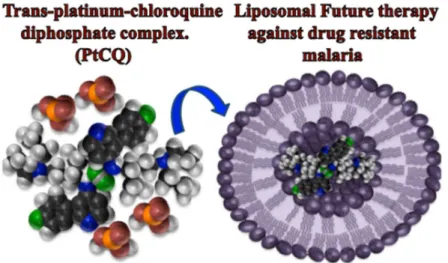
In the current study, two nano drug delivery systems were developed and characterized. One is for malaria, which is among the biggest killer diseases in the developing world. Platinum Chloroquine Diphosphate Dichloride complex (PtCQ) showed therapeutic effects againt chloroquine resistant malaria parasites. However and for the first time to encapsulate PtCQ into cationic liposomes with high encapsulation efficiency and high stability using remote loading method employing ammonium sulfate gradient. These PtCQ cationic liposomes hold promise for the future treatment of malaria and serious diseases. The other nano drug delivery system is for cancer, which is among
14
the leading killers in the developed countries. To the best of our knowledge, it is the first time to use krill lipid based liposomes to encapsulate a highly hydrophobic compound to prepare a low cost controlled drug delivery medicine to target cancer and other inflammatory diseases. These curcumin-loaded marinosomes exhibit high encapsulation efficiency, high antioxidant activity, and high physicochemical and oxidative stability. These curcumin-loaded marinosomes exhibited an effective cytotoxic effect on lung cancer (A549) and HUVEC cells. Hope these two drug delivery systems to be two feathers in the wings of the healing bird that fly on both worlds.
15
1.3. References
Bangham, A.D., Standish, M.M., Watkins, J.C., 1965. Diffusion of univalent ions across the lamellae of swollen phospholipids. J. Mol. Biol 13, 238–252.
Bangham, A.D., Standish, M.M., Watkins, J.C., Weissmann, G., 1967. The diffusion of ions from a phospholipid model membrane system. Protoplasma 63, 183–187.
Batzri, S., Korn, E.D., 1973. Single bilayer liposomes prepared without sonication. Biochim. Biophys. Acta. 16, 1015–1019.
Brannon-Peppase, L., Blanchette, J.Q., 2004. Nanoparticle and targeted systems for cancer therapy. Adv. Drug. Deliv. Rev. 56, 1649–1659.
Deamer, D.W., 2010. From “banghasomes” to liposomes: a memoir of Alec Bangham, 1921–2010, FASEB. J 24, 1308–1310.
Ferlay, J., Soerjomataram, I., Dikshit, R. 2015. Cancer incidence and mortality worldwide: sources, methods and major patterns in GLOBOCAN 2012. Int. J. Cancer 136, E359–E386.
GLOBOCAN: Global Burden of Cancer Study. Agency for Research on Cancer (2012). Greenwood, B.M., Fidock, D.A., Kyle, D.E., Kappe, S.H., Alonso, P.L., Collins, P.L.F.H.,
P. E. Duffy, P.E., 2008. Malaria: progress, perils, and prospects for eradication. J. Clin. Invest. 118, 1266–1276.
Gregoriadis, G., Ryman, B.E., 1971. Liposomes as carriers of enzymes or drugs: a new approach to the treatment of storage diseases. Biochem. J. 124, 58P.
16
Huang, C., 1969. Studies on phosphatidylcholine vesicles: formation and physical characteristics. Biochemistry 8, 344–352
Kimelberg, H.K., Tracy, T.F., Biddlecome, S.M., Bourke, R.S., 1976. The effect of entrap-ment in liposomes on the in vivo distribution of H-methotrexate in a primate. Cancer Res. 36, 2949–2957.
Li, Y., Zhang, D., Capoglu, I., Hujsak, K.A., Damania, D., Cherkezyan, L., Roth, E., Bleher, R., Wu, J.S., Subramanian, H., Dravid, V.P., Backman, V., 2017. Measuring the autocorrelation function of nanoscale three-dimensional density distribution in individual cells using scanning transmission electron microscopy, atomic force microscopy, and a new deconvolution algorithm. Microsc. Microanal. 23, 661–667. Miller, L.H., Ackermen, H.C., Su, X-Z, Wellems, T.E., 2013. Malaria biology and
disease pathogenesis: insights for new treatments. Nature Med. 19, 156–167.
Moles, E., Urbán, P., Jiménez-Díaz, M.B., Viera-Morilla, S., Angulo-Barturen, I., Busquets, M.A., Fernàndez-Busquets, X., 2015. Immunoliposome-mediated drug delivery to Plasmodium-infected and non-infected red blood cells as a dual therapeutic/prophylactic antimalarial strategy. J. Control. Release. 210, 217–229. Olson, F., Hunt, C.A., Szoka, F.C., Vail, W.J., Papahadjopoulos, D., 1979. Preparation of
liposomes of defined size distribution by extrusion through polycarbonate membranes. Biochim. Biophys. Acta 557, 9–23.
Papahadjopoulos, D., Watkins, J.C., 1967. Phospholipid model membranes. II.
Permeability properties of hydrated liquid crystals, Biochim. Biophys. Acta. 135, 639–652.
17
Peeters, P.A.M., Huiskamp, C.W.E.M., Eling, W.M.C. and Crommelin, D.J.A, 1989. Chloroquine containing liposomes in the chemotherapy of murine malaria. Cambridge J., Parasitol. 98, 381–386.
Pison, U., Welte, T., Giersig, M., and Groneberg, D.A., 2006. Nanomedicine for respiratory diseases. Eur. J. Pharmacol. 533, 341–350.
Poste, G., Papahadjopoulos, D., 1976. Lipid vesicles as carriers for introducing materials into cultured cells: influence of vesicle lipid composition on mechanism(s) of vesicle incorporation into cells. Proc. Natl. Acad. Sci. U.S.A. 73, 1603–1607.
Santos-Magalhães, N. S., Mosqueira, V. C. F, 2010. Nanotechnology applied to the treatment of malaria. Adv. Drug Deliv. Rev. 62, 560–575.
Schatzlein, A.G., 2006. Delivering cancer stem cell therapies – A role for nanomedicines? Eur. J. Cancer 42, 1309–1315.
Shi, J., Votruba, A.R., Farokhzad O.C., and Langer, R., 2010. Nanotechnology in drug delivery and tissue engineering: from discovery to applications. Nano Aalett 10, 3223–3230.
Stylios, G.K., Giannoudis, P.V., and Wan, T., 2005. Applications of nanotechnologies in medical practice. Injury 36, S6–S13.
Suri, S.S., Fenniri, H., Singh, B., 2007. Nanotechnology-based drug delivery systems, J. Occup. Med. Toxicol. 1, 2–16.
Urbán, P., Estelrich, J., Cortés, A., Fernàndez-Busquets, X., 2011. A nanovector with complete discrimination for targeted delivery to Plasmodium falciparum-infected versus non-infected red blood cells in vitro. J. Control. Release. 151, 202–211.
18
WHO: World Malaria Report. World Health Organization (2015).
Yokoyama. M., 2005. Drug targeting with nano-sized carrier systems. J. Artif. Organs 8, 77–84.
Zhigaltsev, I. V., Belliveau, N., Hafez, I., Leung, A.K., Huft, J., Hansen, C., Cullis, P.R., 2012. Bottom-up design and synthesis of limit size lipid nanoparticle systems with aqueous and triglyceride cores using millisecond microfluidic mixing. Langmuir 28, 3633–3640.
19
CHAPTER 2
Effective loading of platinum–chloroquine into PEGylated
neutral and cationic liposomes as a drug delivery system for
resistant malaria parasites
2.1. Introduction
Malaria is a life-threatening infectious disease that remains a major cause of death, especially in tropical regions. The WHO estimated that 214 million new cases of malaria resulted in 438000 deaths in 2015 (WHO, 2015). The mortality rate of malaria is higher than that caused by AIDS. Malaria is caused by a parasitic protozoan, Plasmodium falciparum, and is transmitted by infected Anopheles mosquitoes. The life cycle of Plasmodium parasites involves being bitten by an infected mosquito, during which the malaria parasite is injected into the bloodstream (Greenwood et al., 2008; Miller et al., 2013). The parasite migrates into hepatocytes and establishes itself in the liver. After a few weeks of dormancy, the activated parasite enters the blood and then infiltrates erythrocytes (red blood cells; RBCs). The parasite grows and divides in the erythrocytes by absorbing the nutrients from the blood via transporters, and these transporters are currently a topic of investigation by medical researchers.
The amphiphilicity of most current antimalarial drugs lead them to distribute widely into body tissues after systemic or oral administration, and can be easily
20
metabolized in the liver (Greenwood et al., 2008), and thus a high dose that may cause toxic side effects is needed to provide an antimalarial effect. The need to lower the dose to avoid toxicity is likely a main factor contributing to the development of resistance; this low dose also means that an ineffective dose is delivered to Plasmodium-infected RBCs. There is therefore an urgent need to develop new strategies to treat malaria and overcome the resistance of the parasite to current antimalarial drugs, especially in regions where Plasmodium falciparum is endemic.
Lipid-based drug nano-carriers have various advantages for drug delivery (Baruah et al., 2017). For example, liposomal formulations can increase drug bioavailability and thus increase therapeutic activity, decreasing side effects due to reduced dosing. Plasmodium-infected RBCs are the main chemotherapeutic target because several life stages of the parasite occur in the blood and give rise to the symptoms and pathologies of malaria. Consequently, several liposomal formulations encapsulating antimalarial drugs have been developed over the past several years in order to test their utility in malaria chemotherapy (Peeters et al., 1989; Santos-Magalhães et al., 2010). Additionally, immunoliposomes have been widely investigated and showed significant therapeutic effects (Urbán et al., 2011; Moles et al., 2015). However, one of the most drawbacks of immunoliposomes is that the antigens from different subspecies of malarial parasites are different. Therefore novel and universal drug delivery systems, which have effective targeting ability against various kinds of malaria parasites have been pursuing.
21
The ability of cationic liposomes to be adsorbed onto the cell surface and fuse with the negatively charged cellular membranes and thus deliver their cargo has been previously reported and shown in Fig. 2 (Li et al., 1997; Stebelska et al., 2006; Obata et al., 2010; Campbell et al., 2009; Zhao et al., 2011; Abu Lila et al., 2009). Anionic lipids such as phosphatidylserine (PS) are typically located within the inner monolayer of erythrocytes. The infection of erythrocytes with the malaria parasite leads to erythrocyte apoptosis, and this suicidal erythrocyte death is called eryptosis. The main characteristic stage of eryptosis is lipid scrambling and exposure of negatively charged PS on the outer surface of erythrocytes (Tagami et al., 2015; Föller et al., 2009), which increase the ability of erythrocytes to fuse with cationic liposomes (Stebelska et al., 2006). However, the use of cationic liposomes in vivo is limited due to their short circulation time, which leads to their recognition by the immune system including macrophages (Levchenko et al., 2002; Johnstone et al., 2001; Campbell et al., 2002). It is well known that polyethylene glycol (PEG) prevents the interaction with the biological in vivo environment and extend the circulation time of liposomes in blood. Cationic liposomes are therefore promising candidates for drug delivery targeting malaria-infected RBCs, but currently there is little information regarding the use of cationic liposomes. Additionally, cationic liposomes have not applied yet towards a working strategy for the treatment of malaria.
22
Fig. 2 Targeting of malaria-infected erythrocytes by cationic liposomes Cationic liposomes have the ability to fuse with the negatively charged cellular membranes and thus deliver its cargo.
23
Hemoglobin catabolism is an essential process that provides the parasite with amino acids it needs to grow resulting in the formation of heme. For Energy production purpose, the parasite converts heme into hematin. As hematin is toxic, the parasite converts it into hemozoin, which is chemically identical to β-hematin. The mechanism of action of chloroquine (CQ) is based on the inhibition of detoxification of hematin products by the parasite through interaction with hematin and preventing the formation of hemozoin causing the parasite to be poisoned by its own metabolic products. Although promising were the CQ antimalrial effects, the emergence and spread of CQ-resistant parasites is a growing global health problem (Navarro et al., 2011; Urbán et al., 2011). PtCQ is a trans platinum-chloroquine complex that shows therapeutic effects against the chloroquine-resistant malaria parasite. The high lipophilicity of PtCQ and the structural modification of CQ imposed by the presence of Plattinum lead to enhanced activity and ability to lower CQ resistance. Consequently, the main mechanism of anti-malarial action of PtCQ was the interaction of this metal compound with the specific target ferriprotoporphyrin IX (Fe (III) PPIX) and inhibition of β-haematinin in order to increase anti-malarial activity and fight the parasite resistance (Navarro et al., 2011).
The present study focuses on generating PtCQ-loaded PEGylated cationic liposomes exhibiting high drug encapsulation and high drug retention. Foremost, many fundamental properties of PtCQ, such as drug solubility, remain poorly investigated, although it was reported that PtCQ dissolves in dimethylsulphoxide (DMSO). We therefore investigated methods for efficiently loading PtCQ into liposomes and first
24
characterized the handling properties of PtCQ. Next; PtCQ was loaded with high encapsulation efficiency into cationic liposomes for the first time using the remote-loading method employing an ammonium sulfate gradient as shown in Fig. 3. The liposome formulations showed good drug retention, minimum leakage of PtCQ after two months’ storage at 4˚C, and further exhibited little release under in vitro culture conditions at 37˚C for 72 hours.
Fig. 3 The efficient drug encapsulation into PEGylated cationic liposomes by using remote-loading method
25
2.2. Materials and Methods
2.2.1 Reagents
Chloroquine diphosphate (CQDP), potassium tetrachloroplatinate (K2[PtCl4]),
hydrogenated soy phosphatidylcholine (HSPC), 1,2-dioleoyl-3-trimethylammonium-propane (DOTAP) and cholesterol (CHOL) were purchased from Wako Pure Chemical Industries, Ltd. (Osaka, Japan). 1,2-Distearoyl-sn-glycero-3-phosphoethanolamine-N-[methoxy (polyethylene glycol)-2000] (DSPE-PEG2000) was generously donated by NOF Corporation (Tokyo,
Japan).
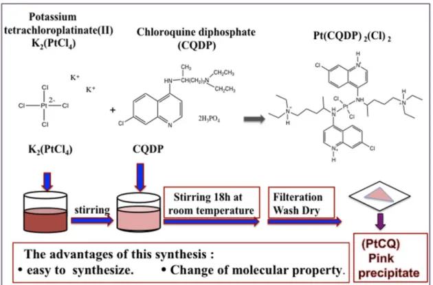
2.2.2 Synthesis of the trans-Pt(CQDP)
2(Cl)
2complex (
Navarro et al., 2011)The trans-Pt(CQDP)2(Cl)2 complex was synthesized as shown in Fig. 4. A
solution of K2[PtCl4] (100 mg, 0.24 mmol) in water (30 ml) was stirred until it
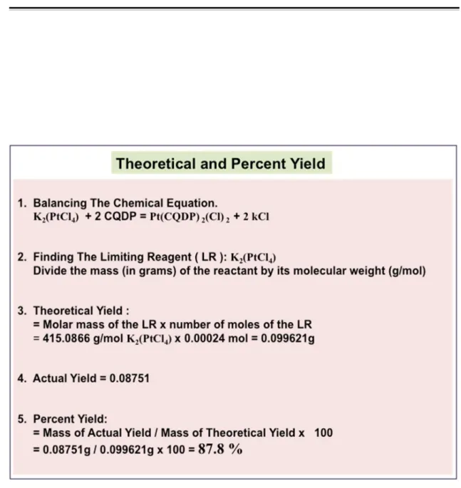
completely dissolved and then CQDP (250 mg, 0.48 mmol) was added. Stirring was continued for 18 hours at room temperature and a pink precipitate was obtained. This precipitate was collected by filtration, washed with water, and dried under vacuum. Yield 87.8% (calculated as in Fig. 5); elemental analysis (%) Calc. for C36H64N6Cl4O16P4Pt (1297.65 g.mol−1): C 33.3; N 6.5; H 4.9. Found: C 31.7; N 6.7; H
4.4. IR υ (N-H) 3305 cm−1; υ (C=C) 1616 cm−1; υ (C=N) 1581 cm−1; υ (Pt-cl) 341 cm−1; υ
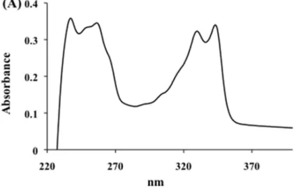
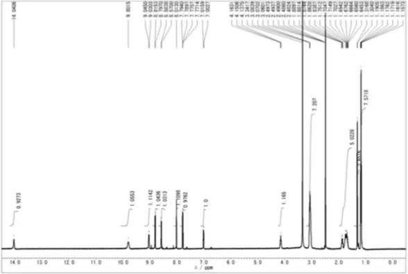
(Pt-N) 420 cm−1. UV-vis 238 and 344 nm. 1H-NMR (DMSO-d6; δ ppm): 9.05 (1H; d; J,
6.09 Hz; NH); 8.82 (1H; d; J, 9.13 Hz; H5); 8.58 (1H; d; J, 7.01 Hz; H2); 8.02 (1H; d; J, 1.83 Hz; H8); 7.8 (1H; dd; J1, 1.83 and J2, 7.01 Hz; H6); 7.02 (1H; d; J, 7.01 Hz; H3);
26
4.17 (1H; m; H1 ́); 3.15 (6H, m, H4 ́ and H5 ́); 1.79 (4H; m; H2 ́ and H3 ́); 1.3 (3H; d; J, 6.39 Hz; H1 ́ ́); 1.18 (6H, t, H6 ́).
Fig. 4 Synthesis of platinum chloroquine diphosphate dichloride ( PtCQ )
27
28
2.2.3 Preparation of PtCQ-loaded liposomes using the thin drug-lipid film
method
Liposomes were prepared by the thin drug-lipid film hydration method, followed by membrane extrusion as shown in Fig. 6 and described previously, with minor modifications (Isacchi1 et al., 2011). Briefly, neutral PtCQ-loaded liposomes were prepared by dissolving each lipid powder (HSPC, CHOL and DSPE-PEG2000) with
isopropanol and mixed at the molar ratio 55:45:5 (HSPC: CHOL: DSPE-PEG2000). PtCQ
(1 mg) was dissolved in dichloromethane (DCM) and a trace of DMSO (The details in Fig. 7 and Fig. 8) and then added to the lipid mixture. The drug-lipid mixture was evaporated to give a thin drug-lipid film that was placed under high vacuum for at least overnight to remove residual solvent. The dried drug-lipid film was hydrated in HBS (20 mM HEPES, 150 mM NaCl, pH 7.5) to form multilamellar vesicles (MLVs), sonicated for 10 min, and then extruded (Mini-extruder, Avanti Polar Lipids, Alabaster, AL, USA) 10 times through Whatman Nuclepore Track-Etched polycarbonate membranes (GE Healthcare Life Sciences; Chicago, IL, USA) with 200 nm pores and then with 100 nm pores at 70˚C to transform the MLVs into unilamellar vesicles. The formulation was cooled on ice for 15 min and unencapsulated PtCQ was removed by ultracentrifugation (2.1104 g, 4˚C, 30 min) as previously reported (Laouini et al., 2012). Cationic
PtCQ-loaded liposomes were prepared by dissolving the lipids (HSPC: DOTAP: CHOL: DSPE-PEG2000) in isopropanol, then mixing in the molar ratio 50:5:45:5, and 35:20:45:5.
PtCQ (1 mg) was added to the lipid mixture at different weight ratios (1:7, 1:14) and then the vesicles were prepared as described above. The mean size and polydispersity index
29
(PDI) were determined using a dynamic light scattering instrument (ZetaSizer Nano-ZS; Malvern Instrument Ltd., Malvern, U.K). Zeta potentials were measured using Zetasizer Nano ZS90 (Malvern). The zeta potentials were measured by dispersing the liposome solution in 5% dextrose. The encapsulation efficiency (EE) was calculated as described in “Determination of the EE.”
30
Fig. 7 Fundamental properties of PtCQ
31
2.2.4 Preparation of PtCQ-loaded liposomes using the remote-loading
method
PtCQ-loaded liposomes were prepared as as shown in Fig. 9 and previously reported, with minor modifications (Haran et al., 1993; Tagami et al., 2015; Tagami et al., 2014; Qiu et al., 2008). Briefly, neutral PtCQ-loaded liposomes were prepared by mixing the lipids (HSPC: CHOL: DSPE-PEG2000) at the molar ratio 55:45:5. Cationic
PtCQ-loaded liposomes were prepared by mixing the lipids (HSPC: DOTAP: CHOL: DSPE-PEG2000) with different molar ratios of the cationic lipid DOTAP (0, 5, 10, 15 and
20 mol%) and dissolving in organic solvent. The solvent from the lipid mixtures was evaporated to provide thin lipid films that were placed under high vacuum for at least overnight to remove residual solvent. The thin films were hydrated with 250 mM ammonium sulfate (pH 4.0), sonicated for 10 min, and size-controlled liposomes were prepared using the extruder as described above. Next, the external liposomal phase was replaced with HBS via dialysis (Slide-A-Lyzer 10 kDa MWCO, Pierce Biotechnology, Rockford, IL, USA) for 24 h against 1000 volumes of HBS. The concentration of CHOL was determined using a cholesterol E-Wako kit (Wako Pure Chemical) and a microplate reader (Wallac 4000 ARVO multi-label counter; PerkinElmer, Waltham, MA, USA) in order to estimate the concentrations of the various liposome preparations. Neutral PtCQ-loaded liposomes were prepared by mixing PtCQ solution in HBS (0.65 mg/ml) with the neutral liposomal solution at three drug-to-lipid ratios (1:2.5, 1:5 and 1:7 (w/w) and incubated at 60˚C for 60 min for drug loading. The cationic PtCQ-loaded liposomes were prepared by using a drug/lipid ratio of 1:7 (w/w). The mean size, PDI, and zeta
32
potential were measured using Zetasizer Nano ZS90 (Malvern) as described above and the EE was calculated as described in “Determination of the EE.”
33
2.2.5 Determination of the EE
For PtCQ-loaded liposomes prepared using the thin drug-lipid film method, the sample was ultracentrifuged (2.1104 g, 4˚C, minimum 30 min) and the pellet
was resuspended in HBS buffer. The concentration of the encapsulated drug was measured by absorbance at 343 nm using a UV-visible spectrophotometer (UV-1800, Shimadzu, Kyoto, Japan) after completely disrupting the liposomes using 2% Triton X-100. The absorbance was converted into drug concentration using a standard curve. The EE was calculated using the previously reported equation (Ramana et al., 2010):
EE = (Drug Encapsulated / Total Drug) × 100%
For PtCQ-loaded liposomes prepared using the remote-loading method, the samples were cooled and applied onto a gel filtration column (CL-4B, Sigma Aldrich, Saint Louis, MO, USA) equilibrated with HBS to remove unencapsulated PtCQ. The liposome fraction of the eluate was collected, the concentration of lipid was determined again, and the concentration of the encapsulated drug was measured as described above.
2.2.6. Measurement of drug release from PtCQ-loaded liposomes under
storage conditions
All PtCQ-liposomal formulations were stored at 4˚C. As a typical experiment, aliquots (100 μl) were withdrawn at specified time points (0, 1, 2, 4 and 8 weeks),
34
diluted to 1 ml with HBS buffer, and ultracentrifuged (2.1104 g, 4˚C, 30 min) using
Amicon filters (MWCO, 10K; Millipore, Bedford, MA, USA) to separate the released PtCQ. The released PtCQ was measured by absorbance at 343 nm as described above. The percentage of PtCQ released from the liposomes was calculated using the previously reported formula (Aditya et al., 2012):
Drug release (%) = (PtCQ released / Total PtCQ) × 100
Where “PtCQ released” indicates the amount of released PtCQ collected at a specified time point t and “Total PtCQ” indicates the total amount of PtCQ entrapped in the liposomes.
2.2.7 Measurement of drug release from liposomes under in vitro culture
conditions (release test)
As a typical experiment, 1 ml of liposomal solution was mixed with 1 ml of RPMI 1640 medium (Wako Pure Chemical) supplemented with 10% heat-inactivated fetal bovine serum (Invitrogen, Carlsbad, CA, USA) and the samples were incubated at 37˚C in a humidified incubator in an atmosphere of 5% CO2/95% air. Aliquots (100 µL)
were withdrawn at specified time points (0, 1, 3, 6, 24, 48 and 72 hours), diluted to 1 ml with HBS buffer, and ultra-centrifuged to separate the released PtCQ (2.1104 g, 4˚C, 30
min). The percentage of PtCQ released was determined as in section 2.6. The chloroquine absorbance at 343 nm was unaffected by the culture medium, and the standard curve was unaffected, as shown in Fig. 10.
35
2.2.8 Statistical Analysis
All data shown here are represented in “mean value ± standard deviation (S.D.)”. An ordinary one-way ANOVA (followed by Tukey's or Dunnett's multiple comparison test), and a two-way ANOVA with (Bonferroni post-test or Dunnett's multiple comparison test) were used to assess statistical significance by using GraphPad Prism (GraphPad Software Inc., CA, USA). P < 0.05 was regarded as statistically significant.
36
2.3. Results and discussion
2.3.1 Synthesis and characterization of the Pt(CQDP)2(Cl)2 complex
The platinum-chloroquine diphosphate complex was synthesized and isolated in water at room temperature. A mixture of K2[PtCl4] and CQDP was prepared to displace
two chloride ligands, leading to the new PtCQ complex that was isolated in good yield as a pink solid precipitate (Fig. 11). The complex was characterized by elemental analysis, and UV-visible, IR and proton-NMR spectroscopy (for analysis data details, see section 2.2 and Fig. 12). The complex is slightly soluble in water and insoluble in organic solvents except DMSO. The elemental analysis of this complex is in agreement with the molecular formula proposed. The IR spectrum of the complex shows peaks clearly associated with the presence of coordinated CQDP. In the 1H NMR spectrum, the 1H
chemical shift variation of each signal with respect to that of the free ligand reported previously (Navarro et al., 2011)was similar and used to deduce the mode of bonding of CQDP to the metal. The largest shift
with respect to the free ligand (CQDP) was observed for NH and H1 ́ as they are the nearest protons to the N-atom bonded to the metal, indicating that CQDP is bound to platinum through the
nitrogen of the secondary amine. Fig. 11 Chemical structure of platinum
37
38
39
2.3.2. PtCQ-loaded liposomes prepared using the thin drug-lipid film
method
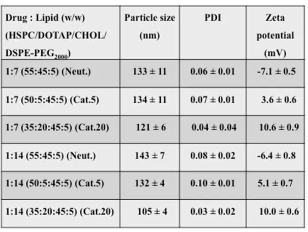
The high lipophilicity of metal chloroquine complexes has been previously reported (Navarro et al., 2014) and the preparation of highly stable lipophilic cisplatin complex/liposomes with high encapsulation ratios has been achieved (Khokhar et al., 1991). Consequently, in this study we succeeded in loading the PtCQ complex into neutral and cationic liposomes at reasonable drug-to-lipid ratios by incorporating the drug into the lipid phase using the thin drug-lipid film method. As shown in Table 1, the PtCQ-loaded liposomes had uniform sizes ranging from about 105 nm to 143 nm and a narrow size distribution (PDI < 0.097). Furthermore, the zeta potential of PEGylated liposomes was changed from low negative (-7.1 ± 0.5 mV) of the neutral liposomes (1:7) to low positive (10.6 ± 0.9 mV, Cat.20) of the cationic liposomes (1:7). The negative charge of the neutral liposomes is due to the presence of negatively charged phosphate group in DSPE-PEG2000 (Woodle et al., 1992). In addition, Fig. 13 reveals that the
cationic lipid content clearly affects the drug loading in the thin drug-lipid film method. Based on the statistical study, the addition of 5 mol% of the cationic lipid DOTAP at low liposome concentration (7 mg) has a significant decrease (p = 0.0076) on the EE% of Cat.20 compared to Cat.5, however it has no significant effect (p > 0.05) on the EE% of Cat.5 and Cat.20 compared to neutral liposomes. On the other hand, at higher liposome concentration (14 mg), there is a significant decrease (p < 0.0001) on the EE% of Cat.20 compared to Cat.5 and the neutral liposomes, however no significant effect (p > 0.05) was noticed in the EE% of Cat.5 compared to the neutral liposomes. The decrease in the
40
EE% with the increase of the cationic lipid content could be due to the electrostatic repulsion between the cationic drug and the cationic lipid such as DOTAP.
Table 1. PtCQ-liposomes loaded by thin drug–lipid film method
Neutral and cationic formulations with drug–lipid (1:7, 1:14 w/w) have been prepared. The neutral lipids have been mixed with the molar ratio (55:45:5). The cationic lipids have been mixed with the molar ratio (5:50:45:5) and (35:20:45:5). Data represent the mean ± S.D. (n = 3).
41
Fig. 13. The EE of liposomes prepared using the thin drug–lipid film method Three formulations were prepared, at drug to lipid ratios of 1:7, 1:14 (w/w). Data represent the mean ± S.D. (n = 3), (**** p < 0.0001, ** p = 0.0076). The information about liposomal composition is written in Table 1.
42
2.3.3 PtCQ-loaded liposomes prepared using the remote-loading method
To our knowledge, this is the first report describing the encapsulation of the PtCQ complex into liposomes using an ammonium sulfate gradient and the results are promising. The rate of loading and the stability of the loaded drug in the intra-liposomal medium mainly depend on the properties of i) the loaded drug, ii) the intra-liposomal medium, and iii) the liposome bilayer, as discussed below.
(i) Properties of the loaded drug:
Chemical compounds can be categorized as hydrophilic compounds, amphiphilic compounds, and hydrophobic compounds. Hydrophilic compounds do not interact with the liposome bilayer and can be encapsulated in the aqueous phase of the liposome interior. Hydrophobic compounds integrate into the liposome bilayer. Amphiphilic compounds may be amenable to high loading due to their possible high affinity for the liposomal membrane or may be remotely loaded into the intra-liposomal aqueous phase by using a pH gradient. CQ is a diprotic amphiphilic weak base with two proton-binding groups: the side chain terminal amine (pKa1 = 10.2) and the quinoline nitrogen atom
(pKa2 = 8.4). Accordingly, unprotonated CQ can be driven by a pH gradient across a
parasite’s food vacuole membrane and thus accumulate in the acidic food vacuole. CQ can be actively encapsulated by applying a pH gradient (Qiu et al., 2008; Madden et al., 1990), during which it is protonated to the diproton form following exposure to the intra-liposomal acidic medium, then captured inside the liposomes due to the low permeability of the liposomal bilayer membrane to the protonated drug (Qiu et al., 2008). In the present study we used the ammonium sulfate gradient to load PtCQ into
43
liposomes composed mainly of neutral or cationic saturated phospholipids with an EE of almost 100%, as shown in Figure 14 and Figure 15.
Fig. 14. Effect of increasing the liposomal content on the EE of PtCQ loaded inside neutral liposomes prepared using the remote loading method
Three formulations were prepared, at drug to lipid ratios of 1:2.5, 1:5 and 1:7 (w/w). Data represent the mean ± S.D. (n = 3). Statistical data infers that each group is significantly different (****p < 0.0001, ***p = 0.0008). The information about liposomal composition is written in Table 2.
44
Fig. 15. Effect of increasing the cationic lipid content (DOTAP) on the EE of PtCQ loaded inside cationic liposomes prepared using the remote loading method
Five formulations were prepared (0, 5, 10, 15, 20 mol% as cationic lipid). The drug-to-lipid ratio is 1:7 (w/w). Data represent the mean ± S.D. (n = 4). The information about liposomal composition is written in Table 3.
45
(ii) Intra-liposomal medium properties:
Various chemical gradients have been used to create a trans-membrane potential as a driving force to facilitate the loading of a drug through an active loading mechanism. For example, ammonium sulfate induces a potential gradient for weak bases and acids and this gradient remains stable for over six months for liposomal formulations stored below the transition temperature of the phospholipids (Haran et al., 1993). Furthermore, lipophilic amino-containing drugs can be partitioned into the lipid bilayer, resulting in a drug gradient through the lipid bilayer that aids the influx of the drug from the outside to the inside (Harrigan et al., 1993).
In the present study, we loaded the PtCQ complex into neutral liposomes using a pH gradient and the trans-membrane potential generated by an ammonium sulfate gradient as driving forces. Following hydration of the PtCQ-loaded liposomes using an ammonium sulfate solution, an equal concentration of ammonium sulfate is found outside and inside the liposomes. Removal of the outside ammonium sulfate solution by dialysis leads to an ammonium sulfate gradient in which the concentration of ammonium sulfate inside the liposomes is greater than that outside and the gradient is due to the large difference in the permeability coefficient through lipid bilayer as mentioned before (Haran et al., 1993). The higher concentration of ammonium inside the liposome causes the efflux of neutral ammonium, leaving the protons inside the liposome. This ammonium gradient results in the influx of the PtCQ into the acidic intra-liposomal medium and thus its encapsulation. The drug may be tetra-protonated due to the presence of two chloroquine molecules in the complex. The outside-inside pH gradient was 7.5–4
46
to give a pH gradient equal to 3.5 units (∆pH = (external pH − internal pH) = 3.5) since this produces a much higher inside concentration of the weak base compared to the outside concentration. As shown in Fig. 14 and Table 2, PtCQ-loaded liposomes were prepared with different drug-to-lipid weight ratios and the maximum EE was obtained at a drug-to-lipid ratio of 1:7 (w/w). In a preliminary experiment, the loading of PtCQ was compared using ammonium sulfate or citrate buffer and the EE was found to be higher with ammonium sulfate, perhaps due to the high permeability coefficient of the NH3
molecule, which causes fast diffusion of neutral ammonia to the external medium of the liposomes. Each diffusing NH3 molecule leaves one proton inside the liposome, thus
acidifying the intra-liposomal medium and resulting in a pH gradient and influx of the drug (Haran et al., 1993). The pH gradient had a greater effect on CQ, which has two amino moieties, than on a weak base with one amino moiety (Qiu et al., 2008).
(iii) Liposome bilayer properties
It was previously demonstrated that drug-loaded liposomes prepared by the remote loading method and comprising saturated lipids can be maintained at 4˚C for at least six months without deterioration (Haran et al., 1993). Liposomes composed of saturated lipids can maintain a pH gradient due to the relatively high transition temperature of the bilayer. In the present work, we used the saturated lipid HSPC to construct the membrane of PtCQ-loaded liposomes generated using the remote loading method. The demonstrated stability of the PtCQ-loaded liposomes under storage and culture conditions verifies the ability of this saturated lipid bilayer to maintain a specified pH gradient and retain the drug in the interior phase of the liposome as long as the
47
temperature does not exceed the transition temperature of the lipid, thus preventing thermal mobility that could disrupt the membrane required for maintaining the pH gradient (Qiu et al., 2008). We studied the effect of temperature on drug loading at two temperatures below the transition temperature 60˚C, after incubation for one hour and obtained maximum loading at 60˚C as described below.
Table 2. PtCQ-liposomes loaded by ammonium sulfate gradient
The neutral lipids have been mixed with the molar ratio (HSPC/CHOL/DSPE- PEG2000 = 55:45:5). Three different formulations with drug–lipid (1:2.5, 1:5, 1:7 w/w)
48
2.3.4 Cationic PtCQ-loaded liposomes produced using the
remote-loading method
Five types of cationic PtCQ-loaded liposomes with a drug-to-lipid ratio of 1:7 (w/w) but differing in the ratios of the constituent lipids were prepared and characterized, and the results are shown in Table 3. The PtCQ-loaded cationic liposomes has uniform sizes about 130 nm and a narrow size distribution (PDI < 0.05). It was reported that the cationic liposomes demonstrate an enhanced cellular uptake compared to the neutral liposomes, it could be due to the electrostatic interaction between the positive charge and the cell (Zhao et al., 2011). Additionally, the increase of cationic lipids in liposomes could lead to a higher affinity for the anionic sites, however it could result in their aggregation in the bloodstream through electrostatic interactions with the anionic species in the blood and thus increase their uptake by the RES (reticuloendothelial system)(Zhao et al., 2011). Liposomes are typically modified with PEG (PEGylation) to form a water layer that could prevent protein adsorption onto the liposome surface and decrease the uptake of PEG-liposomes by macrophages andthus help in prolonging the lifetime of liposomes in the blood circulation (Kraft et al., 2014). The partial coating of cationic particles with PEG masks the values of zeta potential (electric potential across a double membrane surface), however it can’t prevent them from binding to the cells and the biodistribution completely. Moribe et al., reported that the inclusion of 0 to 10% PEG sharply decreases zeta potential of the liposome (Moribe et al., 1997). For this reason typically 5–10 mol% of PEG is enough to delay the recognition of liposomes by immune system cells.This concentration couldretain a significant cationic charge and binding to
49
the cells. Consequently, All formulations prepared in this study were PEGylated with 5 mol %. Additionally, it has been reported that theadhesion of the liposome membrane to the cell depends on the ratio of the liposome–cell charge, thus the probability of adhesion is maximized at a finite polymer layer thickness in case of the cationic liposomes are overcharged compared to the cell (Dan et al., 2002). Accordingly, in this study, several PtCQ-loaded liposomes with different molar ratio of the cationic lipid (DOTAP, 0, 5, 10, 15 and 20 mol%) were prepared to provide stable cationic PtCQ-loaded liposomes that could selectively bind to erythrocytes. Table 3 reveals that the zeta potential of PEGylated cationic liposomes changes from low negative (-7.5 ± 1.3) for (Cat.0) to neutral (-0.4 ± 0.7, Cat.10), and to low positive (10.7 ± 3.0, Cat.20).
The addition of negatively charged lipids to liposomes generated using the passive loading method can increases EE due to electrostatic interaction between the positively charged chloroquine and the negatively charged liposomes. However, characterization of the PtCQ-loaded liposomes prepared using the thin drug-lipid film method presented in section 3.2 confirmed that the EE of cationic liposomes decrease with increasing the cationic lipid content and this could be due to the electrostatic repulsion between the cationic drug and the cationic lipid. In contrast, Fig. 15 reveals that the different cationic PtCQ-loaded liposomes prepared by ammonium sulfate gradient (Cat.5, Cat.10, Cat.15, and Cat.20) have no significant effect (p > 0.6559) on EE% compared to Cat.0 (96.9 ± 5.0%). This result confirms that the most of PtCQ loaded by
50
ammonium sulfate gradient is not bilayer-associated, in good agreement with a previous report (Haran et al., 1993).
Table 3. PtCQ-liposomes loaded by ammonium sulfate gradient
The cationic lipids have been mixed with different molar ratios. Five different formulations with HSPC–DOTAP (55:0, 50:5, 45:10, 40:15, 35:20) have been prepared. Data represent the mean ± S.D. (n = 3, 4).
51
2.3.5 Drug release from cationic PtCQ-loaded liposomes under storage
conditions (storage test)
The stability of cationic PtCQ-loaded liposomes (cat.0, cat.5, cat.10, cat.15, cat.20) loaded using the ammonium sulfate gradient (drug-to-lipid ratio 1:7 (w/w)) was studied during eight weeks’ storage at 4˚C. Fig. 16 reveals that there is no significant release (p > 0.05) of the drug at different cationic PtCQ-loaded liposomes after eight weeks’ storage at 4˚C, compared to Cat.0 (5.7 ± 4.9 %). This demonstrates the utility of the ammonium sulfate gradient method for incorporating amphiphilic drugs into stable liposomes containing a saturated lipid (HSPC). It was reported that the addition of PEG stabilizes liposomes due to repulsive barrier resulted from steric pressure forces and charged phosphate moieties (Woodle et al., 1992; Needham et al., 1992). Additionally, the presence of CHOL stabilizes liposomal formulations, as reported previously (Briuglia et al., 2015; Lee et al., 2005).
52
Fig. 16. Release pattern of PtCQ from cationic liposomes, Prepared by Remote Loading Method, in HEPES Buffer during Storage at 4°C. PtCQ was loaded into the liposomes at a drug-to-lipid ratio of 1:7 (w/w). Data represent the mean ± S.D. (n = 3). Cat. 0, Cat. 5, Cat. 10, Cat. 15 and Cat. 20 are the five formulations of cationic PtCQ-loaded liposomes prepared using different molar ratio of the cationic lipid (DOTAP, 0, 5, 10, 15 and 20 mol %, respectively)
53
2.3.6 Drug release from cationic PtCQ-loaded liposomes under culture
conditions (release test)
A drug release study was carried out using medium containing FBS to mimic in vivo conditions. Figure 17 reveals that the PtCQ liposomes loaded by thin drug-lipid film method (drug to lipid ratio 1:7 (w/w)) have initial burst release equal to 8.9 ± 0.5% of neutral liposomes and equal to 13.2 ± 2.1% of cationic liposomes (Cat.5) at 0 hour. The cause for initial burst release may be due to the adsorption of some drug molecules on the liposome surface during formation of the thin film. Furthermore, after 72 hours, there is a significant drug release (p = 0.0001) of Cat.5 compared to neutral liposomes’cumulative release equal to 17.8 ± 2.6%. The cationic liposomes showed a maximum release which is slightly higher than that of neutral liposomes and this could be due to electrostatic repulsion between the cationic drug and the cationic lipid.
The five formulations of cationic PtCQ-loaded liposomes (cat.0, cat.5, cat.10, cat.15, cat.20) loaded using an ammonium sulfate gradient (drug to lipid ratio 1:7 (w/w) described above didn't show initial burst release and showed no significant release (p > 0.05) of the drug after 72 hours compared to Cat.0 (4.0 ± 2.3%), as shown in Figure 18.
This demonstrates that the ammonium sulfate gradient is a promising method for the encapsulation of amphiphilic drugs into liposomes containing a saturated lipid (HSPC). Furthermore, the amount of drug released was sustainably released indicating that liposomes loaded with amphipathic weak bases using an ammonium sulfate gradient can provide controlled release of the loaded molecule. Increasing the cationic lipid from 0% to 20% mole percent has no significant effect on the release of the drug; showing that the
54
surface charge has no effect on the release of amphipathic weak bases loaded using ammonium sulfate and pH gradients. The slow drug release observed in case of ammonium sulfate gradient loaded PtCQ-liposomes is necessary for long storage and to avoid drug leaking before reaching their target (Moles et al., 2015). Afterward the liposomes would be adsorbed on the cell surface followed by a depletion of the liposomal proton gradient due to the body temperature or liposomes-cell interaction episodes. This depletion leads to accumulation of the weak basic drugs inside the RBC due to the electrochemical gradient resulted from the phospholipid asymmetry in RBC membranes, which maintains negatively charged membranes.
Fig. 17. Release pattern of PtCQ from neutral and cationic liposomes, Prepared by thin drug–lipid film method, under culture conditions. PtCQ-loaded liposomes were incubated in medium containing 10 % FBS at
37°C. PtCQ was loaded into the liposomes at a drug-to-lipid ratio of 1:7 (w/w). Data represent the mean ± S.D. (n = 3). (**** p < 0.0001, *** p = 0.0007, ** p = 0.0024) compared to neutral liposomes.
55
Fig. 18. Release pattern of PtCQ from cationic liposomes, Prepared by
PtCQ-loaded liposomes were incubated in medium containing 10 % FBS at 37°C. PtCQ was loaded into the liposomes at a drug-to-lipid ratio of 1:7 (w/w). Data represent the mean ± S.D. (n = 3). Cat. 0, Cat. 5, Cat. 10, Cat. 15 and Cat. 20 are the five formulations of cationic PtCQ-loaded liposomes prepared using different molar ratio of the cationic lipid (DOTAP, 0, 5, 10, 15 and 20 mol %, respectively)
56
2.4. Conclusion
This chapter described the synthesis and characterization of the trans-platinum-chloroquine complex (PtCQ) and the application of two methods to generate PtCQ-loaded liposomes: the thin drug-lipid film and the remote loading methods. Liposomes of uniform diameter were prepared using the thin film hydration and extrusion technique and the PtCQ was encapsulated in the liposomes. The most efficient encapsulation was obtained at a drug-to-lipid ratio of 1:7 (w/w) using the ammonium sulfate gradient active loading method. This approach provided liposomal formulations capable of sustaining a proton gradient for the weak basic drug PtCQ and the encapsulation efficiency was approximately 100%. High intra-liposomal retention levels of 93% the drug inside different cationic liposomes for several hours under culture and storage conditions were maintained, suggesting that such liposomes hold promise for the future treatment of malaria by targeting Plasmodium falciparum. We will study the in vitro and in vivo effects of cationic PtCQ-loaded liposomes on this parasite and the encapsulation of other metal-CQ complexes in the future.
57
2.5. References
Abu Lila, A.S., Kizuki, S., Doi, Y., Suzuki, T., Ishida, T., Kiwada, H., 2009. Oxaliplatin encapsulated in PEG-coated cationic liposomes induces significant tumor growth suppression via a dual-targeting approach in a murine solid tumor model. J. Control Release. 137: 8–14.
Aditya, N.P., Chimote, G., Gunalan, K., Banerjee, R., Patankar, S., Madhusudhan, B., 2012. Curcuminoids-loaded liposomes in combination with arteether protects against Plasmodium berghei infection in mice. Exp. Parasitol. 131, 292–299.
Baruah, U.K, Gowthamarajan, K., Vanka, R., 2017. Malaria treatment using novel nano-based drug delivery systems. J. Drug Target. 25,567–581.
Briuglia, M-L., Rotella, C., McFarlane, A., Lamprou, D. A., 2015. Influence of cholesterol on liposome stability and on in vitro drug release. Drug Delivery Translat. Res. 5, 231–242.
Campbell, R.B, Ying, B., Kuesters, G.M., Hemphill, R. 2009. Fighting cancer: from the bench to bedside using second-generation cationic liposomal therapeutics. J. Pharm. Sci. 98, 411–429.
Campbell, R.B., Fukumura, D., Brown, E.B., Mazzola, L.M., Izumi, Y., Jain, R.K., Torchilin, V.P., and Munn, L.L., 2002. Cationic charge determines the distribution of liposomes between the vascular and extravascular compartments of tumors. Cancer Res. 62, 6831–6836.
58
Dan, N., 2002. Effect of liposome charge and PEG polymer layer thickness on cell-liposome electrostatic interactions. Biochim. Biophys. Acta. 1564, 343–348. Föller, M., Bobbala, D., Koka, S., Huber, S. M., Gulbins, E. and Lang, F., 2009. Suicide
for survival–death of infected erythrocytes as a host mechanism to survive malaria. Cell Physiol. Biochem. 24, 133–140.
Greenwood, B.M., Fidock, D.A., Kyle, D.E., Kappe, S.H., Alonso, P.L., Collins, P.L.F.H., Duffy, P.E, 2008. Malaria: progress, perils, and prospects for eradication. J Clin Invest. 118, 1266–1276.
Haran, G., Cohen, R., Bar, L.K. and Barenholz, Y., 1993. Transmembrane ammonium sulfate gradients in liposomes produce efficient and stable entrapment of amphipathic weak bases. Biochim. Biophys. Acta 1151, 201–215.
Harrigan, P.R., Wong, K.F., Redelmeier, T.E., Wheeler, J.J. and Cullis, P.R., 1993. Accumulation of doxorubicin and other lipophilic amines into large unilamellar vesicles in response to transmembrane pH gradients. Biochim. Biophys. Acta. 1149, 329–338.
Isacchi1, B., Arrigucci, S., la Marca, G., Camilla Bergonzi, M., Vannucchi, M.G., Novelli, A., and Bilia, A.R., 2011. Conventional and long-circulating liposomes of artemisinin: preparation, characterization, and pharmacokinetic profile in mice. J. of Liposome Res. 21, 237–244.
Johnstone, S.A., Masin, D., Mayer, L., Bally, M.B., 2001.Surface-associated serum proteins inhibit the uptake of phosphatidylserine and poly (ethylene glycol) liposomes by mouse macrophages. Biochim. Biophys. Acta. 1513, 25–37.
59
Khokhar, A. R., Al-Baker, S., Brown, T. and Perez-Soler, R., 1991. Chemical and biological studies on a series of lipid-soluble (trans-(R,R)- and -(S,S)-1,2-diaminocyclohexane) platinum(II) complexes incorporated in liposomes. J. Med. Chem. 34, 325–329.
Kraft, J. C., Freeling, J. P., Wang, Z. and Ho, R. J. Y., 2014. Emerging research and clinical development trends of liposome and lipid nanoparticle drug delivery systems. J. Pharm. Sci. 103, 29–52.
Laouini, A., Jaafar-Maalej, C., Limayem-Blouza, I., Sfar, S., Charcosset, C. and Fessi, H., 2012. Preparation, Characterization and Applications of Liposomes: State of the Art. J. Colloid Sci. and Biotech. 1, 147–168.
Lee, S-C, Lee, K-E, Kim, J-J and Lim, S-H, 2005. The effect of cholesterol in the liposome bilayer on the stabilization of incorporated retinol. J. Liposome Res. 15, 157–166.
Levchenko, T.S, Rammohan, R., Lukyanov, A.N, Whiteman, K.R, Torchilin, V.P, 2002. Liposome clearance in mice: the effect of a separate and combined presence of surface charge and polymer coating. Int. J. Pharm. 240, 95–102.
Li, L.H., Hui, S.W., 1997. The effect of lipid molecular packing stress on cationic liposome-induced rabbit erythrocyte fusion. Biochim. Biophys. Acta. 1323, 105–16. Madden, T.D., Harrigan, P.R., Tai, L.C.L., Bally, M.B., Mayer, L.D., Redelmeier, T.E.,
Loughrey, H.C, Tilcock, C.P, Reinish, L.W, Cullis, P.R., 1990. The accumulation of drugs within large unilamellar vesicles exhibiting a proton gradient: a survey. Chem. Phys. Lipids. 53, 37–46.
60
Miller, L.H., Ackermen, H.C., Su, X-Z, Wellems, T.E., 2013. Malaria biology and disease pathogenesis: insights for new treatments. Nat. Med. 19, 156–167.
Moles, E., Urbán, P., Jiménez-Díaz, M.B., Viera-Morilla, S., Angulo-Barturen, I., Busquets, M. A., Fernàndez-Busquets, X., 2015. Immunoliposome-mediated drug delivery to Plasmodium-infected and non-infected red blood cells as a dual therapeutic/prophylactic antimalarial strategy. J. Control Release. 210, 217–229. Moribe, K., Maruyama, K, Iwatauru, M., 1997. Estimation of surface state of
Poly(ethylene glycol)-coated liposomes using an aqueous two-phase partitioning technique. Chem. Pharm. Bull. 45, 1683–1687.
Navarro, M., Castroa, W., Higuera-Padillaa, A.R, Sierraaltab, A., Abadc, M.J., Taylorc, P., and Roberto, A., 2011.Sánchez-Delgadod, Synthesis, characterization and biological activity of trans- platinum (II) complexes with chloroquine. J. Inorg. Biochem. 105, 1684–1691.
Navarro, M., Castro, W., Madamet, M., Amalvict, R., Benoit, N., Pradines, B., 2014. Metal-chloroquine derivatives as possible anti-malarial drugs: evaluation of anti-malarial activity and mode of action. Malar. J. 13, 471.
Needham, D., Mclntosh, T.J. and Lasic, D.D., 1992. Repulsive interactions and mechanical stability of polymer-grafted lipid membranes. Biochim. Biophys. Acta. 1108, 40–48.
Obata, Y., Tajima, S., Takeoka, S., 2010. Evaluation of pH-responsive liposomes containing amino acid- based zwitterionic lipids for improving intracellular drug delivery in vitro and in vivo. J. Control Release.142, 267–276.
61
Peeters, P.A.M., Huiskamp, C.W.E.M., Eling, W.M.C. and Crommelin, D.J.A., 1989. Chloroquine containing liposomes in the chemotherapy of murine malaria. Cambridge J., Parasitol. 98, 381–386.
Qiu, L., Jing, N., Jin, Y., 2008. Preparation and in vitro evaluation of liposomal chloroquine diphosphate loaded by a transmembrane pH-gradient method. Int. J. Pharm. 361, 56–63.
Ramana, L.N., Sethuraman, S., Ranga U., and Krishnan, U.M., 2010. Development of a liposomal nanodelivery system for nevirapine. J. Biomed. Sci. 17, 57.
Santos-Magalhães, N. S., Mosqueira, V. C. F., 2010. Nanotechnology applied to the treatment of malaria. Adv. Drug Deliv. Rev. 62, 560–575.
Stebelska, K., Wyrozumska, P., Sikorski, A.F., 2006. PS exposure increases the susceptibility of cells to fusion with DOTAP liposomes. Chem. Biol. Interact. 160, 165–174.
Tagami, T., Yanai, H., Terada, Y. and Ozeki, T. 2015. Evaluation of Phosphatidylserine-Specific Peptide-Conjugated Liposomes Using a Model System of Malaria-Infected Erythrocytes. Biol. Pharm. Bull. 38, 1649–1651.
Tagami, T., Kubota M., and Ozeki, T., 2015. Effective remote loading of doxorubicin into DPPC/Poloxamer 188 hybrid liposome to retain thermosensitive property and the assessment of carrier-based acute cytotoxicity for pulmonary administration. J. Pharm. Sci. 104, 3824–3832.