INTRODUCTION
Neurofibromatosis type 1 (NF1), previously termed as von Recklinghausen’s disease, is a com-mon neurocutaneous condition with an autosomal dominant pattern of inheritance. Its birth incidence is approximately of one in 2500 and the minimum prevalence one in 4000 - 5000 (1). The disease has variable clinical expression and severity. Diverse as-sociated complications have been described, involv-ing any organ system and ranginvolv-ing from body and facial disfigurement, scoliosis and distinctive bony dysplasias with or without pseudoarthrosis and
vasculopathies (cardiovascular and cerebrovascular diseases) to cognitive function impairment and ma-lignant conditions (peripheral nerve sheath tumors, central nervous system gliomas, leukaemia, rhab-domyosarcoma and pheochromocytoma) (2).
In particular, NF1 affects the respiratory system in various ways : chest wall abnormalities (cutane-ous and subcutane(cutane-ous neurofibromas, kyphoscolio-sis, rib notching from intercostal neurofibroma) ; primary thoracic or metastatic neural tumors ; in-terstitial fibrosis with bibasilar predominance with or without asymmetric upper lobe cystic and bullous disease ; pulmonary arterial hypertension, spontane-ous massive haemothorax ; central alveolar hypov-entilation ; bilateral diaphragmatic paralysis ; medi-astinal involvement (lateral meningocele) (3 - 9).
Eosinophilic lung diseases have not so far been reported as associated complications of NF1. A case of chronic eosinophilic pneumonia in a patient with
CASE REPORT
Chronic eosinophilic pneumonia associated with
neurofi-bromatosis type 1 : an unusual complication
Stamatis Katsenos
1,3, Melita Nikolopoulou
1, Efstathios Rallis
2, and
Stavros H Constantopoulos
3 1Department of Pneumonology, and2
Department of Dermatology, Army General Hospital of Athens, Athens, Greece ; and 3
Department of Pneumonology, University Hospital of Ioannina, Ioannina, Greece
Abstract : Neurofibromatosis type 1 (formerly known as von Recklinghausen’s disease) is an autosomal dominant disorder, which results from the proliferation of the neural crest cells, thus affecting any organ system. Several pulmonary manifestations have hitherto been reported, including chest wall deformities, diffuse lung disease, thoracic neoplasms, pulmonary arterial hypertension, central hypoventilation, diaphragmatic paralysis and meningocele. However, eosinophilic lung disorders have not been described. An unusual case of chronic eosinophilic pneumonia in a patient with neurofibromatosis type 1, is reported herein. He had a propitious outcome, following corticosteroid treatment. This is the first well-documented case of chronic eosinophilic pneumonia and neurofibroma-tosis type 1 in the same patient. These clinical entities might share common pathogenic mechanisms, as suggested by the present study, that could explain their co-existence. J. Med. Invest. 56 : 64-69, February, 2009
Keywords : bronchoalveolar lavage, chronic eosinophilic pneumonia, neurofibromatosis-1, peripheral eosinophilia
Received for publication August 1, 2008 ; accepted October 7, 2008.
Address correspondence and reprint requests to Stamatis Katsenos, MD, 3, Ierarhou Panaretou St, 45445, Ioannina, Greece and Fax : +302651029432.
NF1 is described herein ; the patient recuperated with appropriate corticosteroid treatment.
CASE REPORT
A 25-year-old male non-smoker was referred to us because of dry cough and exertional breathless-ness. The onset of symptoms dates from the previ-ous four weeks with gradual worsening. His past medical history was unremarkable.
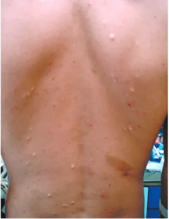
On physical examination, multiple café-au-lait macules (!6), oval with fairly smooth margins meas-uring 1.5 cm or greater in diameter, were distrib-uted over the trunk and extremities (Figure 1). Freckles were also seen in both the axillary regions. On palpation, several firm subcutaneous papules scattered on the trunk were identified (Figure 1). A painful subcutaneous nodule on the right thigh was also detected. Chest auscultation showed sparse in-spiratory squeaks throughout the upper and middle lung fields. The remainder of the clinical examina-tion was unremarkable.
Basic laboratory tests were normal except for pe-ripheral blood eosinophilia (2910 cells/μl), slightly increased erythrocyte sedimentation rate (25 mm/
h), elevated serum C-reactive protein (9 mg/dl) and eosinophilic cationic protein (ECP) levels (196 μg/ l). Arterial blood gases while breathing room air showed mild hypoxemia (PaO2: 70 mmHg, PaCO2:
34 mmHg, pH : 7.44 and bicarbonate 24.5 mmol/l). Further investigations, which included antinuclear antibodies, C- and P-antineutrophilic cytoplasmatic antibodies, immunoglobulin levels, IgE antibody for specific allergens and serum complement analysis were unremarkable.
Skin tests for both M. tuberculosis and atypical mycobacteria were negative. Repetitive serologic tests and stool examination had no effect on the de-tection of parasites ova. Pulmonary function tests in conjunction with pulmonary diffusing capacity (sin-gle breath CO test) were within normal limits.
Chest X-ray revealed peripheral opacities in the left upper lung field, which represented alveolar con-solidation (Figure 2). A high resolution chest com-puted tomography (HRCT) disclosed bilateral air space consolidations located in the periphery of the upper lobes as well as ground-glass opacities in the lingula (Figure 3a, b). A second chest X-ray, three days later demonstrated a nodular infiltrate in the right lower lung field with attenuation of left upper lung field opacities ; findings compatible with a mi-gratory-like pattern.
The patient underwent fiberoptic bronchoscopy which showed only slight mucosal redness in the whole tracheobronchial tree. Bronchoalveolar lavage (BAL) was performed from the left upper lobe and lingula. BALF differential cell counts revealed 48% eosinophils, 32% alveolar macrophages, 16% lympho-cytes and 3.8% neutrophils. All the microbiology
Figure 1. Café - au - lait spot and subcutaneous papules dis-persed over the trunk.
Figure 2. Chest radiograph showing peripheral alveolar opaci-ties in the left upper lobe.
studies of BAL samples provided no evidence for bacterial, fungal or viral infection. Cytologic exami-nation was also negative.
Bone marrow aspiration and biopsy were normal. Slit lamp examination revealed reddish brown spots within the lower pole of the irises, compatible with Lisch nodules (Figure 4). Excisional biopsy of the subcutaneous nodule on the right thigh was typical of neurofibroma. Additional work-up, including com-puted tomography of the abdomen, magnetic reso-nance imaging of the brain, long bones plain radio-graphs, as well as Doppler echocardiography dis-closed no abnormalities.
The diagnosis of chronic eosinophilic pneumonia and NF1 was eventually established. The patient was started on prednisone 40 mg/day with rapid im-provement of his clinical picture and chest imaging features normalization within two weeks. The initial prednisone dose was effectively tapered down over a period of several months, with a schedule of the steroid therapy completion in a year.
DISCUSSION
We report a case of a patient with known neurofi-bromatosis type 1 (NF1), who presented with chronic eosinophilic pneumonia, suggesting that NF1 may promote the development of chronic eosinophilic pneumonia.
NF1 is a comparatively common autosomal domi-nant neurocutaneous syndrome with a prevalence rate of about 1 per 4000 - 5000 (1). There is no fam-ily history of this disorder in approximately half of the patients, suggesting that spontaneous muta-tions are tangible. The gene liable for the NF1, called NF1 gene, has been identified on chromo-some 17q11.2. Its protein product, neurofibromin, is a negative regulator of cell proliferation and differ-entiation, functioning as a tumor suppressor (10). Although recent developments in molecular genetic testing confirm the diagnosis of NF1 in over 95% of patients, its diagnosis is still based mostly on pub-lished clinical criteria (11), (Table 1). In the present case, four major criteria were considered sufficient to support the diagnosis of NF1.
Chronic eosinophilic pneumonia is a distinct dis-ease of unknown aetiology often accompanied by allergic diatheses, mainly asthma, which can ante-date the condition by many years or can develop
Figure 3a. High resolution chest computed tomography
(HRCT) illustrating bilateral air space consolidations located in the periphery of the upper lobes.
Figure 3b. HRCT depicts ground - glass opacities in the lingual.
concurrently (12). Although firm diagnostic criteria are not available, the diagnosis can be derived from the combination of : (i) non - specific pulmonary symptoms (usually over two weeks duration), (ii) pulmonary opacities that are in most cases bilateral, peripheral and migratory, (iii) pronounced blood and/or alveolar eosinophilia (blood eosinophilic count!1000/mm3, bronchoalveolar lavage
eosino-philis!40%) and (iv) absence of any known cause of pulmonary eosinophilia (13). The hallmark of chronic eosinophilic pneumonia is a rapid and re-markable resolution of clinical, radiological and bio-logical features with oral corticosteroids. In our pa-tient this resolution occurred in two weeks.
Neurofibromatosis type 1 is an extremely variable condition in terms of its features occurrence and severity. Some afflicted patients have mild manifes-tations, whereas others experience more severe complications reducing life expectancy (1, 2). Mild cases may be missed, since affected individuals sel-dom seek medical attention. The present case was revealed by a compulsory physical examination as part of health evaluation for military service. The presenting respiratory symptoms, just before join-ing the army, forced our patient to ask further medi-cal assistance, thus leading to the definite diagnosis. A multitude of phenotypic features has been de-scribed in NF1, as virtually any organ can be af-fected by this neurocutaneous disorder. Especially, NF1 has a variety of manifestations in the thorax and lungs, as mentioned previously (3 - 9). Never-theless, pulmonary eosinophilic disorders have not been reported to date in association with NF1. Search of the literature revealed only a report of NF1 complicated by acute lymphoblastic leukaemia (ALL) and hypereosinophilia (14).
The authors postulate that the occurrence of chronic eosinophilic pneumonia may be facilitated by neurofibromatosis type 1. It is firmly thought that
nerve growth factor (NGF) plays a pivotal role in this relationship. Compelling circumstantial evidence suggests that NGF is involved in the pathogenesis of neurofibromatosis and its neurological compli-cations (mainly neural tumors) (15 -17). Indeed, high NGF plasma levels were detected in patients with NF1 (18).
However, several lines of evidence have also im-plicated NGF in the pathophysiology of various al-lergic diseases (19). Strictly speaking, NGF has been shown to interact with non-neuronal cells in-volved in innate and adaptive immune systems, such as lymphocytes, mast cells, macrophages, basophils and eosinophils themselves. Therefore, it intervenes in allergy-related immune cell functional activities, such as mast-cell degranulation, Th2 cytokine syn-thesis and release, antibody production from B cells and eosinophil survival (20).
Mast cells and eosinophils, the major key effector cells in allergic inflammation, are a source of NGF and influenced by it in their differentiation, survival and activation (21, 22). This mast cell-eosinophil cross-talk, through NGF and other specific pre-formed mediators, can perpetuate and even inten-sify allergic inflammatory process with possible de-velopment of tissue damage.
The magnitude of NGF involvement in the inflam-matory stages of allergy has not only been shown in animal models or in vitro studies, but also in pa-tients with allergic diseases and especially bronchial asthma, the best studied eosinophilic lung disorder. The expression of NGF is highly up-regulated and significantly correlated with the increased airways mast cells in asthmatic patients (23). Furthermore, enhanced NGF plasma and BAL levels are strongly correlated with atopy markers, disease severity and the development of bronchial hyperresponsiveness, suggesting that NGF mediates activation of bron-chial eosinophils and substantially regulates eosino-philic inflammation in asthma (24). Similar immuno-pathogenic mechanisms have been observed in chronic eosinophilic pneumonia, lacking however any evidence on NGF involvement (12).
Recent studies in mice models demonstrate that
Nf1 deficiency results in increased numbers of
im-mature and im-mature T cell subsets, as well as mast cells ; the latter ones might contribute to neurofibro-mas development through the release of several cy-tokines (10, 25). Antigenic stimulation may elicit T-lymphocytes and mast cells activation, which may be primed by Nf1 gene mutations. As mentioned earlier, these cells are a source of NGF, which in
Table 1. Diagnostic criteria for neurofibromatosis type 1 (NF1) (adapted from ref.11)
!Six or more café-au-lait spots!0.5 cm in prepubertal children and!1.5 cm in postpubertal individuals
!Axillary or inguinal freckling
!Two or more cutaneous neurofibromas !One plexiform neurofibroma
!Two or more iris Lisch nodules !An optic glioma
!A characteristic bony lesion (pseudarthrosis, hypoplasia of sphenoid wing, severe kyphoscoliosis)
!First degree relative with NF1
In order to make the diagnosis, at least two major criteria are required.
turn is the major key mediator that might orches-trate the eosinophilic airway inflammation, thus lead-ing to the occurrence of chronic eosinophilic pneu-monia (26). This immunological response could also be subserved by genetic factors that are hinted by a history of asthma and atopy in patient’s brother.
Based on the aforementioned data concerning the role of NGF in NF1 and allergic inflammatory dis-eases, we measured NGF levels in the patient’s se-rum and BAL using a highly sensitive NGF-spe-cific two - site enzyme - linked immunosorbent assay (ELISA) -kit (Promega, Madison, WI, USA). Both NGF plasma and BAL levels were significantly in-creased (157 pg/ml and 235 pg/ml respectively with normal levels 3.8!1.7 pg/ml and 53.9!20.2 pg/ ml). This may demonstrate a possible association among NF1, NGF and chronic eosinophilic pneumo-nia.
In conclusion, these observations suggest that chronic eosinophilic pneumonia could be included among potential pulmonary manifestations of NF1. Although a causal association between NF1 and chronic eosinophilic pneumonia cannot be explicitly ascertained from this descriptive study, we suggest that priming of specific immune cells by NF1 may generate chronic eosinophilic pneumonia, supported by further triggering factors (e.g. drugs, environ-mental exposure, bacterial and viral infections). These remarks may prompt studies on NGF involve-ment in chronic eosinophilic pneumonia.
REFERENCES
1. Ferner RE : Neurofibromatosis 1. Eur J Hum Genet 15 : 131 - 138, 2007
2. Tonsgard JH : Clinical manifestations and man-agement of neurofibromatosis type 1. Semin Pediatr Neurol 13 : 2 - 7, 2006
3. Aughenbaugh GL : Thoracic manifestations of neurocutaneous diseases. Radiol Clin North Am 22 : 741 - 756, 1984
4. Zamora AC, Collard HR, Wolters PJ, Webb WR, King TE : Neurofibromatosis-associated lung disease : a case series and literature re-view. Eur Respir J 29 : 210 - 214, 2007
5. Simeoni S, Puccetti A, Chilosi M, Tinazzi E, Prati D, Corrocher R, Lunardi C : Type 1 neurofibromatosis complicated by pulmonary artery hypertension : a case report. J Med In-vest 54 : 354 -358, 2007
6. Baldo X, Ortiz MR, Sebastian F, Bernado L :
Fatal right spontaneous haemothorax in Von Recklinghausen’s disease. Interact Cardiovasc Thorac Surg 2 : 35 - 37, 2003
7. Sforza E, Colamaria V, Lugaresi E : Neurofibro-matosis associated with central alveolar hypov-entilation syndrome during sleep. Acta Paedi-atr 83 : 794 - 796, 1994
8. Hassoun PM, Celli BR : Bilateral diaphragm pa-ralysis secondary to central von Reckling-hausen’s disease. Chest 117 : 1196 -2000, 2000 9. Rainov NG, Heidecke V, Burkert W : Thoracic and lumbar meningocele in neurofibromatosis type 1 : report of two cases and review of the literature. Neurosurg Rev 18 : 127 - 134, 1995 10. Theos A, Korf BR : Pathophysiology of
neurofibromatosis type 1. Ann Intern Med 144 : 842 -849, 2006
11. Neurofibromatosis. Conference statement. Na-tional Institutes of Health Consensus Develop-ment Conference. Arch Neurol 45 : 575 - 578, 1988
12. Alam M, Burki NK : Chronic eosinophilic pneu-monia : a review. South Med J 100 : 49 -53, 2007
13. Cottin V, Cordier JF : Eosinophilic pneumonias. Allergy 60 : 841 - 857, 2005
14. Horigome H, Sumazaki R, Iwasaki N, Imoto N, Kinugasa H, Saito M, Matsui A : Fatal eosinophilic heart disease in a child with neurofi-bromatosis-1 complicated by acute lymphoblas-tic leukaemia. Heart Vessels 20 : 120 -122, 2005 15. Riccardi VM : Genetic alterations and growth factors in the pathogenesis of von Reckling-hausen neurofibromatosis. Neurofibromatosis 2 : 292 - 298, 1989
16. Carroll SL, Stonecypher MS : Tumor suppres-sor mutations and growth factor signaling in the pathogenesis of NF1-associated peripheral nerve sheath tumors : II. The role of dysregu-lated growth factor signaling. J Neuropathol Exp Neurol 64 : 1- 9, 2005
17. Riopelle RJ, Riccardi VM : Neuronal growth factors from tumours of Von Recklinghausen neurofibromatosis. Can J Neurol Sci 14 : 141 -144, 1987
18. Fabricant RN, Todaro GJ : Increased serum levels of nerve growth factor in von Reckling-hausen’s disease. Arch Neurol 38 : 401- 405, 1981
19. Nockher WA, Renz H : Neurotrophins in al-lergic diseases : From neuronal growth fac-tors to intercellular signalling molecules.
J Allergy Clin Immunol 117 : 583 - 589, 2006 20. Micera A, Puxeddu H, Aloe L, Levi-Schaffer
F : New insights on the involvement of nerve growth factor in allergic inflammation and fibrosis. Cytokine Growth Factors Rev 14 : 369 -374, 2003
21. Leon A, Buriani A, Dal Toso R, Fabris M, Romanello S, Aloe L, Levi-Montalcini R : Mast cells synthesize, store and release nerve growth factor. Proc Natl Acad Sci USA 91 : 3739 - 3743, 1994
22. Solomon A, Aloe L, Pe’er J, Frucht-Pery J, Bonini S, Bonini S, Levi-Schaffer F : Nerve growth factor is preformed and activates human peripheral blood eosinophils. J Allergy Clin Im-munol 102 : 454 - 460, 1998
23. Kassel O, De Blay F, Duvernelle D, Olgart-Höglund C, Israel-Biet D, Krieger P, Moreau L, Muller C, Pauli G, Frossard N : Local in-crease in the number of mast cells and expres-sion of nerve growth factor in the bronchus of asthmatic patients after repeated inhalation
of allergen at low-dose. Clin Exp Allergy 31 : 1432 - 1440, 2001
24. Olgart-Höglund C, De Blay F, Oster J-P, Duvernelle C, Kassel O, Pauli G, Frossard N : Nerve growth factor levels and localization in human asthmatic bronchi. Eur Respir J 20 : 1110 -1116, 2002
25. Ingram DA, Zhang L, Mc Carthy J, Wenning MJ, Fisher L, Yang FC, Clapp DW, Kapur R : Lymphoproliferative defects in mice lacking the expression of neurofibromin : functional and biochemical consequences of NF1 deficiency in T-cell development and function. Blood 100 : 3656 -3662, 2002
26. Lambiase A, Bracci - Laudiero L, Bonini S, Bonini S, Starace G, Milco D, Elios M, De Carli M, Aloe L : Human CD4+ T cell clones pro-duce and release nerve growth factor and ex-press high-affinity nerve growth factor recep-tors. J Allergy Clin Immunol 100 : 408 - 414, 1997