博 士 学 位 論 文
Identification and Physiological Activities of Organic Compounds
Produced from Natural Product Resources, and Effective
Utilization of the Resources
近 畿 大 学 大 学 院
システム工学研究科システム工学専攻
博 士 学 位 論 文
Identification and Physiological Activities of Organic Compounds
Produced from Natural Product Resources, and Effective
Utilization of the Resources
平成 28 年 3 月
近 畿 大 学 大 学 院
システム工学研究科システム工学専攻
i
PREFACE
The work in this thesis was conducted under the guidance of Professor Masato Nomura at the
Department of Natural Products Chemistry of Kinki University from 2013 to 2016.
The inherent function of natural products is to maintain ecological balance. Advances in
technology have allowed the extraction and separation of these resources and have allowed for their
structures to be elucidated. These advances have presented the opportunity to use the active
ingredient from plant resources in their pure form. In the 21st century, this has led to the discovery
of new bioactive natural products and has stimulated drug development and advances in health
maintenance and the treatment of disease. In recent years, degradation of the environment and
changes in lifestyle has contributed to the emergence of new diseases. Consequently, new
pharmaceutical products and functional materials are necessary. In this respect, finding new plant
resources and assessing their functions is important.
This study aims to evaluate the physiological activity of organic compounds from natural
product resources and to assess their usefulness to engineering and industry. I shall be happy if this
work contributes in some way to natural products chemistry.
Shuhsien Wu
Natural Products Chemistry, Kinki University
ii
LIST OF PUBLICATIONS
Contents in thesis are published in following papers.
1. Physiological Activity of Chinese Lichen (Gyrophora esculenta) Component, Methyl
2,4-Dihydroxy-6-methylbezoate and the Related Compounds
Shuhsien Wu, Zhendong Zhao, Yoshiharu Okada, Yoshiyuki Watanaba, Toshiyuki Takahata,
Toshio Inoue, Eiji Otsubo, Jing Wang, Yanju Lu and Masato Nomura
Asian Journal of Chemistry, 26 (3), 702-708, 2014.
2. Evaluation of the Fatty Acid Composition of the Seeds of Mangifera indica L. and Their
Application
Shuhsien Wu, Megumi Tokuda, Ayaka Kashiwagi, Atsushi Henmi, Yoshuiharu Okada,
Shinya Tachibana and Masato Nomura
Journal of Oleo Science, 64 (5), 479-484, 2015.
3. Deodorizing effect of mango seed kernel oil for 10 odorous substances
Shuhsien Wu, Atsushi Henmi, Shinya Tachibana and Masato Nomura
Research reports of the Faculty of Engineering, Kinki University, 49, 1-6, 2015.
4. Novel physiological roles of mango seed oil using proteomic analysis of differentially
expressed proteins
Shuhsien Wu, Kenichi Matsumoto, Shinya Tachibana, Toshio Inoue and Masato Nomura
iii
CONTENTS
Introduction --- 1
References --- 3
Chapter 1. Chinese lichen: the physiological activity of methyl 2,4-dihydroxy-6-methylbenzoate and its derivatives Introduction Results and Discussion Experiment section Conclusion References Chapter 2. Mango seed oil: the composition of Mangifera indica L. seed oil and its cosmetic and pharmaceutical applications Introduction Materials and Methods Results and Discussion Conclusion References Summary --- 25 --- 7 --- 20 --- 45 --- 54 --- 24 --- 32 --- 45 --- 7 --- 14 --- 19
- 1 - INTRODUCTION
In ancient China, the legend of Shennong avers that the emperor tasted hundreds of herbs to
distinguish medicinal varieties; in so doing, he initiated the study of pharmaceuticals. Plants have
long occupied a mystical and functional role in human society. A famous Japanese tale, The Hare of
Inaba (Kojiki version), conveys how the god of medicine healed a hare’s wound using the ear of
reedmaces. During the Dragon Boat Festival (May 5) in China, calamus or mugwort is customarily
hung up for the prevention of disease. Egypt is considered to be the origin of western medicine and
pharmacy. Aromatic plants were incorporated into daily life, notably for preserving dead bodies 1)
and blending a cosmetic perfume known as kyphi 2). In the latter half of the 18th century, the
Swedish chemist Carl Wilhelm Scheele began separating organic acids from plants 3). The
extraction, isolation, and structural elucidation of natural organic compounds progressed rapidly
due to improved analytical instruments and measurement techniques.
Plants have been important resources for preserving the global environment. The advance of
civilization has influenced the environment; conversely, the environment has affected civilization
and necessitated a reconsideration of human values. Plants provide the food, clothing, and shelter
required for humans and animals, and they serve a vital role in daily life and culture.
Plant-based therapies have a well-established history. Forest bathing has been observed to
mitigate stress by inducing relaxation 4-6). Forest trees and flowers co-evolved naturally over time.
Monoterpenoids (16 hydrogen atoms with 10 carbon atoms) are a class of volatile compounds
present in various forest plants; they have been used as relaxants, antibacterials 7), insect
repellants 8), and antioxidants 9). Dr. Boris P. Tokin coined the word phytoncide in 1928 to describe
a volatile substance that plants exude to kill competing organisms. Plants deodorize the forest 10) by
neutralizing malodorous compounds or by masking the odors with other compounds.
Lipids are a significant disease factor due to life styles that involve drinking, smoking, lack of
exercise, and social stress 11). Lipids store energy in vivo; they feature as components of the cell
membrane and are biosynthesized into physiologically active precursors like prostaglandin
12)
- 2 -
triacylglycerol, phospholipids, and cholesterol are mainly introduced into humans from ingested
food.
Diseases related to diet and lifestyle (high blood pressure, diabetes, and obesity) have been
increasing in frequency 15). Hyperlipidemia, excess levels of lipids in the bloodstream, occurs when
the biological metabolism of lipids is insufficient 16). For example, arteriosclerosis may be caused
by the excess consumption of cholesterol: the concentration of LDL increases in the bloodstream,
cholesterol accumulates on the vessel walls, and blood flow decreases. Lipid signaling molecules
have also been implicated in cell proliferation, differentiation, and immune system activity 17).
Foods that contain chemical functionality have gained attention for preventative and therapeutic
treatments.
Foods have been classified according to function: nutrition (primary), sense (second), and
biological regulation (third) 18). The functional components in food have been related to immunity
19)
, antitumor behavior 20), suppression of blood glucose and blood pressure 21), and antioxidant
effects 22). The oral administration of evening primrose oil, a lipid that contains γ-linolenic acid, was shown to be effective in treating atopic dermatitis 23). Low-grade autoimmune disorders and
chronic inflammatory disease have been observed in Eskimo populations, due to the excessive
intake of eicosapentaenoic acid (EPA) 24, 25). β-carotenoid, biosynthesized from the isoprenoid pathway, is a secondary metabolite that has been reported to inhibit the proliferation of colon
cancer cells 26).
Oxidative stress is caused by the excess production of active oxygen and oxygen free radicals
by lipid peroxidation. These free radicals damage proteins, enzymes, and nucleic acids; they are
related to aging and disease. Antioxidants are important because they counter the effects of free
radicals in the body. Antioxidants are found in natural foods and fermentation products 27),
including enzymes (superoxide dismutase, catalase), vitamins (vitamin E, ubiquinol), fat-soluble
low–molecular weight molecules (bilirubin), and water-soluble molecules (glutathione).
Plants have been present on earth for over 250,000 years. Many of the biochemical
- 3 -
evaluating physiological activity (antioxidation 28), antiallergenic behavior 29), and
immunomodulation 30) have been discovered. In recent years, the influence of the environment and
lifestyle has contributed to the emergence of new diseases. Research has exploited the inherent
functions of plant-based compounds to solve human-health problems.
This paper examines the potential commercial uses of selected plants. Rock tripe (Gyrophora
esculenta) belongs to the umbilicariaceae family and is the subject of Chapter 1. The samples for
this study were grown on the sandstone cliffs at Zhangjiajie, Hunan province, China. The active
components were identified, and the physiological activity of the natural and synthetic compounds
was investigated. The seed of mango fruit (Mangifera indica L.) is usually discarded;
however, Chapter 2 explores its use as a value-added commodity chemical. Mango seed oil (MSO)
samples from Miyazaki, Japan and Taiwan were compared; their performance was assayed for use
in cosmetics, deodorants, and detergents. The antitumor activity of MSO was examined for HeLa
cells.
REFERENCES
1) Inoue, S., Abe, S., An invitation to anti-infectious aromatherapy. pp 4-6 (2011).
2) Hamm, S., Bleton, J., Connan, J., Tchapla A., A chemical investigation by headspace SPME
and GC–MS of volatile and semi-volatile terpenes in various olibanum samples.
Phytochemistry, 66 (12), 1499-1514 (2005).
3) Galassi, F. M., Borghi, C., A brief history of uric acid: From gout to cardiovascular risk factor,
Eur J Intern Med., 26 (5), 373 (2015).
4) Mao, G. X., Cao, Y. B., Lan, X. G., He, Z. H., Chen, Z. M., Wang, Y. Z., Hu, X. L., Lv, Y. D.,
Wang, G. F., Yan. J., Therapeutic effect of forest bathing on human hypertension in the elderly.
J. Cardiology, 60 (6), 495-502 (2012).
5) Lee, J., Park, B. J., Tsunetsugu, Y., Ohira, T., Kagawa, T., Miyazaki, Y., Effect of forest
bathing on physiological and psychological responses in young Japanese male subjects. Public
- 4 -
6) Morita, E., Fukuda, S., Nagano, J., Hamajima, N., Yamamoto, H., Iwai, Y., Nakashima, T.,
Ohira, H., Shirakawa, T., Psychological effects of forest environments on healthy adults:
Shinrin-yoku (forest-air bathing, walking) as a possible method of stress reduction. Public
Health, 121 (1), 54-63 (2007).
7) Carrillo-Hormaza, L., Mora, C., Alvarez, R., Alzate, F., Osorio, E., Chemical composition and
antibacterial activity against Enterobacter cloacae of essential oils from Asteraceae species
growing in the Páramos of Colombia. Industrial Crops and Products, 77, 108-115 (2015).
8) Sumarah, M. W., Puniani, E., Sørensen, D., Blackwell, B. A., Miller, J. D., Secondary
metabolites from anti-insect extracts of endophytic fungi isolated from Picea rubens.
Phytochemistry, 71 (7), 760-765 (2010).
9) Fu, R., Zhang, Y., Guo, Y., Chen F., Chemical composition, antioxidant and antimicrobial
activity of Chinese tallow tree leaves. Industrial Crops and Products, 76, 374-377 (2015).
10) Abe, T., Nomura, M., Chemical constituents of phytoncides and their antioxidative activities.
Aroma Research, 7 (1), 56-62 (2006).
11) Larsson, S. C., Drca, N., Jensen-Urstad, M., Wolk. A., Combined impact of healthy lifesyle
factors on risk of atrial fibrillation: Prospective study in men and women. Int. J. Cardiol.,
203, 46-49 (2016).
12) Akihisa, S., Koike, K., Konoshima, T., Hano, Y., Hotita, K., Masuda, K., Miyazawa, M.,
Yasukawa, K., Chemistry of organic natural resources. pp 69-80 (2007).
13) Lupi, F. R., Gentile, L., Gabriele, D., Mazzulla, S., Baldino, N., de Cindio B., Olive oil and
hyperthermal water bigels for cosmetic uses. J. colloid interface sci., 459, 70-78 (2015).
14) Ake. A., Pharmaceutical containing oils for preventing or ameliorating snoring. U.S. Pat. Appl.
Publ. US 20140363528 A1 20141211 (2014).
15) Lee, W. W. M., Choi, K. C., Yum, R. W. Y., Yu, D. S. F., Chair. S. Y., Effectiveness of
motivational interviewing on lifestyle modification and health outcomes of clients at risk or
diagnosed with cardiovascular diseases: A systematic review. Int. j. nurs. studies., 53, 331-341
- 5 -
16) Hosomi, R., Fukunaga, K., Arai, H., Kanda, S., Nishiyama, T., Kanada, T., Yoshida, M., Effect
of phospholipid n-3 polyunsaturated fatty acids on rat lipid metabolism. European Journal of
Lipid Science and Technology, 112 (5), 537-544 (2010).
17) Wymann, M. P., Schneiter, R., Lipid signalling in disease. Nature Reviews Molecular Cell
Biology,9 (2), 162-176 (2008).
18) Shimizu, T., Health claims on functional foods: the Japanese regulations and an international
comparison, Nutr Res Rev., 16 (2), 241-52 (2003).
19) Sirdaarta, J., Matthews, B., Cock, I.E., Kakadu plum fruit extracts inhibit growth of the
bacterial triggers of rheumatoid arthritis: Identification of stilbene and tannin components. J.
Functional Food, 17, 610-620 (2015).
20) Zhang, W., Chen, C., Shi, H., Yang, M., Liu, Y., Ji, P., Chen, H., Tan, R.X., Li, E., Curcumin is
a biologically active copper chelator with antitumor activity. Phytomedicine, 23 (1), 1-8
(2016).
21) Ueshima, H., Stamler, J., Elliott, P., Chan, Q., Brown, I.J., Carnethon, M.R., Daviglus, M.L.,
He, K., Moag-Stahlberg, A., Rodriguez, B.L., Steffen, L.M., Van Horn, L., Yarnell, J., Zhou,
B., Food omega-3 fatty acid intake of individuals (total, linolenic acid, long-chain) and their
blood pressure. Hypertension. 50 (2), 313-319 (2007).
22) Hur, S. J., Lee, S. Y., Kim, Y. C., Choi, I., Kim. G.B., Effect of fermentation on the antioxidant
activity in plant-based foods. Food Chem., 160, 346-356 (2014).
23) Simon, D., Eng, P. A., Borelli, S., Kagi, R., Zimmermann, C., Zahner, C., Drewe, J., Hess, L.,
Ferrari, G., Lautenschlager, S., Wuthrich, B., Schmid-Grendelmeier, P., Gamma-Linolenic
Acid Levels Correlate with Clinical Efficacy of Evening Primrose Oil in Patients with Atopic
Dermatitis. Adv. Ther., 31 (2), 180-188 (2014).
24) de Camargo Talon, L., de Oliveira, E.P., Moreto, F., Portero-McLellan, K.C., Burini, R.C., Omega-3 fatty acids supplementation decreases metabolic syndrome prevalence after lifestyle
modification program. J. Functional Foods, 19 (part B), 922-928 (2015).
- 6 -
Yamamoto, A., Ishino, H., Matsuyama, M., Yoshimura, R., Yoshikawa, T., Eicosapentaenoic
acid suppresses the proliferation of synoviocytes from rheumatoid arthritis. J. Clin. Biochem.
Nutr., 43 (2), 126-128 (2008).
26) Niranjana, R., Gayathri, R., Mol, S.N., Sugawara, T., Hirata, T., Miyashita, K., Ganesan,
P., Carotenoids modulate the hallmarks of cancer cells. J. Functional Foods, 18 (part B),
968-985 (2015).
27) Majumdar, R. K., Bejjanki, S. K., Roy, D., Shitole, S., Saha, A., Narayan, B., Biochemical and
microbial characterization of Ngari and Hentaak - traditional fermented fish products of India.
J. Food Science and Technology (New Delhi, India), 52 (12), 8284-8291 (2015).
28) Malar, J., Chairman, K., Singh, A.R.J., Shifa Vanmathi, J., Balasubramanian, A., Vasanthi K.,
Antioxidative activity of different parts of the plant Lepidium sativum Linn. Biotechnology
Reports, 3, 95-98 (2014).
29) Choi, Y. W., Lee, K. P., Kim, J. M., Kang, S., Park, S.J., Lee, J. M., Moon, H. R., Jung, J. H.,
Lee, Y. G., Im D. S., Petatewalide B, a novel compound from Petasites japonicus with
anti-allergic activity. J. Ethnopharmacol., 178, 17-24 (2015).
30) Sun, L., Chu, J., Sun, Z., Chen, L., Physicochemical properties, immunomodulation and
antitumor activities of polysaccharide from Pavlova viridis. Life Sciences, 144, 156-161
7
CHAPTER 1 Chinese lichen: the physiological activity of methyl 2,4-dihydroxy-
6-methylbenzoate and its derivatives
1. INTRODUCION
In modern society, stress has been cited as one of the main obstacles to mental and physical health 1).
Lifestyle-related diseases (cancer, hardening of the arteries, hypertension, diabetes, etc.) are mainly caused
by active oxygen and free radicals 2, 3), which are produced in excess in the body. They induce various
diseases by damaging cells through protein denaturation, lipoperoxidation, nucleolytic degradation, and
enzyme inactivation 4, 5). Recent trends in health-consciousness have promoted the consumption of
traditional foods; these contain beneficial active compounds linked to the prevention of disease or antiaging
6)
. Lichen has been consumed by humans since antiquity; its unique composition (it is an homobium of
fungi and algae) is believed to confer medicinal qualities 7, 8). Lichen has been reported to effectively inhibit
lifestyle-related diseases, such as hypertension and hypercholesteremia 9).
Gyrophora esculenta, also known as Umbilicaria esculenta, (Miyoshi) minks, and iwatake, grows on
the surface of rocks or on tree trunks; it is distributed widely from the extratropical belt of eastern Asia
(including China, Japan and Korea) to mountainous areas such as the Himalayas. Members of the family
Umbilicariaceae look like rounded, thin leaves: they have a gray top surface, black back surface, and thick
spines. Umbilicariaceae grow slowly at a rate of 1 cm every two or three years 7, 8, 10). Hashimoto et al.
reported that iwatake exhibits physiological activity as an anticancer agent, anti-inflammatory, and
glycuresis prophylactic 11, 12). This study identified and investigated an antioxidant compound contained in
the oily extract of Chinese lichens Gyrophora esculenta (Miyoshi). Several analogous chemicals were
derived from the natural compound and likewise investigated. The physiological activity of the compounds
differs based on their chemical substituents. The effects of these compounds on antioxidation, cytotoxicity,
the suppression of cytokine production, and the inhibition of histamine release are reported herein.
2. RESULTS AND DISCUSSION
8
The crude oil was obtained from a methanol extraction of the Chinese lichens and analysis of the
composition of the oil was carried out with gas chromatography mass spectroscopy (GC-MS). The results
are shown in Fig. 1. 3,5-Dihydroxytoluene (orcinol) (1) and methyl 2,4-dihydroxy-6-methylbenzoate
(methyl orsellinate) (2) are identified as the primary components (GLC%, 44:56). The crude oil was
extracted sequentially with hexane, diethyl ether, and ethyl acetate. The antioxidant activities of these
fractions were assessed on the basis of a 2,2-diphenyl-1-picrylhydrazyl (DPPH) radical scavenging assay
and a superoxide dismutase-like (SOD-like) assay. The results are shown in Table 1.
The ethyl acetate fraction shows the highest DPPH radical scavenging activity, and the diethyl ether
and ethyl acetate fractions show high activity in the SOD-like assay. At low concentration (0.1 mg/mL),
the diethyl ether and ethyl acetate fractions show higher SOD-like activities (22.7% and 18.8%,
respectively) than that of ascorbic acid (12.6%). The diethyl ether fraction was chromatographed on silica
gel to separate the effective antioxidant compounds. As shown in Table 2, fraction (A-4) shows the highest
35.9 370.9 22.2 23.5 25.4 >400 9.7 10.7 63.3 113.7 22.7 23.9 84.5 43.9 18.8 29.5 23.0 >400 8.7 9.3 100.0 6.0 - 12.6 88.9
a) Concentration : 1 mg / mL b) Corrected concentration : 0.2 mg / mL c) 50% Scavenging Concentration ( mg / mL ) d) Concentration : 1 mM
Extract a)
DPPH Radical Scavenging Assay SOD-like Activity Assay
Scavenging Rate (%) b) SC50 c) Inhibition Rate (%) 0.1 mg / mL 1 mg / mL - - Ascorbic acid - Methanol Hexane Diethyl ether Ethyl acetate Residue α-Tocopherol d)
Table 1 DPPH radical scavenging assay and SOD-like assay of Gyrophora esculenta. CH3 HO OH COOCH3 OH H3C OH Fig. 1 Structure of compounds (1) and (2).
9
inhibition of the four fractions (A-1)–(A-4), equal to that of ascorbic acid (12.6%) in the SOD-like assay. In
addition, fraction (A-4) was re-chromatographed on silica gel to give pure methyl orsellinate (2) as a white
crystalline solid. Its active oxygen inhibition was determined at 0.1 and 1.0 mg/mL and was found to
increase from 11.1% to 14.9%, respectively. Therefore, compound 2, an important component identified in
the methanol extract of Chinese lichens, has great potential as an antioxidative agent.
2.2. Synthesis and activity of compounds related to (2)
Following the results for the physiological activities of methyl orsellinate (2), derivatives of 2 were
synthesized, as shown in Scheme 1. Four compounds, methyl 2,4-dihydroxybenzoate (4), methyl
2-hydroxy-4-methoxybenzoate (5), methyl 2,4-dimethoxybenzoate (6), and methyl 4-hydroxy-2-methoxy-
benzoate (7), were synthesized from 2,4-Dihydroxybenzoic acid (3). As discussed previously, the
physiological activities of compound 2 and its derivatives, such as DPPH radical scavenging effects,
antitumor activity, and antibacterial activity, have been reported 13-15). In this chapter, the antioxidant
activities of compound 2 and its derivatives evaluated on the basis of SOD-like assays at different
concentrations are reported. The results are shown in Table 3. Compounds 4 and 7 were exhibited medium
inhibition activities (10.9%–15.8%) at a concentration of 1.0 mg/mL. Furthermore, at a concentration of
0.1 mg/mL, compound 7 shows a 9.5% inhibition rate, which is close to that of ascorbic acid (10.2%) as a
reference compound. A-1 17.7 24.7 A-2 18.1 24.5 A-3 16.0 26.0 A-4 23.3 26.0 A4-1 2.4 2.4 A4-2 11.1 14.9 A4-3 11.6 12.5 A4-4 6.5 16.1 A4-5 9.9 15.8 Ascorbic acid 12.6 98.7 Fraction 0.1 mg / mL
SOD-like Activity Assay Inhibition Rate (%)
1 mg / mL
10
Scheme 1
These results suggest that the hydroxyl group at the para position of the aromatic ring plays an
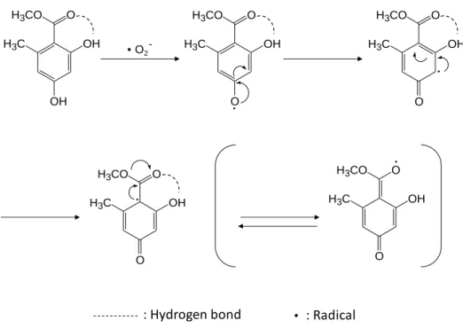
important role in antioxidant activity. My proposed mechanism for the antioxidant activity of compound 2
is shown inFig. 2. The hydroxyl group of compound 2 reacts with the active oxygen, and the phenoxy
radical formed is transformed to an oxy radical.
The structure of compound 2 changes into a para quinoid structure 16). The active radical is stabilized
via the resonance effect. Since the hydroxyl group at the ortho position of compound 2 forms a hydrogen
bond to the ester carbonyl group, the hydroxyl group slightly influences the antioxidant activity. COOH OH OH COOCH3 OH OH O COOCH3 OH O O COOCH3 OCH3 O COOCH3 OCH3 OH (3) (4) (4a) (4b) (7) H2SO4/ CH3OH 60-70 ºC overnight NaH / CH3I THF overnight COOCH3 OH OCH3 COOCH3 OCH3 OCH3 (5) (6) + C6H5COCl / (C2H5)3N / CH2Cl2 overnight (CH3)2SO4/ KOH 60-70 ºC overnight CH3ONa CH3OH overnight 4 2.7 15.8 5 4.4 6.5 6 1.0 1.5 7 9.5 10.9 Ascorbic acid 10.2 97.1 a) Concentration (mg/mL) Compound 0.1 a) 1.0 Inhibition rate (%)
11
Next, the antioxidant capacity of compounds 2 and 4–7 were assessed using a biological antioxidant
potential (BAP) test kit. The capacities of ascorbic acid and α-tocopherol are shown in Table 4, which were
determined at different concentrations (0.1–1.0 mg/mL) to calculate their equivalent amounts as standards.
The BAP value of 7 is 1119 μM, which is the same antioxidant activity as that of a 0.07 mg/mL
solution of ascorbic acid or a 0.15 mg/mL solution of α-tocopherol (Table 5). While compound 2, the main
component in fraction A4-2, exhibits high SOD-like activity, the BAP value of compound 2 is only 752
mM. The BAP values of compounds 4, 5, and 6 are also below 890 μM. These results indicate that a
structure bearing a hydroxyl group at the para position with no hydrogen bonded methoxy group at the
ortho position is important for antioxidant activity. Therefore, compound 7 shows high antioxidant activity.
OH H3C O H3CO O OH H3C OH H3CO O OH H3C O H3CO O OH H3C O H3CO O OH H3C O H3CO O O2
-: Hydrogen bond : Radical
0.01 a) 0.1 0.3 0.6 1.0
Asocorbic acid 785 1335 2241 4115 6412
α-Tocopherol 780 1012 1368 2237 3148
a) Concentration (mg/mL)
Compound Antioxidant potential (μm)
Fig. 2 Plausible structure of methyl-2,4-dihydroxy-6-methylbenzoate radical generated by active oygen.
12
According to the immunofluorescent method 17), cytotoxicity testing of compounds 2 and 4–7 was
performed with A549 cells from an alveolar epithelial cell line derived from humans. As shown in Table 6,
the cell survival rates for all compounds are higher than 80%, which indicates a lack of cytotoxicity. At a
concentration of 100 μM, the cell survival rates for compounds 4 and 7 are 86% and 88%, respectively. The hydroxyl group at the para position, which was shown earlier to affect the antioxidant activity, also
affects cell cytotoxicity. In contrast, the cell survival rate for compound 2 is very high (97%) at the same
concentration. Therefore, the toxicity of a compound bearing a hydroxyl group at the para position is
mitigated when a methyl group is located at the ortho position.
Alveolar epithelial cells are known to produce IL-8, an inflammatory cytokine, in response to
oxidative stress. Consequently, anti-inflammation activity tests on A549 cells were performed with an
enzyme-linked immunosorbent assay (ELISA) kit. The results are summarized in Table 7. Although the
IL-8 production rates for all compounds are more than 80%, which indicates no significant inhibitory effect,
compound 2 shows slightly lower rates regardless of the concentration. Thus, the presence of the hydroxyl
group at the para position also seems to affect the production of cytokines.
(μM) (mg/mL) (mg/mL) 2 752 0.006 0.011 4 726 N.D. a) 0.006 5 887 0.062 0.035 6 830 0.038 0.025 7 1119 0.158 0.075 a) N.D. : Not detect
BAP value α-Tocopherol Ascorbic acid Sample
2 4 5 6 7
1 103 97 110 101 93
10 103 90 110 103 91
100 97 86 129 104 88
IL-8 production, the percentage of control
Concentration (μm) Compound
Table 6 A549 cell toxicity for compounds (2) and (4-7).
13
The antiallergenic properties of compounds 2–7 were assessed on the basis of a histamine-release
assay. An itch-inducing agent (compound 48/80) 18), which is known to cause degranulation of rat
peritoneal mast cells, was added to the cells, and the released histamine contents were determined using an
amino acid analyzer. The results are summarized in Table 8. Compared with a control, compounds 2 and 4
show histamine-release inhibitory effects of 24.21% ± 2.08% and 21.08% ± 3.76%, respectively. These
results indicate that hydroxyl groups at both the ortho and para positions are important for
histamine-release inhibition.
Furthermore, the dependence of histamine-release suppression on sample concentration was
investigated. As shown in Table 9, histamine-release suppression is significant at a concentration of 30
μg/mL. The structures of compounds 2 and 4 are similar to those of quercetin and resveratrol 19, 20)
, which
are well-known antihistamines. The hydroxyl groups at the ortho and/or para positions of compounds 2
and 4 are considered to be involved in the suppression of histamine release 21, 22).
2 4 5 6 7
1 92 107 112 88 104
10 93 101 104 117 100
100 91 101 110 94 95
IL-8 production, the percentage of control
Concentration (μm) Compound Compound * Control 35.23 ± 4.53 2 24.21 ± 2.08 ** 4 21.08 ± 3.76 ** 5 34.42 ± 5.14 6 40.01 ± 3.71 7 33.42 ± 8.27 * Concentration (100 μg/mL).
** p < 0.05 compared with the control group (Dunnett's test).
Histamine release (%)
Table 7 Anti-inflammation activity for compounds (2) and (4-7).
14 3. EXPERIMENTAL METHODS
1H and 13C NMR spectra were obtained on a JEOL JNM-EX 400 spectrometer in CDCl3 operating at
400 and 100 MHz respectively, with Me4Si as internal standard. IR spectra were recorded with Shimadzu
FTIR-8100A spectrometer. Melting points were measured in open capillary tubes and are uncorrected. Gas
liquid chromatography and gas chromatograph-mass spectroscopy were carried out with Hewlett Packard
HP 6890 GC and Hewlett Packard HP 5972 MSD, equipped with capillary column (TC-1, id 0.25 mm ×
30 m, I.D. 1.0 μm), respectively. The conditions of GC-MS were column temperature at 50 °C for 5 min
then to 270 °C at 3 °C/min, injection temperature at 240 °C, interface temperature at 230 °C, ionization
voltage 1.3 kV, He as carrier gas, flow rate at 2 mL/min. And the structures were estimated through Library
NIST (National Institute of Standards and Technology) WebBook.
3.1. Solvent fractionation and activity tests
Chinese lichens (283 g) were collected during May 2008 at Zhangjiajie, Hunan province, China.
Dried lichens were ground and extracted with methanol (3.0 L) at room temperature (20 ± 2 °C) for 1
month. After filtering, the methanol extract (400 mL) was evaporated in vacuo to give a brown oily residue
(3.0 g).
The oily residue (3.0 g) was sequentially extracted with hexane, diethyl ether, and ethyl acetate to
obtain 0.681, 0.788, and 0.037 g of oily extracts, respectively. The diethyl ether extract (0.770 g) was
separated on silica gel using hexane-ethyl acetate (1:9) to afford four fractions: A-1 (0.237 g), A-2 (0.041
g), A-3 (0.021 g), and A-4 (0.146 g). Fraction A-4 (0.1 g) was chromatographed using preparative thin
layer chromatography (TLC, silica gel 60, hexane-ethyl acetate (3:2)) to give five fractions A4-1 (Rf = 0.76, 2 31.2 ± 3.11 27.4 ± 4.96 14.9 ± 2.97 ** 18.2 ± 1.35
4 36.6 ± 2.67 30.2 ± 4.57 17.4 ± 7.24 ** 14.0 ± 5.14 * Concentration (μg/mL).
** p < 0.05 compared with the control group (Dunnett's test). Compound *
Control 10 30 100
Histamine release (%)
15
0.014 g), A4-2 (Rf = 0.55, 0.052 g), A4-3 (Rf = 0.45, 0.020 g), A4-4 (Rf = 0.39, 0.010 g), and A4-5 (Rf =
0.27, 0.028 g).
3.2. 3,5-Dihydroxytoluene (Orcinol) (1)
Fraction (A4-1) was re-chromatographed using preparative TLC (silica gel 60, hexane-ethyl acetate
(2:3)) to obtain pure 3,5-Dihydroxytoluene (orcinol) (1) as a white crystalline solid.
Yield 0.006 g; m.p. 56–59 °C (lit. °C) 23, 24); GC-MS m/z (%): 124 [M]+ (100 %), 123 (48), 95 (11), 78
(5), 67 (6), 55 (12), 51 (14), 41 (14), 39 (24), 18 (6).
3.3. Methyl 2,4-dihydroxy-6-methylbenzoate (Methyl Orsellinate) (2)
Fraction (A4-2) was re-chromatographed using preparative TLC (silica gel 60, hexane-ethyl acetate
(1:1)) to obtain pure methyl 2,4-dihydroxy-6-methylbenzoate (methyl orsellinate) (2) as a white crystalline
solid.
Yield 0.040 g; m.p. 139–143 °C (lit. °C) 24,25); IR (KBr) 3370 (OH), 3316 (OH), 1651 (C=O), 1616
(C=C) cm-1; 1H NMR (CDCl3) δ 2.49 (3H, s, CH3), 3.93 (3H, s, OCH3), 5.37 (1H, s, OH), 6.24 (1H, s,
ArH), 6.29 (1H, s, ArH), 11.79 (1H, s, OH); 13C NMR (CDCl3) δ 24.25, 51.78, 100.97, 105.36, 110.99,
143.62, 159.76, 164.86, 171.66; MS m/z (%): 182 [M]+, 150 (100), 122 (63), 94 (20), 69 (30), 66 (27), 53
(25).
3.4. Syntheses
3.4.1. Synthesis of methyl 2,4-dihydroxybenzoate (4) 26, 27)
A solution of 2,4-dihydroxybenzoate (3) (1.54 g, 10 mmol) in methanol (50 mL) containing sulfuric
acid (10 mL) was refluxed for 24 h at 70 °C. The reaction mixture was then poured into ice water (100 mL).
The mixture was extracted with diethyl ether, washed with saturated aqueous NaHCO3, and dried over
anhydrous magnesium sulfate. After evaporation of solvent in vacuo, the residue was chromatographed on
preparative TLC (diethyl ether-chloroform (1:1)) to give methyl 2,4-dihydroxybenzoate (4).
16
= 8.6 Hz, ArH), 7.74 (1H, d, J = 8.61 Hz, ArH), 10.97 (1H, s, OH); 13C NMR (CDCl3) δ 51.98, 103.01,
105.85, 107.59, 131.74, 161.53, 163.46, 170.08; MS m/z (%): 168 [M]+, 137 (35), 136 (100), 108 (44), 95
(6), 81 (12), 80 (14), 69 (15), 53 (22).
3.4.2. Synthesis of methyl 2-hydroxy-4-methoxybenzoate (5) 28) and methyl 2,4-dimethoxy-benzoate (6)
To a suspension of sodium hydride (60% dispersion in mineral oil, 0.05 g, 1.25 mmol) in THF (5.0
mL) was added a solution of 4 (0.17 g, 1.0 mmol) in THF (10 mL) under an argon atmosphere with stirring
for 24 h at room temperature (20 ± 2 °C). A solution of iodomethane (0.1 g, 0.7 mmol) in THF (3.0 mL)
was added to the reaction mixture, which was then continuously stirred for more than 24 h at room
temperature. After the reaction mixture was quenched with 2 M HCl solution (5.0 mL), the mixture was
extracted with diethyl ether, washed with water and brine, and dried over anhydrous magnesium sulfate.
After evaporating of solvent in vacuo, the residue was chromatographed on preparative TLC
(chloroform-methanol (9:1)) to give compound A (Rf = 0.78) and compound B (Rf = 0.50).
The compound A was re-chromatographed on preparative TLC (hexane-ethyl acetate (8:2)) to give
pure methyl 2-hydroxy-4-methoxybenzoate (5). The compound B was re-chromatographed using
preparative TLC (diethyl ether-chloroform (1:9)) to give pure methyl 2,4-dimethoxybenzoate (6).
Yield 0.018 g (9.8%); 1H NMR (CDCl3) δ 3.85 (3H, s, OCH3), 3.92 (3H, s, COOCH3), 6.44 (2H, d, J
= 8.18 Hz, ArH), 7.74 (1H, d, J = 9.2 Hz, ArH), 10.99 (1H, s, OH); 13C NMR (CDCl3) δ 51.92, 55.41,
100.50, 105.28, 107.43, 131.04, 163.55, 165.37, 170.20; MS m/z (%): 182 [M]+, 151 (28), 150 (100), 122
(39), 107 (25), 95 (7), 79 (16), 63 (11), 51 (20).
Yield 0.025 g (12.5%); 1H NMR (CDCl3) δ 3.85 (3H, s, OCH3), 3.86 (3H, s, OCH3), 3.90 (3H,
s, COOCH3), 6.50 (2H, d, J = 7.8 Hz, ArH), 7.86 (1H, d, J = 8.91 Hz, ArH); 13C NMR (CDCl3) δ 51.66,
55.41, 55.90, 98.79, 104.35, 112.05, 133.70, 161.10, 164.03, 165.91; MS m/z (%): 196 [M]+, 166 (10), 165
(100), 163 (18), 150 (6), 135 (12), 122 (13), 107 (14), 92 (6), 77 (12), 63 (13), 51 (16), 44 (17).
3.4.3. Synthesis of methyl 4-hydroxy-2-methoxybenzoate (7) 29)
17
0 °C with stirring. A solution of benzoyl chloride (0.77 g, 5.4 mmol) in chloromethane (2.0 mL) was added
to the mixture. The reaction mixture was stirred for 24 h at room temperature. After the reaction mixture
was quenched with 2 M HCl solution (5.0 mL), the mixture was extracted with chloroform, washed with
water, and dried over anhydrous magnesium sulfate. After evaporation of solvent in vacuo, the residue was
chromatographed on preparative TLC (chloroform) to give methyl 4-benzoyloxy-2-hydroxybenzoate (4a)
(1.13 g, 83.1%): 1H NMR (CDCl3) δ 3.97(3H, s, COOCH3), 6.80 (1H, dd, J = 2.3 and 8.3 Hz, ArH), 6.89
(1H, d, J = 2.2 Hz, ArH), 7.53 (2H, t, J = 7.7 Hz, ArH), 7.66 (1H, t, J = 7.5 Hz, ArH), 7.91 (1H, d, J = 8.7
Hz, ArH), 8.19 (2H, d, J = 7.1 Hz, ArH), 10.94 (1H, s, OH); MS m/z (%): 272 [M]+, 105 (100), 77 (35), 51
(18).
To a solution of 4a (0.66 g, 2.4 mmol) in THF (30 mL) was added KOH (0.44 g, 7.8 mmol). The
mixture was stirred for 30 min at room temperature. Methyl sulfate (0.7 mL) was added to the mixture at
room temperature and stirred for 12 h. After the reaction mixture was quenched with water and 2 M HCl
solution (20 mL), the mixture was extracted with ethyl acetate, washed with brine, and dried over
anhydrous magnesium sulfate. After evaporation of solvent in vacuo, the residue was chromatographed on
preparative TLC (diethyl ether-chloroform (5:95)) to give methyl 4-benzoyloxy-2-methoxybenzoate (4b)
(0.61 g, 88.6%). 1H NMR (CDCl3) δ 3.87 (3H, s, OCH3), 3.88 (3H, s, COOCH3), 6.86 (1H, dd, J = 2.2
and 8.4 Hz, ArH), 6.89 (1H, d, J = 6.2 Hz, ArH), 7.49 (2H, t, J = 7.7 Hz, ArH), 7.63 (1H, t, J = 7.5 Hz,
ArH), 7.90 (1H, d, J = 8.4 Hz, ArH), 8.18 (2H, dd, J =1.3 and 8.4 Hz, ArH); MS m/z(%): 286 [M]+, 255
(2), 107 (2), 106 (10), 105 (100), 77 (41), 63 (3), 51 (19).
A mixture of 4b (0.088 g, 0.31 mmol) and sodium methoxide (0.026 g) in methanol (6.0 mL) was
stirred for 12 h at room temperature. After the reaction mixture was quenched with 2 M HCl solution (5.0
mL), the mixture was extracted with ethyl acetate, washed with brine, and dried over anhydrous
magnesium sulfate. After evaporation of solvent in vacuo, the residue was chromatographed on preparative
TLC (hexane-ethyl acetate (2:3)) to give methyl 4-hydroxy-2-methoxybenzoate (7) (0.041 g, 73.0%). 1H
NMR (CDCl3) δ 3.74 (3H, s, OCH3), 3.86 (3H, s, COOCH3), 6.47 (1H, s, ArH), 6.49 (1H, d, J = 2.3 Hz,
ArH), 7.61 (1H, s, OH), 7.79 (1H, d, J = 8.91 Hz, ArH); 13C NMR (CDCl3) δ 51.80, 55.71, 99.38, 107.16,
18
(13), 108 (17), 93 (10), 80 (7), 65 (16), 52 (15), 51 (15).
3.5. Physiological activity
3.5.1. DPPH-radical scavenging assay 30)
The evaluation of the DPPH-radical scavenging effect was performed according to the established
procedure. Sample compounds were dissolved in ethanol to a concentration of 0.1 mg/mL. A DPPH free
radical was dissolved in ethanol to a concentration of 0.2 mM.
3.5.2. Superoxide dismutase (SOD) assay 30)
SOD-like activity was determined by the nitroblue tetrazolium (NBT) reduction method with a SOD
Activity Detection Kit (Wako Pure Chemical Ind. Ltd). Sample compounds were dissolved in DMSO to a
concentration of 3.0 mM.
3.5.3. BAP test
The antioxidant activity was determined with a BAP test kit (Uisuma) at 37 °C. Spectrophotometry
(FRAS 4, Uisuma) at 505 nm was used to determine the Fe3+ concentration according to a procedure
reported in literature 17).
3.5.4. Cytotoxicity test
Human-derived type-II alveolar epithelial cells (A549) (ATCC: American type cell culture) were used.
For cell cultures, the following reagents were obtained from Gibco: fetal bovine serum (FBS), Dulbecco’s Modified Eagle Medium (DMEM), phosphate buffer saline (PBS), 0.05%–trypsin-EDTA solution, and
DMSO. Cells were passaged and incubated for 24 h, according to a procedure reported in the literature 31), and stored at room temperature for 0.5 h. 100 μL of Cell Titer-Glo Luminescence reagent (Promega Corporation) were added for the measurement of living cells. Samples were vibrated for 2 min to
drive reaction with ATP in the cells, and the number of viable cells was calculated by measuring the color
19
3.5.5. Cytokine-production suppression test
The A549 cells (2000 cells/mL), which were subconfluent after the first passage, were placed into 96
well plates and incubated overnight. The stiction of cells was confirmed and the medium was removed.
100 μL of DMEM containing 10% FBS IL-1beta (R & D System Co.) was adjusted to a concentration of 200 μM and added to 100 μL of the test substance (concentrations varied from 1–100 μM). The samples were incubated for 24 h. Finally, the concentration of IL-8 was determined with an ELISA kit according to
a procedure reported in literature 32).
3.5.6. Histamine-release assay
Male Wistar rats (7 weeks age, weight 190–220 g, Saitama Experimental Animal Supply Co.) were
fed in a room with air-conditioning equipment at room temperature (24 ± 2 °C) and humidity (45% ± 15%)
and were then treated according to procedures in literature 33, 34). Histamine content in the supernatant was
determined by an amino-acid analyzer (Autoanalyzer II, Blanc LOUBET Co., Ltd.).
4. CONCLUSIONS
Methyl 2,4-dihydroxy-6-methylbenzoate (methyl orsellinate) (2), which has a high antioxidant
activity, was isolated from the Chinese lichen Gyrophora esculenta grown in a harsh environment. The
physiological activities of 2 and its synthesized related compounds (4-7) were assessed on the basis of a
SOD-like assay, cytotoxicity assay, cytokine inhibitory assay, and histamine-release inhibition assay. In the
SOD-like assay, the inhibition rate of 2 (0.1 mg/mL, 11.1%) was higher than that of ascorbic acid (0.1
mg/mL, 12.6%) as a standard antioxidant. Results for the inhibition rates of 4–7 indicated that the hydroxyl
group at the 4-position played an important role in antioxidative activity. Compound 2, which exhibited
inhibition of active oxygen, showed no cell toxicity, with a cell survival rate of over 80% in a cytotoxicity
assay with A549 cells (type II alveolar epithelial cells). In the cytokine inhibitory assay, the family of
lichen-based compounds did not show significant inhibition of IL-8 (i.e., inflammatory cytokine)
production. In contrast, compounds 2 and 4 exhibited histamine release in rat peritoneal mast cells. Thus,
20
histamine-release inhibition assay.
New biological activities of lichen-derived compounds were revealed. Thus, the lichens show
potential as a functional food for maintenance and promotion of good health as well as for prevention of
lifestyle-related diseases.
5. REFERENCES
1) Tsuchiya T., Hosoi, J., Psychoneuroendocrinological and psychoneuroimmunological effects of stress
on skin functions, Fragrance J., 24, 26-34 (1996).
2) Inoue, S., Kawanishi, S., Oxidative DNA damage induced by simultaneous generation of nitric oxide
and superoxide, FEBS Lett., 371 (1), 86-88 (1995).
3) Skulachev, V. P., Cytochrome c in the apoptotic and antioxidant cascades, FEBS Lett., 423 (3),
275-280 (1998).
4) Yoshikawa, T., Kokura, S., Tainaka, K., Itani, K., Oyamada, H., Kaneko, T., Naito, Y., Kondo, M.,
The role of active oxygen species and lipid peroxidation in the antitumor effect of
hyperthermia, Cancer Res., 53 (10), 2326-2329 (1993).
5) Yoshikawa, T., Kokura, S., Oyamada, H., Iinuma, S., Nishimura, S., Kaneko, T., Naito Y., Kondo,
M., Antitumor effect of ischemia-reperfusion injury induced by transient embolization, Cancer Res.,
54 (19), 5033-5035 (1994).
6) Ohta, A., “Chapter 1. Kenko shiko Jidai”, in Ohta, A. (ed.), Materials for Functional Food, CMC,
Tokyo, pp 3-36 (2006).
7) Yamamoto, Y., Screening of biological activities and isolation of biological-active compounds from
lichens, Regulation of Plant Growth & Development, 35 (2), 169-179 (2000).
8) Ohmura, Y., Moon K.H., Kashiwadani, H., Notes on the evolutionary relationship between lichenized
fungus and its photobiont inferred from analyses of their genetic diversity, Bunrui, 8 (2), 123-128
(2008).
9) Ismed, F., Lohezic-Le Devehat, F., Delalande, O., Sinbandhit, S., Bakhtiar, A., Boustie, J., Lobarin
21
10) Fernando, V., Leopoldo, G.S., Carmen, A., Functional Analysis of the Intrathalline and
Intracellular ChlorophyllConcentrations in the Lichen Family Umbilicariaceae, Annals of
Botany, 78, 471–477 (1996).
11) Hashimoto, T., Yoshikawa, K., Kokudo, N., Asakawa, Y., Kimura, T., Inose, T., A. Hirasawa and G.
Tsujimoto, Search for Antidiabetogenic Principles from Edible Mushrooms and Lichen, 3rd
Symposium on Pharmaceutical Food Science, Osaka, Japan, pp 98-100 (2009).
12) Hashimoto, T., Kokudo, N., Yano, H., Yoshikawa, K., Umeyama A., Arihara, S., Proceedings of the
6th annual meeting of the Japanese society for Lichenology, Lichenology, 6 (2), 168 (2007).
13) Gomes, A. T., Honda, N. K., Roese, F. M., Muzzi R. M., Sauer, L., Cytotoxic activity of orsellinates,
Z. aturforsch C, 61 (9/10), 653-657 (2006).
14) Lopes, T. I. B., Coelho, R. G., Yoshida N. C., Honda, N. K., Radical-scavenging activity of
orsellinates, Chem. Pharm. Bull., 56 (11), 1551-1554 (2008).
15) Sanchez, J. F., Chiang, Y. M., Szewczyk, E., Davidson, A. D., Ahuja, M., Elizabeth Oakley, C., Woo
Bok, J., Keller, N., Oakley, B. R., Wang, C. C. C., Molecular genetic analysis of the orsellinic acid
/F9775 gene cluster of Aspergillus nidulans, Mol. Biosyst., 6 (3), 587-593 (2010).
16) Sawai, Y., NMR analytical approach to clarify the molecular mechanisms of the antioxidative and
radical-scavenging activities of antioxidants in tea- reaction of polyphenils with stable free redicals,
Nogyo Shokuhin Sangyo Gijutsu Kenkyu Sogo Kiko, Seibutsukei Tokutei Sangyo Gijutsu Kenkyu Shien Center, Yasai Chagyo Kenkyusho, 6, 23-58 (2007).
17) Ode, S., The relationship between free-radical and prognosis of sudden deafness, Dokkyo J. Med. Sci.,
37 (2), 69-76 (2010).
18) Katsu, T., Ono, H., Tasaka K., Fujita, Y., Isolation of the trimer of compound 48/80 and
determination of its histamine-releasing activity, Chem. Pharm. Bull., 32 (10), 4185-4188 (1984).
19) Quílez, A. M., Saenz, M. T., García M. D., de la Puerta, R., Phytochemical analysis and anti-allergic
study of Agave intermixta Trel. and Cissus sicyoides L, J. Pharm. Pharmacol., 56 (9), 1185-1189
(2004).
22
Food Ingred. J. Jpn, 211 (7), 568-675 (2006).
21) Marinova, M. E., Yanshlieva V. N., Totseva, R. I., Anti-oxidant activity and mechanism of action of
trans-resveratrol in different lipid systems, J. Food Sci. Technol., 37 (2), 145-152 (2002).
22) Rizvi, S. I., Pandey, K. B., Activation of the erythrocyte plasma membrane redox system by
resveratrol: a possible mechanism for antioxidant properties, Pharmacol. Reports, 62 (4), 726-732
(2010).
23) Repinskaya, I. B., Koptyug, V. A., Reaction of phenols and their derivatives with aromatic
compounds in the presence of acidic agents. IV. Condensation of substituted resorcinols with
benzene and chlorobenzene, Zh. Organicheskoi Khim., 16 (7), 1508-1514 (1980).
24) Just, G., Sacripante, G., Zamir, L., Methyl 4-carbomethoxy-3-hydroxy-5-methylphenylacetoacetate
from the disilylated dianion of methyl acetoacetate, Synth. Commun., 15 (11), 1007-1012 (1985).
25) Nakatani, N., Kim, C. H., Kikuzaki, H., Muroi T., Matsumura Y., Constituents of Gyrophora
esculenta, Chem Express, 6 (8), 587-590 (1991).
26) Tangdenpaisal, K., Sualek, S., Ruchirawat S., Ploypradith, P., Factors affecting orthogonality in the
deprotection of 2,4-di-protected aromatic ethers employing solid-supported acids, Tetrahedron, 65
(22), 4316-4325 (2009).
27) Ploypradith, P., Cheryklin, P., Niyomtham, N., Bertoni D. R., Ruchirawat, S., Solid-supported acids
as mild and versatile reagents for the deprotection of aromatic ethers, Org. Lett., 9 (14), 2637-2640
(2007).
28) Matos M. C., Murphy, V. P., Synthesis of macrolide-saccharide hybrids by ring-closing metathesis of
precursors derived from glycitols and benzoic acids, J. Org. Chem., 72 (5), 1803-1806 (2007).
29) Danishefsky, S., Yan, C. F., Singh, R. K., Gammill, R. B., McCurry, P. M., Fritsch N., Clardy, J.,
Derivatives of 1-methoxy-3-trimethylsilyloxy-1,3-butadiene for Diels-Alder reactions, J. Am. Chem.
Soc., 101 (23), 7001-7008 (1979).
30) Abe, T., Nomura, M., Chemical constituents of phytoncides and their antioxidative activities, Aroma
Res., 7 (1), 56-62 (2006).
23
of Luminescent Enzyme Luciferase, Low Temperature and Materials Sciences (Kyoto Univ.),
11 , 44-51 (2007).
32) Woldehiwet, Z., Horrocks, B.K., Antigenicity of ovine strains of Anaplasma phagocytophilum grown
in tick cells and ovine granulocytes, J. Comp. Pathol., 132 (4), 322-328 (2005).
33) Inoue, T., Sugimoto, Y., Masuda, H., Kamei, C., Antiallergic effect of flavonoid glycosides obtained
from Mentha piperita L, Biol. Pharm. Bull., 25 (2), 256-259 (2002).
34) Hossen, M. A., Inoue, T., Shinmei, Y., Minami, K., Fujii, Y., Kamei, C., Caffeic acid inhibits
24
CHAPTER 2. Mango seed oil: the composition of Mangifera indica L. seed oil and its cosmetic
and pharmaceutical applications
1. INTRODUCTION
Mango (Mangifera indica L.) is one of the numerous species of tropical fruit that belong to the
Anacardiaceae family. Mango originated in North India and the Malay Peninsula. It is cultivated
extensively in tropical regions and is grown worldwide. The mango was introduced in Japan early
during the Meiji period (1868–1912). In terms of production, about 80% of mangoes are grown in
Okinawa and the rest are grown in the Miyazaki, Fukuoka, and Wakayama Prefectures. There are
over 500 varieties of mangoes that are selected for specific production areas and local preferences.
Many fragrant functional groups have been identified in mangoes, including alcohols, terpenes,
carbonyls, and esters 1-7). Mangoes contain antioxidants, such as carotenoids, polyphenols, and
tannins. Mangiferin is a characteristic component of mango with physiological behavior, such as
anti-inflammation 8), anti-diabetic effects 9, 10), immunoregulation 11), antitumor activity 12, 13), and
antioxidation 14, 15); furthermore, mangoes can be used as astringents for mucous inflammation 16-18).
In Brazil, mango leaves have traditionally been used in the treatment of backaches and bronchitis.
They have been reported to have analgesic, anti-inflammatory, and immunoregulatory activities 19,
20)
. Mango bark has been used in dyes 21, 22). The seed mainly comprises long-chain fatty acids like
oleic acid and stearic acid 23); this indicates the presence of unsaponifiable matter, such as higher
alcohols and sterol. These natural products have not yet been commercialized 24).
Oils are used daily in personal-care products. Plant-based, organic cosmetics are desirable
because they are gentle on the skin and have no adverse effects 25-27). For example, olive extract is a
principal ingredient in cosmetics, anti-aging products, and whitening agents 28-30). Shea butter has a
low viscosity and can be absorbed by osmosis; it is often used as a moisturizer in cosmetics
because of these beneficial interactions with skin 31-33). In Japan, commodities related to odor
mitigation have increased in popularity: aromatic, antibacterial air fresheners; fabric softeners that
25
become increasingly problematic as a result of over 40,000 types of compounds near human living
spaces. Nitrogen- and sulfur-containing compounds and low-molecular-weight fatty acids, emanate
from sources such as excreta, rotten food (onions, fish), gasoline, and paint thinners 34, 35). A law
was passed in Japan (no. 91, June 1, 1971) to preserve the environment and protect public health by
mandating physical deodorization. Numerous products have been developed to reduce the odors of
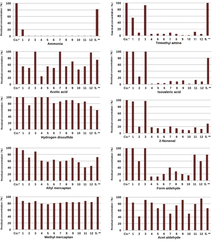
sweat (n-butyric acid), musty socks (isovaleric acid), and aging (2-nonenal) 36-38). These odors are
generated by chemical reactions, such as oxidation; consequently, functional compounds in natural
foods, such as antioxidants, have been commercialized.
Typical oils, like olive oil, are mainly composed of oleic acid. A case study in Greece has
shown an inverse correlation between olive oil intake and the risk of breast cancer 39). A similar
result was reported by the European Prospective Investigation into Cancer and Nutrition (EPIC);
the risk of breast cancer in a cohort of postmenopausal women decreased with olive oil intake 40). A
correlation was suggested between decreased breast-cancer risk and the traditional, olive-oil-rich
Mediterranean diet. Mechanistically, olive oil may be inhibiting the formation of malignant tumors.
Mango seeds are often discarded as waste after the extraction of mango juice. This study
primarily aims to estimate the fatty acid composition of oil extracted from mango seeds; specimens
from different cultivation areas were examined. Their potential use in cosmetics was evaluated by
producing lotions and soaps, conducting deodorization tests, and analyzing the impact of these
results. The secondary aim is to examine the antitumor activity of mango seed oil (MSO) extract.
Differential expression proteomics was performed to assess the effects of MSO on HeLa cell
growth and examine its safety for therapeutic applications.
2. MATERIALS AND METHODS
2.1. Preparation of mango seed kernel oil
Mango seeds were purchased from Miyazaki, Japan, and Taiwan (2012) from Konan chemical
industry (Osaka, Japan). The kernels were dried and ground to a powder using a metallic mill
26
Taiwan, 300.8 g) were extracted in n-hexane (2,500 mL) for 1 week at room temperature (20 ±
2 °C). The supernatant was filtered, and the solvent was evaporated in vacuo, yielding yellow oil.
The mango seeds from Miyazaki and Taiwan comprised 9.78% and 4.02% oil, respectively. The
yellow oil was decolorized using activated clay, producing colorless seed oil (Miyazaki, 67.5 g and
Taiwan, 11.2 g).
2.2. Determining the composition of fatty acids and lipid classes
Following the conventional method, MSO was esterified using boron trifluoride-methanol
(Tokyo Chemical Industry, Japan) to produce volatile methyl esters. The composition of the fatty
acids was determined by gas chromatography (GC; GC-2104; Shimadzu) using a TC-70 column (Φ
0.25 mm × 60 m) equipped with a flame ionization detector. The temperatures of the column,
injector, and detector were 190 °C, 250 °C, and 260 °C, respectively. Helium was used as the
carrier gas.
The methyl ester analogs of the fatty acids were used for peak identification. The retention
times were then compared with those of a standard solution of fatty acid methyl esters.
The composition of lipid classes was determined by GC under the same experimental
conditions used to characterize fatty acids. A commercial standard was used for peak identification.
The retention times were compared with those of a standard solution of glycerol.
2.3. Chemical properties
The neutralization number, unsaponifiable matter, saponification number, acid number, iodine
number, and ester value were determined for each MSO sample and measurements were performed
in triplicate. These values were determined using conventional methods 41-42).
Neutralization number: MSO (0.25 g) was weighed in triplicate and dissolved in a solution of
dimethyl ester and ethanol (25 mL, 1:1).
Unsaponifiable matter: MSO (1.0 g) was dissolved in 1 M solution of potassium hydroxide
27
Saponification number: MSO (0.8 g) was dissolved in 0.5 M solution of potassium hydroxide
in ethanol (25 mL) and refluxed for 1 h.
Acid number: MSO (2.0 g) was dissolved in a solution of dimethyl ester and ethanol (20 mL,
2:1).
Ester value: the ester value was calculated as the saponification number minus the acid
number.
Iodine number: MSO (0.3 g) was dissolved in cyclohexane (10 mL). Iodine monochloride
solution (Wijs’ solution, 25 mL) was added, and the solution was kept in the dark for 30 min at
room temperature (18–20 °C).
2.4. Deodorizing effect
2.4.1. Preparation of odorous substances
Chemicals with offensive odors were purchased from Kishida Chemical Co. (Osaka, Japan):
aqueous ammonia (28%), trimethylamine, methyl mercaptan, acetic acid, formaldehyde (37%),
acetaldehyde, isovaleric acid, and allyl mercaptan; trans-2-nonenal was purchased from
Sigma-Aldrich (Tokyo, Japan). To prepare a hydrogen disulfide solution, sodium sulfide
nonahydrate and hydrochloric acid (35%) were purchased from Kishida Chemical Co. (Osaka,
Japan).
2.4.2. Determination of the deodorization rate of MSO with a gas detector tube 43, 44)
Six odorous solutions were diluted with deionized water to obtain the correct concentrations:
0.5% aqueous ammonia, 0.3% trimethylamine, 10% hydrogen disulfide, 3% acetic acid, 3%
isovaleric acid, and 5% acetaldehyde. Methyl mercaptan was diluted three-fold with benzene, and
formaldehyde was diluted 300-fold with deionized water. Hydrogen disulfide was mixed with a
10% solution of sodium sulfide and hydrochloric acid (diluted 20-fold with deionized water). These
were used as the eight standard solutions.
28
trimethylamine (20 ppm), hydrogen disulfide (20 ppm), methyl mercaptan (5 ppm), acetic acid (50
ppm), isovaleric acid (50 ppm), formaldehyde (30 ppm), and acetaldehyde (100 ppm).
Each standard solution was placed in a flask and charged with 1 g of MSO. The flask was
sealed (airtight) for 30 min at room temperature (20 ± 2 °C). Thereafter, the concentration of the
odorous substance was determined by a Kitagawa AP-20 gas detector (Komyo Rikagaku Kogyo,
Kawasaki, Japan) with a Kitagawa gas detector tube. A 100-mL sample of the headspace gas was
analyzed. A control test was performed without fatty acids.
The deodorizing rate (%) was calculated according to the following equation:
2.4.3. Determination of the deodorizing rate of MSO with a flame ionization detector (FID) 45, 46)
The concentration of a 0.2% solution of trans-2-nonenal was adjusted with ethanol. The
concentration was determined by GC (GC-2014AF, Shimadzu) equipped with an FID (200 °C) and
a Unisole F-200 30/60 column (Φ 3.2 mm × 2.1 m). The column temperature was maintained at
120 °C. Helium (50 mL/min) was used as a carrier gas.
Furthermore, 1 g of MSO was placed in a flask and an adjusted solution of trans-2-nonenal (5
μL) was added. The flask was sealed (airtight) for 30 min at room temperature. A 2-mL sample of headspace gas was used for analysis. A control test was performed without fatty acids.
The deodorizing rate (%) was calculated according to the following equation:
2.4.4. Determination of the deodorizing rate of MSO with a flame photometric detector (FPD) 47,
48)
A 0.001% solution of allyl mercaptan was prepared in ethanol. 1 g of MSO was placed in a
29
(airtight) for 30 min at room temperature. A 2-mL sample of the headspace gas was used for
analysis. A control test was performed without fatty acids.
The remaining concentration of ally mercaptan compound was determined by a GC
(GC-2014AF, Shimadzu) equipped with an FPD (200 °C) and a 1,2,3-tris(2-cyanoethoxy)propane
column (Φ 3.2 mm × 2.1 m). The column temperature was maintained at 50 °C. Helium (40 mL/min) was used as a carrier gas.
The deodorizing rate (%) was calculated according to the following equation:
2.5. Experimental production of cosmetics
2.5.1. Experimental production of soap
Sodium hydroxide (2.36 g) was dissolved in purified water (0.88 mL) and kept isothermal
at 40 °C. MSO (6.76 g) was heated and added dropwise to the NaOH solution 28-29). The pH of the
MSO soap dissolved in water was determined.
2.5.2. Foaming and anti-foam testing of soaps
The foaming of MSO soap, olive oil soap, and commercial soap (Cow Brand Soap Blue
Box, Cow Brand Soap Kyoshisha Co., Ltd.) were compared. 15 mg of soap was added to a
graduated cylinder followed by warm water (2 mL, 40 °C). The cylinder was shaken
vigorously 400 times, and the height of the foam was measured immediately and after 1 to 7 h.
2.5.3. Detergency of soap
The detergency of MSO-soap was tested lipstick and sauce stains on clothing. The soap was
applied to the stain and scrubbed 20 times using a toothbrush 30).
30
MSO (4.50 g) was added to vegetable emulsifying wax (1.65 g) and dissolved in hot water at
60–70 °C. Purified water was added drop wise, and the solution was mixed for 20 min.
2.6. Differential expression proteomics
2.6.1. Cell culture
HeLa cells derived from human cervical cancer cells (DS Pharma Biomedical, Japan) were
used in this study. The cells were grown at 37 °C in a 5% CO2 atmosphere; the culture dishes
comprised Dulbecco’s Modified Eagle Medium (DMEM, Nihon Pharmaceutical, Japan), 10% (v/v)
fetal bovine serum (FBS, Biowest, USA), 50 U/mL penicillin–streptomycin (Invitrogen, USA),
75% NaHCO3, and L-glutamine. Cells were passaged using a trypsin–EDTA solution
(0.05%–0.02%) every 2 days.
2.6.2. Water-soluble tetrazolium salt-1 (WST-1) assay
HeLa cells (at concentration of 0.5 × 10⁵ and 0.25 × 10⁵ cells/mL) were added into each well
of a 96-well plate and then incubated for 24 h. Dimethyl sulfoxide (DMSO) was dissolved in MSO
at a DMSO concentration of 1% (v/v). Next, cells were exposed to concentrations of MSO ranging
from 0–500 μg/mL and incubated for 24 h. After treatment, premixed WST-1 (Takara Bio., Japan)
was added to each well and incubated for 3 h at 37 °C. The absorbance was measured at 450 nm
using a spectrophotometer (Molecular Devices Co.).
2.6.3. Proteomic analysis
HeLa cells (6.36 × 10⁴ cells/cm2) in DMEM/10%-FBS were added to a 10-cm dish and incubated at 37 °C for 24 h. The final concentration of MSO was adjusted to 75 μg/mL with DMSO; it was added to the cells and incubated at 37 °C for 24 h. DMSO was used as a control. The
medium was removed and washed twice with phosphate buffer saline (PBS).
31
The cells (control and MSO-treated) were collected and charged with 500-mM
triethylammonium bicarbonate (TEAB, Sigma, Tokyo, Japan) containing 7 M urea and
0.1%-NP-40. The solids were dissolved using sonication and the solution was rotated overnight in a
cold room. After centrifugation, the eluate was exchanged with 50-mM TEAB using spin
concentrators (Corning, Tokyo, Japan). The protein concentration was determined using a
bicinchoninic acid protein assay (Thermo Fisher Scientific, Rockford, IL, USA).
2.6.3.2. iTRAQ labeling
iTRAQ labeling was performed according to the method reported by Matsumoto 49). Each
sample was denatured, reduced, and digested by trypsin (AB Sciex). Each digest was labeled with a
different iTRAQ tag using an iTRAQ Reagent Multiplex kit (AB Sciex). iTRAQ label 116 was
used for labeling control samples, and iTRAQ label 117 was used for MSO-treated samples. The
labeled samples were combined and fractionated into six fractions using strong cation exchange
(SCX) chromatography, according to the manufacturer’s instructions (AB Sciex).
2.6.3.3. Analysis by chromatography and mass spectroscopy
Chromatography and spectroscopy measurements were performed based on the methods
reported by Matsumoto 50). One fraction from SCX chromatography was fractionated to 171 spots
with a liquid chromatography (LC) system (DiNa nanoLC, KYA Technologies Co., Tokyo, Japan).
Each fraction was diluted in a matrix (4 mg/mL α-cyano-4-hydroxycinnamic acid, Wako Pure Chemical Industries Ltd., Osaka, Japan). Peptide samples were analyzed on a matrix assisted
laser desorption ionization-time of flight (MALDI-TOF) mass spectrometer (MS, 5800
MALDI-TOF/TOF MS/MS) with TOF/TOF series software (AB Sciex). MS/MS data was analyzed
by ProteinPliot TM software (AB Sciex). A statistical analysis was performed to compare the control
and MSO-treated samples; p < 0.05 was considered statistically significant.
32
Panther software was used for the protein-classification analysis.
2.6.3.5. Statistics
The results of WST-1 assay were expressed as the mean ± the standard error of the mean.
Statistical analysis was performed using an ANOVA and Dunnett’s test. Results with p < 0.05 were considered statistically significant. Statistical tests were performed using EZR (Satitama
Medical Center, Jichi Medical University, Saitama, Japan).
3. RESULTS AND DISCUSSION
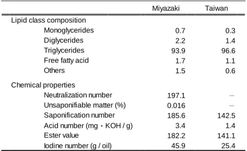
3.1. Composition of lipid classes and fatty acids
GC analysis revealed that triacyl glycerol was the main type of glycerol present in MSOs from
both Miyazaki and Taiwan (Table 10). The main fatty acids in MSO were palmitic acid (C16:0),
stearic acid (C18:0), oleic acid (C18:1), linoleic acid (C18:2), and linolenic acid (C18:3) (Table 11). For
comparison, mangoes from Thailand, the Philippines, and Mexico were purchased from a market in
Japan; seed oil was extracted from these mangoes to determine the fatty acid composition. The
major fatty acids determined experimentally agreed with the findings of Muchiri 23): stearic acid
(Thailand, 42.2%; the Philippines, 57.5%; and Mexico, 34.2%) and oleic acid (Thailand, 44.8%;
the Philippines, 34.3%; and Mexico, 55.4%). The other fatty acids were palmitic acid (Thailand,
5.8%; the Philippines, 4.7%; and Mexico, 5.9%) and linoleic acid (Thailand, 4.5%; the Philippines,
1.0%; and Mexico, 3.1%). Arachidic acid was absent from the seed oil of mangoes from Miyazaki
and Taiwan; it was present in the seed oil of mangoes from Thailand, the Philippines, and Mexico
(2.7%, 2.4%, and 1.3%, respectively). The fatty acid composition differed according to the country
of production and mango variety. Oleic acid comprised 46% of the fatty acid composition in the
Miyazaki MSO but only 44% of the fatty acids in the Taiwan MSO. In contrast, the seed oil from
Taiwan mangoes had a slightly higher amount of the other fatty acids as compared with the
Miyazaki MSO. The fatty acid composition of MSO is nearly the same as that of shea butter, which