Effects of sensorimotor cortex stimulation on
nociceptive neurons of nucleus ventralis
posterolateralis in cats
その他の言語のタイ
トル
ネコ視床後外側腹側核侵害受容ニューロンに及ぼす
大脳皮質体性感覚運動野刺激の効果
ネコ シショウ コウガイソク フクソクカク シンガ
イ ジュヨウ ニューロン ニ オヨボス ダイノウヒ
シツ タイセイ カンカク ウンドウヤ シゲキ ノ コ
ウカ
著者
Kizuki Noyuri, Matsushita Mikiko, Koyama Natsu
journal or
publication title
滋賀医科大学雑誌
volume
12
page range
31-49
year
1997-05
URL
http://hdl.handle.net/10422/3164
滋賀医大誌12, 3卜49, 1997
Effects of Sensorimotor Cortex Stimulation
on Nociceptive Neurons of Nucleus Ventrahs
Posterolateralis in Cats
Noyuri Kizukill, Mikiko Matsushital', Natsu Koyama2)
1 ) First Department of Surgery, Shiga University of Medical Science
2 ) First Department of Physiology, Shiga University of Medical Science
Abstract: In order to clarify functional significance of the cerebral cortex in endogenous central modulation of visceral nociception, the effects of electrical stimulation of the sensorimotor cortex
on nociceptive specific (NS) and wide dynamic range (WDR) neurons in the nucleus ventralis posterolateralis (VPL) of the thalamus were tested in urethane-chloralose anesthetized cats. Thirty six NS and 14 WDR units receiving high threshold greater splanchnic (SPL) afferent input were recorded from the shell region of the caudal VPL. An arterial injection of an algogenic substance, bradykinin, into the mesenteric artery excited 4 NS and 3 WDR units tested, suggest-ing they are subservsuggest-ing visceral pain. The remainsuggest-ing 32 NS and ll WDR units were subjected to the study of effects of cortical stimulation. They were antidromically excited by electrical stimulation of the primary somatosensory cortex (SI) indicating that they were thalamocortical projection neurons. Stimulation of the SI from which the antidromic response could be evoked,
excited 16 NS and 4 WDR units at an intensity lower than the threshold for antidromic
excita-tion. In the remaining, 16 NS and 7 WDR units, responses to visceral and spinothalamic inputs
were inhibited by SI stimulation at an intensity subthreshold for antidromic excitation. Both excitation and inhibition could be elicited also from surrounding sensorimotor cortex. Mapping
of the inhibitory cortical field revealed that the lowest threshold inhibitory point for a given neuron coincided with the point from which antidromic excitation could be elicited. Chemical stimulation of the SI somatosensory cortex with glutamate also inhibited both NS and WDR neurons. The results indicate that in the VPL, there is a corticothalamic modulation of thalamic nociception originating from the SI somatosensory cortex, in addition to recurrent inhibition.
Key ¥M〕rds: sensorimotor cortex, thalamus, nociception, analgesia, nucleus ventralis
posterolater-alis.
introduction
how the central nervous system modulates nocice-ptive information is the development of the theory pertaining to the existence of an endogenous pain One of the recent advances in the concept of modulation system. The discoveries of opiate
Received September 26, 1996: Accepted after revision November 1, 1996
Correspondence: Noyuri Kizuki, First Department of Surgery, Shiga University of Medical Science, Seta, Otsu, Shiga, 520-21, Japan
receptors, of endogenous opioid peptides, of stim-ulation produced analgesia and of stress induced analgesia4'9i33)led to the development of this the-ory. The most commonly proposed mechanism of endogenous pain modulation is that the pen-aqueductal gray (PAG) and nucleus raphe dorsalis (NRD) of the midbrain have descending influences on the spinal cord to modulate pain transmission
at the spinal cord level4'11'35'. More recently,
ascending pain modulation systems from the PAG/NRD to the nucleus ventralis posterolater-alis (VPL) 14'20'Z2)and intralaminar nucleil>22)of the thalamus have been proposed. Thus it has been clarified that thalamic relays of ascending pain pathways are subjected to supraspinal inhibition21'25'. This report tests cortical influ-ences which may affect processing of visceral nociceptive information at the level of the VPL.
Materlals and Methods
Experiments were carried out on adult cats. Each animal was initially anesthetized with ketamine hydrochloride (20mg/kg, i.m.), and anesthesia was maintained with an intravenous dose (3.5ml/kg) of urethane-chloralose solution (urethane 125mg/ml and chloralose lOmg/ml) sup-plemented as required. A thermistor probe was placed in the esophagus and the body temperature was maintained at 37±0.5。C by an electric heat-ing pad under the abdomen, and by an infrared lamp.
The left greater splanchnic nerve (SPL) was exposed retroperitoneally through an incision in the lumbosacral fascia at the lateral edge of the erector spmae muscle mass. The exposed SPL was dissected free from surrounding tissues at the level just proximal to the coeliac ganglion. A bipolar platinum hook stimulating electrode was applied to the SPL, and sealed in place with a low melting point paraffin wax.
After preliminary surgery, the prepared
ani-mal was placed in the prone position on a shock proof table. The head was rigidly fixed in a ster-eotaxic instrument. Cramotomies were carried out over the right thalamus to allow access for microelectrode exploration. The right senson-motor cortex was also exposed in order to place a platinum ball stimulating electrode. The dorsal surface of the spinal cord was exposed by a laminectomy at the level of C4-C6. A bipolar concentric stimulating electrode was inserted into the right ventrolateral funiculus (VLF).
Recordings were made from single units in the VPL using glass capillary microelectrodes filled with a solution of 2% Pontamine sky blue in 0.5M sodium acetate. During recordings, the animals were paralyzed with pancuromum bro-mide (0.8mg/kg/h). End-tidal CO, was monitored and maintained at 3.5-4.5%.
The peripheral receptive field properties of VPL neurons were assessed using a variety of mechanical stimuli: gentle brushing of the skin with a soft brush, pressure applied to a fold of skin using a pair of broad-tipped forceps, and pinching with a pair of serrated forceps. All units were tested for SPL input.
All nociceptive units with SPL input were tested for effects of conditioning stimulation of the sensonmotor cortex. Test stimuli were applied either via SPL or by electrical stimulation of VLF.
The locations of units tested were marked by extruding a small amount of Pontamine sky blue from the microelectrode tip electrophoretically
(5wA negative DC current passed for lOmin). At the conclusion of each experiment, the stimulation site in the VLF was lesioned electro-phoretically, by passing a current of 100 /lA DC between the poles of the stimulating electrode for 1 mm. Animals were then deeply anesthetized, and per fused through the beating heart with
1,000 ml of a solution of 0.5% potassium
fer-rocyanide in normal saline, followed by 2,000 ml of 10% formalin. Serial frozen sections (50〟m
thick) were cut and stained with Cresyl violet. Locations of stimulation and recording sites were checked.
Statistical tests were made using analysis of
Non-parametric Willcoxon signed rank test. All
numerical data are presented as mean±SEM. Differences were considered significant if p < 0. 05.
Results
I. Types of units studied
Observations were made on 50 nociceptive VPL units, 36 0f which were classified as nocice-ptive specific (NS) units and 14 as wide dynamic range (WDR) units. NS units were not activated
by innocuous cutaneous stimuli, but were activat-ed by noxious stimuli appliactivat-ed to a restrictactivat-ed area on the contralateral integument. The identifying characteristic of WDR units was that they re-sponded to a wide range of stimulus intensities. In the center of the cutaneous receptive field, they had a graded response to brushing, pressure and noxious pinch, responding best to noxious pinch. This area was surrounded by an area in which they differentially responded to pressure and noxious pinch. This area was further surrounded by an area in which they responded only to noxi-ous pinch40・ The cutanenoxi-ous receptive field of WDR units was confined to the contralateral integument. Both NS and WDR units subjected to the present study were excited not only by cutaneous afferents but also by high threshold visceral afferents m the same manner as has been
reported3'32'34'. Four NS and 3 WDR units were
subjected to the study of effects of arterial injec-tion of an algogenic substance, bradykinin. The remaining 32 NS and ll WDR units were
sub-jected to an examination of effects of cortical stimulation. They were antidromically excited by electrical stimulation of the somatosensory cor-tex with a latency 1.20±0.14 ms. By cortical stimulation, 16 NS and 4 WDR units were
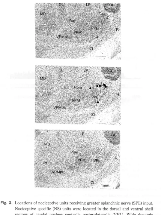
excited, 12 NS and 3 WDR units were inhibited, and 4 NS and 4 WDR units were first inhibited and then facilitated. The distribution of cutaneous receptive fields of these units are summarized in Fig. 1 and 2, while locations of these units were summarized in Fig. 3. NS units were located in
the middle two thirds of both the dorsaユand the
ventral shell region of the caudal VPL, while WDR units were located in the middle two thirds of both the dorsal and the ventral shell region of the VPL just rostral to the NS zone34
II. Effects of injection of bradykinin into the mesentenc artery
Previously, it was reported that VPL nocice-ptive neurons receiving cardiac sympathetic affer-ent inputs regularly exhibited an increased dis-charge in response to intracardiac bradykininl; In order to confirm that the units subjected to the present study are also related to visceral
nocicep-tion, 4 NS and 3 WDR units were tested for
responses to an injection of an algogenic sub-stance, bradykinin. For injection of bradykinin, a cannula was inserted into the anterior mesentenc artery. After the response to the SPL stimulation was recorded, 30^g/kg of bradykinin was inject-ed into the mesenteric artery in about Is, and neuron's response was recorded. An example of responses of WDR units to mesenteric bradykimn injection is illustrated in Fig. 4. The unit was located in the dorsal shell region of the VPL, and had the center of cutaneous receptive field in the back area (Fig. 4A). This neuron responded to
double shock stimulation of the SPL with 0.9-1.5 ms latency (Fig. 4B). When bradykinin was inject-ed into the mesenteric artery, it exhibitinject-ed an increase in discharge rate. The latency of the onset of increased discharge was 40s. The dura-tion of increased discharge outlasted the record-ing period of bmin. The discharge rate was 8.4± 0. 7 spikes/lOs before the injection. It increased t0 91.8±5.7 spikes/10s (Fig. 4C). The mean latency of the onset of increased discharge was 30-50s
Excitation
Inhibition
Inhibition
followed by
Facilitation
Fig. 1. Distribution of cutaneous receptive fields of NS units. Left, NS units excited by stimulation of SI somatosensory cortex (CX). Middle, NS units inhibited by CX stimulation. Right, NS units which showed initial inhibition followed by facilitation due to CX stimulation.
Excitation
i幸一
-tInhibition
Inhibition
followed byFacilitation
#云 _
Fig 2. Distribution of receptive fields of WDR units. Black area indicates low
thresh-old center and shaded area indicates high threshthresh-old surrounding of receptive
field. Left, WDR units excited by SI somatosensory cortex (CX). Middle, WDR
units inhibited by CX stimulation. Right, WDR units which showed initial
inhibition followed by facilitation
(37.9±3.Is, N-7). The mean duration of in-creased discharge was 2.26±0. 76mm. The mean discharge rate was 77.2±33.9 spikes/10s prior to the injection. It increased t0 120.2±39.4 spikes/10s. These results suggest that VPL nociceptive neurons receiving high threshold SPL
due to CX stimulation.
afferent input may be involved in transmission of abdominal visceral nociceptive information.
III. Corticofugal inhibition of nociceptive VPL
units
Fig. 3. Locations of nociceptive units receiving greater splanchnic nerve (SPL) input. Nociceptive specific (NS) units were located in the dorsal and ventral shell regions of caudal nucleus ventralis posterolateralis (VPL). Wide dynamic range (WDR) units were located just rostral to the NS zone.
◇:NS unit excited by SI somatosensory cortex (CX). :NS unit inhibited by CX stimulation. O:NS unit which showed initial inhibition followed by facili-tation due to CX stimulation. □: WDR unit excited by CX stimulation. ▲: WDR unit inhibited by CX stimulation. △: WDR unit which showed initial inhibition followed by facilitation due to CX stimulation. CL: nucleus centralis lateralis; GL: corpus geniculatum laterale; LP: nucleus lateralis posterior; MD: nucleus medians dorsalis; Pom: medial region of posterior thalamic nuclear group; R: nucleus reticularis thalami; VPL: nucleus ventralis posterolateralis; VPMpc: nucleus ventralis posteromedialis parvocelluralis; ZI: zona incerta.
IE
Bradykinin30│i,g/kg
サIlla.i--サ蝣<蝣fc-i山Eo
I Hl 川mmmF甜那那柵榔相即欄間間肝附け朋的附都mm醐皿馴耶mⅧ朋那酬耶醐
喝醸t9- -- ・ ro8SiSサォサ*
10S mmHg 200 150 100 50Fig. 4. Effects of an arterial injection of bradykinin. A: cutaneous receptive field. In the black area, the unit had a graded response to brushing, pressure and noxious pinch. In the cross-hatched area, the unit did not respond to brushing, but responded differentially to pressure and noxious pinch. In the shaded area, the unit exclusively responded to noxious pmch. B: a dot raster display of responses to electrical stimulation of the left splanchnic nerve (SPL). C: responses of the unit to a bolus dose of 30^ g /kg bradykinin into the mesenter-ic artery. The upper trace shows a peristimulus time histogram and middle trace shows spike discharges. The lower trace shows the blood pressure record.
the SI somatosensory cortex from which anti-dromic response could be elicited was found to inhibit the responses of the units to SPL and/or VLF stimulation at an intensity subthreshold for the antidromic response.
Results obtained in an NS unit are illustrated in Fig. 5. The unit was located in the ventral shell region of the VPL. A conditioning stimula-tion was delivered to the cortical area (Fig. 5B), stimulation of which evoked antidromic responses of the unit, at an intensity just subthreshold for the antidromic responses. The stimulation consisL ed of a train of 5 pulses at 400Hz. The duration of individual pulses was 0. lms. The conditioning
stimulation at a 40-ms conditioning-test interval, almost completely suppressed the responses of the unit to test stimuli applied either to the SPL or to the VLF. The time courses of inhibition are plot-ted in Fig. 5E. The inhibition lasplot-ted about 50ms in SPL response and outlasted 120 ms in VLF response.
Inhibitory cortical field was mapped for another NS unit. Figs. 6 and 7 summarize the results obtained. The unit followed a lOOHz stim-ulation (Fig. 6C) delivered to an area in the SI somatosensory cortex in the sigmoid gyrus (Fig. 6 B). The antidromic nature of the response was further confirmed by a collision test, in which
orthodromic stimuli delivered to the VLF were followed by antidromic stimuli at various inter-vals (Fig. 6D). Responses to both VLF and SPL stimulation were completely inhibited by a train of conditioning stimuli applied to the same somatosensory cortex at an intensity just subthre-shold for antidromic responses (Fig. 6E). A condi-tioning stimulation at 0.5 times the threshold for antidromic responses was used for mapping. Inhi-bition of responses to VLF stimulation at a 30-ms conditioning-test interval was obtained from 12 spots in the sensorimotor cortex tested. The dis-tance between each spot was 2mm. The maximum inhibition was produced by stimulation of the area from which antidromic responses were
elicit-ed(Fig. 7). Of 16 NSand 7 WDRunitsinwhich
inhibition could be observed, 4 NS and 4 WDR
units showed an initial inhibition followed by facilitation. Results obtained in an NS unit and in a WDR unit are illustrated in Fig.8 and in Fig.9, respectively. The unit illustrated in Fig.9 was located in the ventral shell region of the VPL, and had a cutaneous receptive field in the abdomen (Fig. 9A). Electrical stimulation of the SI somatosensory cortex (CX), from which anti-dromic responses could be elicited (Fig. 9B) inhib-ited the responses of the unit both to SPL and to VLF stimulation. The maximum inhibitions of both responses were observed at a 20-ms conditionmg-test interval. The SPL response and the VLF response began to recover with pro-longed latencies at a 30-ms conditioning-test interval, and at a 50-ms conditioning-test inter-val, respectively. In about 60ms, both SPL and VLF responses recovered to the control level. Subsequently facilitation was observed. The num-ber of spikes in responses to both SPL and VLF stimulations with conditioning stimuli increased
to 200-300% of those without conditioning stimu-lation (Fig. 9C). The time courses of changes in responses are plotted m Fig. 9D.
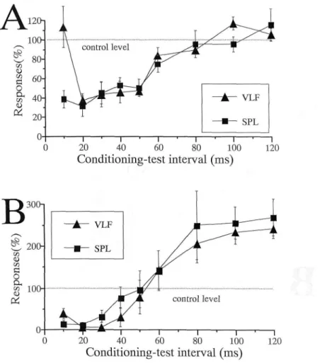
The mean time courses of the inhibition ob-served in 12 NS and 3 WDR units, and of the
inhibition followed by facilitation observed in 4 NS and 4 WDR units are summarized in Fig. 10 A and Fig. 10B, respectively.
IV. Effects of glutamate application to the SI somatosensory cortex on unit responses
Nine NS and one WDR units were tested for
effects of application of glutamate after confirm-ing an inhibition of responses to SPL and/or VLF stimulation following cortical stimulation. A piece of filter paper (5mmX5mm) soaked with 0.05ml of 0.5M monosodium glutamate was applied to the SI somatosensory cortex. Fig. ll shows the profile of responses to VLF stimulation as a function of time following applications of monosodium glutamate. The mean number of spikes to VLF stimulation gradually reduced after application. The maximum suppression of responses was observed at lOmin after the appli-cation and subsequently responses recovered to the control level. The degree of the maximum inhibition was 21.2±8.3%. The maximum inhibi・ tion was statistically significant (P -0.022<0.05, N-10.
V. Excitation of nociceptive VPL units
Electrical stimulation of the SI somatosensory cortex (CX), from which antidromic responses could be elicited, evoked spike discharges from 16 NS and 4 WDR units at an intensity subthre-shold for the antidromic response. The result obtained in an NS unit is illustrated in Fig. 12. The unit was located in the dorsal shell region of the VPL, and had a cutaneous receptive field in the abdomen (Fig. 12A). The unit responded to single to 5 shock stimulations of the SPL with a minimum latency of 14.8ms and to single shock stimulation of the VLF with a minimum latency of 3.5 ms (Fig. 12C). Electrical stimulation of the SI somatosensory cortex (CX), from which anti・ dromic responses could be elicited (Fig. 12B), at an intensity lower than the threshold for anti-dromic responses evoked spike discharges with
0 0 0 5 1 ( % ) S 3 S U O d s 3 J J 20 40 60 80 1 00 Conditioning-test interval(ms) 120
Fig. 5. Effects of SI somatosensory cortex stimulation on an NS unit. A: receptive field B: stimulation site in the SI somatosensory cortex (indicated by an arrow). C: dot raster displays of the unit responses to SPL stimulation both with and without conditioning stimulation of the SI somatosensory cortex (CX). Conditioning-test intervals are 20ms (Left) and looms (Right). D: dot raster displays of the unit responses to VLF stimulation both with and without conditioning CX stimulation. Conditioning-test intervals are 20ms (Left) and 120ms (Right). E: Time courses of conditioning CX stimulation-produced inhibi-tion of responses to SPL stimulainhibi-tion and to VLF stimulainhibi-tion.
Aォ- ^C鮒酔酔
/・・・s^
-sussKfcr^r空拙
- :-- 蝣w- *" "-VLF CXト5mV
vyIWvォ^WwAy¥viy/"'v'V'YV'u lOmsFig. 6. Antidromic responses of an NS unit to stimulation of the SI somatosensory cortex (CX) A: cutaneous receptive filed. B. stimulation site in CX (indicated by an arrow). C: responses of the unit to 100 Hz stimulation of the CX shown in B. D: collision test using the ipsilateral ventrolateral funiculus (VLF) stimu-lation and CX stimustimu-lation as orthodromic and antidromic stimustimu-lation, respec-tively. E: dot raster displays of the unit responses to SPL stimulation (Left) and to VLF stimulation (Right), both with and without conditioning stimulation.
∴
二
㍉
●
了
∴
︰
.
∴
.
二
㌧
∴
二
.
. 蝣 ' ' i : . : #Fig. 7. Inhibitory field mapping in the sensorimotor cortex. A: stimulation sites (・, ★). The site in the SI somatosensory cortex producing antidromic responses is indicated by a star ★). B: degree of inhibition of responses to VLF stimulation produced by corresponding areas shown in A. The CX stimulations at the area from which antidromic responses were elicited and at the point just
B
Fig. 8. An example of the NS neuron which showed an inhibition followed by facihta-tion. A: cutaneous receptive field. B: stimulation site in the SI somatosensory cortex (indicated by an arrow). C: dot raster displays of the unit responses to SPL stimulation both with and without conditioning stimulation. D: dot raster displays of the unit responses to VLF stimulation both with and without conditioning stimulation. E: time courses of inhibition.
Fig. 9. An example of the WDR neuron which showed an inhibition followed by facilitation. A: cutaneous receptive field. B: stimulation site in the SI somatosensory cortex (indicated by arrow). C: dot raster displays of the unit responses to SPL stimulation and to VLF stimulation both with and without conditioning stimulation. D: time courses of inhibition.
Fig. 10. Mean time courses of conditioning CX stimulation-produced inhibition of responses to SPL stimulation and to VLF stimulation. A: inhibition by CX stimulation. B: inhibition followed by facilitation.
o o o o o o o o o o o a ¥ o o t > 丘 3 > n t m M h ‖H
(
%
)
9
S
U
O
d
s
9
>
I
蝣
,題
題
事
葛
r1
糧
IIIIIIIII
I-ii.
肯
川 IIIIII
‖-農
I(IIIIIII
一
日
IIIIII
一
日
II II I
i U U l l
一
日
= 一
貞
IIIII I川
IN
III H U If
卓
IIIIIII II
一
日
m i
U U U LBJ
pre O 2 4 6 8 1012141618202224262830 time(min.)Fig. 1 1. Mean time courses of glutamate-produced inhibition of responses to VLF stimulation.
A
Fig. 12. An example of the NS neuron excited by CX stimulation. A: cutaneous receptive field. B: stimulation site in the SI somatosensory cortex (indicated by an arrow). C: responses of the unit to VLF stimulation. D: responses of the unit to SPL stimulation. C: responses of the unit to CX stimulation.
15.7 ms latency (Fig. 12C). The mean of mini-mum latencies of the excitations in 20 units was 36.0士8.2ms. The mean number of spikes evoked by 5 shock stimulation was 2.2±0.3.
Discussion
In humans, higher cortical functions such as hypnosis and creative imagery have been shown to effectively reduce pain perception271. It is also known that there is a significant positive correla・ tion between pam level and anxiety in patients with chronic pain24'. Thus, the cerebral cortex may play a role in pain modulation. Clinically, a relief of pain has been demonstrated by
stimulat-ing the internaユcapsule8'10'. In these cases the
relief of pam could be due to the activation of inhibitory pathways arising from the somatosen-sory cortex or of the recurrent inhibitory mecha-nism. In monkeys, stimulation of the sensorimotor cortex has been shown either to excite or to inhibit spinal cord neurons which may be involved in processing noxious stimuli39'. The present study further explored the role of the sensorimotor cortex in the modulation of responses both to visceral afferent and to spinothalamic input recor-ded in the nociceptive VPL neurons of the thalamus.
In the present experiments, electrical stimula-tion of the SI somatosensory cortex which receives nociceptive input relayed through the VPL, inhibited responses of both NS and WDR neurons m the VPL to electrical stimulation not only of the SPL but also of the VLF. The VLF stimulation activates spinothalmic tract fibers arising from the spmal cord to terminate in the thalamus. Hence the present data suggest that in addition to the corticospmal inhibitory pathway, there may be a corticothalamic pain modulation pathway from the SI somatosensory cortex to the VPL to inhibit pam messages from being further transmitted. This hypothesis is supported by the
anatomical evidence that the SI somatosensory cortex has reciprocal connection with the
VPL23,26,39) jn both NS and WDR neurons>
hQw-ever, an antidromic spike was recorded following electrical stimulation of the SI somatosensory cortex. These antidromic spikes were probably initiated by stimulating axons of nociceptive VPL neurons which project to the SI somatosensory cortex. There are at least two explanations for how both antidromic spikes and inhibition may be generated in the same neuron: (1) the electrical stimulation in the cerebral cortex excited thalamocortical axons of VPL nociceptive neur-ons to produce the antidromic spikes and also excited cortical cell bodies which had inhibitory effects on the VPL nociceptive neurons; or (2) thalamocortical axons were antidromically stimulated and their axon collaterals activated inhibitory neurons projecting to the cell bodies of nociceptive VPL neurons to suppress their responses to spinothalamic input. In the present
experiments, it was found that for a g王ven neuron,
the lowest threshold inhibitory point coincided with the point from which antidromic excitation could be elicited. It is also known that thalamocortical axons of low threshold me-chanoreceptive (LTM) neurons in the VPL exerts recurrent inhibitory action not only on the LTM neurons themselves but also on nearby neurons2'301. Hence, the recurrent inhibition may be initiated either by thalamocortical axons of nociceptive VPL neurons or by thalamocortical axons of LTM neurons, or by both.
Electrical stimulation mdiscnminatively excites all neural elements, including fibers which then transmit action potentials both ortho-and antidromically. Thus no firm conclusion can be drawn as to the origin of the inhibitory effect on the VPL. In contrast, the excitatory amino acid glutamate excites selectively the cell bodies and dendrites12'. In the present study, an application of glutamate to the SI somatosensory cortex also resulted in a suppression of responses of
nocice-ptive VPL neurons to VLF stimulation. This result indicates that excitation of glutamate receptor-linked system in the SI somatosensory cortex exerts an inhibitory effect on thalamic processing by activation of a corticothalamic system. However, this does not rule out the possi-bility that a recurrent inhibitory mechanism is additionally included in the inhibitory effects of cortical stimulation reported in the present paper. Nevertheless, the present study has shown that in the VPL, there is a corticothalamic modulation of thalamic nociception originating from the SI somatosensory cortex.
All 4 areas of the SI somatosensory cortex, i.
eリ areas 3a, 3b, 1 and 2 send thin
corticoth-alamic fibers from layer VI neurons to terminate in the ventrobasal (VB) complex consisting of the nucleus ventralis posteromedialis prorius (VPM proper) and VPL18'19'29'. It is probable that many of these layer VI neurons receive monosynaptic thalamocortical synapses3'). En route, the cor-ticothalamic fibers give off collaterals in part of the nucleus reticularis thalami16'. Probably in every neurons in this nucleus, markers for GABA synthesis are present 15). There is sufficient work on the nucleus reticulans thalami that it provides a recurrent inhibition upon thalamocortical relay neurons in their related dorsal thalamic nucleil
Hence, it appears likely that the corticofugal inhibition observed in the present experiments is mediated by this nucleus.
Within the thalamic nuclei of monkey and
cat, the thalamocortical relay neurons constitutes
approximately 75-90% of the total complement
of neurons, and the interneuron (local circuit
neuron) represents approximately 10-25% of the
total neuronal population28'. It is thus possible that the corticofugal inhibition as observed in the present experiments is to some extent mediated by some of these interneurons.
It has been reported that neurons in the VB complex which receive input from medial lemnis-cus are excited by stimulation of restricted areas
of the postcruciate gyrus of the cat '. When these
and adjacent areas of postcruciate cortex were weakly stimulated prior to the medial lemmscal test shock, the VB units were commonly inhibited. Thus excitatory cortical field was surrounded by inhibitory cortical field. These investigators con-eluded that some axon collaterals of pyramidal tract fibers project to the VB complex and pro-duce excitation of small groups of VB neurons, which in turn results in inhibition of surrounding VB neurons. These cortical influences may func-tion to sharpen incoming general sensory impulses. The neurons studied by this group were primarily nonnociceptive neurons, because they received input from the medial lemniscus. In the present experiments, extralemmscal nociceptive neurons in the VPL were studied, and excitation of a significant proportion of both NS and WDR neurons was observed following stimulation of the SI somatosensory cortex. It is possible that the excitatory effects reflect corticofugal excita・ tion of spinothalamic neurons in the spinal dorsal horn39', but we can not rule out possible su-praspinal excitatory mechanism. Since both ex-citation and inhibition could be obtained from the somatosensory cortex, there may exist separate corticofugal feedback loops responsible for adjus-ting the amplification and filtering ascending nociceptive information.
Recent positron emission tomographic (PET) analyses of cerebral blood flow during noxious stimulation in humans have established that the contralateral SI somatosensory cortex is activaL ed along with the cingulate cortex, SII somatosen-sory cortex and insula6'31'. Any damage along the course of the spmothalamocortical pathway lead-ing to the SI somatosensory cortex, i.e., in the lateral system of ascending pain pathways, can produce a central neurogenic pain 5'37). The arche-type of central neurogenic pain is thalamic pain which is part of the thalamic syndrome. The VPL studied m the present experiments is included in this pathway. Interference with the balance
between inhibitory and excitatory feedback from somatosensory cortex may be involved in the manifestation of central pain7'.
Acknowledgments
We wish to express our appreciation to emer-ltus professor Toshikatsu Yokota for his critical reviews and encouragement.
References
1 ) Andersen E, Dafny N: Dorsal raphe stimula-tion reduces responses of parafascicular neur-ons to noxious stimulation. Pain 15, 323-331, 1983.
2) Andersen P, Eccles JC, Sears T: The ventrobasal complex of the thalamus: Types of cells their responses and their functional organization. J Physiol (London) 174: 370-399, 1964.
3 ) Asato F, Yokota TニResponses of neurons in
nucleus ventralis posterolateralis of the cat thalamus to hypogastric input. Brain Res 488: 135-142, 1989.
4) Basbaum AI, Fields HL: Endogenous pain control systems: brainstem spinal pathways and endorphin circuitry. Ann Rev Neurosci 7: 309-338, 1984.
5) Boivie J, Leijon G, Johnson I: Central post-stroke pam-a study of the mechanisms
through analyses of the sensory abnormaエー
ities. Pain 37: 173-185, 1989.
6 ) Casey KL, Minoshima S, Berger KL, Koepe RA, Morrow TJ, Frey KA: Positron emission
tomographic analyses of cerebral structures activated specifically by repetitive noxious heat stimuli. J Neurophysio1 71: 802-807,
1994.
7 ) Cassinari V: Surgical operations on the neur-axis that given rise to central pain, in Pagni
CA (ed): Central Pain, pp 34-93. Cambridge, Ma, Harvard Univ Press, 1969.
8 ) Cooper IS, Upton ARM, Amin I: Reversibility
of chronic neurologic deficits. Some aspects of electrical stimulation of the thalamus and internal capsule in man. Appl Neurophysiol 43: 244-258, 1980.
9 ) Duggan AW, North RA: Electrophysiology of
opioids. Pharmacol Rev 35: 218-281, 1984. 10) Fields HL, Adams JE: Pain after cortical
injury relieved by electrical stimulation of the internal capsule. Brain 97: 169-178, 1974. ll) Fields HL, Heinricher MM, Mason P:
Neur-otransmitters m nociceptive modulatory cir-cuits. Ann Rev Neurosci 14: 219-245, 1991. 12) Goodchild AK, Pampney RAL, Bander RA: A
method for evoking physiological responses by stimulation of cell bodies, but not axons of passage, within localized regions of the
cen-tral nervous system. J Neurosci Method 6:
351-363, 1982.
13) Horie H, Yokota T: Responses of nociceptive VPL neurons to intracardiac injection of
bradykimn in the cat. Brain Res
516二161-164, 1990.
14) Horie H, Pamplin PJ, Yokota T: Inhibition of nociceptive neurons in the shell region of nucleus ventralis posterolateralis following conditioning stimulation of the peria-queductal grey of the cat. Brain Res 561: 35-42, 1991.
15) Houser CR, Vaughn JE, Barber RP, Roberts E: GABA neurons are the major cell type of
the nucleus reticu王aris thalami. Brain Res
200: 341-354,1980.
16) Jones EG: Some aspects of organization of the thalamic reticular nucleus. J Comp Neur-01 162: 285-308, 1975.
17) Jones EG: The Thalamus. pp716-720, New York & London, Plenum Press, 1985. 18) Jones EG, Powell TPS: The projection of the
somatic sensory cortex upon the thalamus in the cat. Brain Res 10: 369-391, 1968.
19) Jones EG, Wise SP, Coulter JD: Differential
thalamic relationships of sensonmotor and parietal cortical fields in monkeys. J Comp Neurol 183: 833-882, 1979.
20) Koyama N, Yokota T: Ascending inhibition of nociceptive neurons in the nucleus ventralis posterolateralis following condition-ing stimulation of the nucleus raphe magnus. Brain Res 609: 298-306, 1993
21) Koyama N, Yokota T: Inhibition of activities of VPL nociceptive neurons by dorsal column stimulation. Pain Res 9: 77-86, 1994. 22) Koyama N, Nishikawa Y, Chua A, Iwamoto
M, Yokota T: Differential inhibition mecha-nism in VPL versus mtralaminar nociceptive neurons of the cat: I. effects of periaqueductal gray stimulation. Jpn J Physiol 45: 1005-1027, 1995.
23) Kusama T, Otani K, Kawana E: Projections of the motor, sensory, auditory and visual cortices in cats. Progr Brain Res 21: 292-322, 1966.
24) Linton SJ, Gotestam KG: Relations between pain, anxiety, mood and muscle tension in chronic pain patients. Psychother Psychosom 45: 78-95, 1985.
25) Matsushita M, Kizuki N, Koyama N:
Ex-citatory effects of vagal afferent input on nociceptive neurons in the nucleus ventralis posterolateralis of the cat thalamus. J Shiga
Univ Med Sci ll: 93-106, 1996.
26) Niimi K, Matsuoka H, Yamazaki Y,
Yanagi-hara M: Anatomical organization of thalamic
projections to the first somatic sensory
cor-tex in the cat. Okajimas FolAnaりpn 58: 997
-1008, 1982.
27) Orne MT: The effect of biofeedback therapy. Ann Rev Med 30: 389-503, 1979.
28) Ralston HJ III: Local circuitry of the somatosensory thalamus in the processing of sensory m formation. Progr Brain Res 87: 13 -28, 1991.
29) Rinvik E: The corticothalamic projection
from the pencruciate and coronal gyn in the cat: An experimental study with silver impregnation method. Exp Brain Res 5: 129-152, 1968.
30) Shimazu H, Yanagisawa N, Garoutte B: Cortico-pyramidal influences on thalamic somatosensory transmission in the cat. Jpn J Physiol 15: 101-124, 1965.
31) TalboりD, Marrett S, Evans AC, Meyer E,
Bushnell MC, Duncan GH: Multiple
represen-tation of pain in human cerebral cortex. Science 251: 1355-1358. , 1991.
32) Taguchi H, Masuda T, Yokota T: Cardiac sympathetic afferent input onto neurons in nucleus ventrahs posterolaterahs of cat thalamus. Brain Res 436: 240-252, 1987. 33) Terenius L: Endogenous peptides and
analge-sia. Ann Rev Pharmacol 18: 189-204, 1978. 34) Umehara H: Splanchnic and somatic afferent
convergence onto neurons in nucleus
ventralis posterolateralis. J Shiga Univ Med
Sci 1:1ト29, 1986.
35) Wang Q・P, Nakai Y: The dorsal raphe: an
important nucleus in pain modulation. Brain Res Bull 34: 575-585, 1994.
36) White EL: Thalamocortical synaptic rela・ tions: A review with emphasis on the proiec-tions of specific thalamic nuclei to the pri-mary sensory areas of the neocortex. Brain Res Rev 1: 275-312, 1979.
37) Willis WD: Central neurogenic pain: Possible mechanisms. Ad Pain Res Ther 19: 81-102, 1991.
38) Wise SP, Jones EG: Cells of origin and
termi-nal distribution of descending projection from the rat somatosensory cortex. J Comp Neurol 17: 129-157, 1977.
39) Yezierski RP, Gebhart KD, Schrock BJ, Willis D: A further examination of effects of cortical stimulation on primate spmoth-alamic tract cells. J Neurophysio1 49, 424-441, 1983.
shell theory of thalamic nociception. Jpn J Physiol 39: 335-348, 1989.
41) Yokota T, Asato F, Koyama N, Masuda T, Taguchi H: Nociceptive body representation in nucleus ventralis posterolateralis of cat thalamus. J Neurophysio1 60: 1714-1727, 1988.