学位論文 博士(医学) 甲
High levels of stromal cell-derived
factor-1α predict secondary cardiac
events in stable patients with a history of
myocardial infarction
(心筋梗塞後安定期の SDF-1α 値で将
来の心イベントを予測できる)
松岡 聡志
山梨大学
1
High levels of stromal cell-derived factor-1α predict secondary cardiac events in stable patients with a history of myocardial infarction
Satoshi Matsuoka (MD)a, Manabu Uematsu (MD)a,Takamitsu Nakamura (MD, PhD)a, Takuya Shimizu (MD)a, Mika Futamata (MD)a, Jun-ei Obata (MD, PhD)a, Daisuke Fujioka (MD, PhD)a, Kazuto Nakamura (MD, PhD)a, Toru Yoshizaki (MD)a, Kiyotaka Kugiyama
(MD, PhD, FJCC)a
a Department of Internal Medicine II, University of Yamanashi, Faculty of Medicine, Chuo,
Yamanashi, Japan
Key words: stromal cell-derived factor-1α, myocardial infarction, prognosis, cardiac events, inflammatory marker.
This paper was published in the following journal: Journal of Cardiology, 69(1),
2 Abstract
Background: We recently showed that stromal cell-derived factor (SDF)-1α, a
proinflammatory mediator, is produced in infarcted myocardium and is associated with left
ventricular (LV) adverse remodeling and progressive dysfunction following acute myocardial
infarction (AMI). The current study examined whether SDF-1α levels in the peripheral vein
can provide prognostic information of outcomes in stable patients with a history of MI.
Methods: Plasma levels of SDF-1α in the peripheral vein were measured by enzyme-linked
immunosorbent assay in 192 stable patients with a history of MI. All patients were followed
prospectively for a period of 90 months or until occurrence of one of the following cardiac
events: cardiac death, non-fatal myocardial infarction, unstable angina requiring unplanned
coronary revascularization, or worsening heart failure requiring hospital admission.
Results: During the follow-up period (77 ± 26 months), 30 patients had cardiac events.
Multivariate Cox analysis revealed that high levels of SDF-1α ( >2162 pg/mL; a cut-off value
determined by receiver-operating characteristic analysis) were a significant predictor of
cardiac events, independent of traditional risk factors (HR: 1.98; 95% CI: 1.38–2.85; p <
0.001). The addition of high levels of SDF-1α to conventional risk factors including brain
natriuretic peptide improved net reclassification improvement (NRI) and integrated
discrimination improvement (IDI) (NRI 0.90, p < 0.000-1αnd IDI 0.05, p = 0.002).
Conclusions: High levels of SDF-1α predicted secondary cardiac events in stable patients
with a history of MI. SDF-1α levels may be a useful risk assessment tool in patients with a
3 Introduction
Acute myocardial infarction (AMI) induces the production of chemokines and
growth factors that recruit neutrophils and phagocytes to the ischemic cardiac tissue, leading
to clearance of dead cells and matrix debris [1,2]. This acute, localized inflammatory response
is transient, and is followed by resolution of the inflammation [1,3]. However, prolonged
inflammation leads to dilative remodeling and progressive dysfunction of the infarcted
myocardium through matrix degradation and cardiomyocyte apoptosis [1,2]. Left ventricle
(LV) adverse remodeling and dysfunction after MI are predictors of poor outcome following
MI, and are associated with ventricular arrhythmias, heart failure, and increased mortality
[4,5]. Stromal cell-derived factor (SDF)-1α/CXCL12 is a CXC chemokine with chemotactic
effects on CXCR4-expressing progenitor cells [6]. We have recently shown that SDF-1α, a
proinflammatory mediator, is produced in the infarcted myocardial lesion and released into
the circulation in patients with AMI [7]. In addition, we showed that the myocardial
production of SDF-1α in the chronic phase (6 months after MI) but not in the early phase (2
weeks after MI) was associated with LV adverse remodeling and progressive dysfunction in
patients with AMI [7]. Thus, prolonged production of SDF-1α in the infarcted myocardium
may exert detrimental effects on post-MI LV function in the chronic phase following MI. The
prognostic value of SDF-1α levels in the peripheral vein was previously evaluated in patients
with AMI/ acute coronary syndrome (ACS) [8,9]. However, it remains unclear whether
SDF-1α levels can provide prognostic information in patients in the chronic phase of MI. The
current study examined the prognostic value of SDF-1α levels in the peripheral vein in stable
4 Methods Study patients
The study screened 386 patients with a history of MI, who underwent cardiac
catheterization in Yamanashi University Hospital between February 2003 and March 2009.
All patients underwent routine blood testing at the time of discharge. The inclusion criteria
were: (1) stable previous MI; (2) no episode of angina at rest and no changes in the frequency
of angina in response to sublingual nitroglycerin in the previous 2 months. Exclusion criteria
included: (1) ACS, stroke, cardiogenic shock, pulmonary edema, major surgery, trauma or
serious infectious disease within 4 weeks prior to enrollment; (2) neoplasm, significant
hepatic or inflammatory disease; (3) chronic renal failure or serum creatinine >2.5 mg/dL,
congestive heart failure, or left main coronary artery disease; (4) other serious diseases.
Finally, a total of 210 patients were enrolled in the study according to these inclusion and
exclusion criteria. The study also included 31 control patients without echocardiographic
findings of significant valvular disease (moderate or more regurgitation, stenosis),
cardiomyopathy [chamber size dilatation, reduced left ventricular ejection fraction (LVEF),
ventricular asynergy, ventricular hypertrophy, aneurysm], and pulmonary hypertension,
chosen from among the 6-1αngiographically normal patients [without percutaneous coronary
intervention (PCI) history] who were evaluated during the study period. Control patients
served as a reference group for plasma SDF-1α concentrations. Each of the control patients
underwent diagnostic coronary angiography for atypical chest pain at rest at Yamanashi
University Hospital during the study period. Control patients fulfilled all of the following
inclusion criteria: (1) no significant ST segment changes on 12-lead electrocardiogram (ECG)
while having chest pain or on ambulatory ECG; (2) neither chest pain nor ST segment
intra-5
coronary infusion of acetylcholine [10]. Clinical characteristics of patients with a history of
MI and control patients are shown in Table -1αll study participants were ethnic Japanese. All
patients gave written, informed consent at the time of enrollment. The study was approved by
the ethics committee of Yamanashi University Hospital. The investigation conformed to the
principles outlined in the 1975 Declaration of Helsinki.
Prospective study
Patients were followed every month in the hospital or with a clinic visit for 90 months, or
until the occurrence of cardiac death, non-fatal MI, refractory unstable angina pectoris (uAP)
requiring unplanned coronary revascularization, or decompensated heart failure. The time to
the first event was evaluated prospectively. Cardiac death was confirmed by hospital records.
Acute MI and uAP were diagnosed by the presence of acute ischemic symptoms lasting >20
min within 48 h prior to hospital admission, and ECG changes. Acute MI was diagnosed
when creatine kinase-MB levels increased to at least 2 times the upper limit of normal or
when troponin T levels were >0.1 ng/mL [11]. The diagnoses of MI and uAP were confirmed
by coronary angiography. Decompensated heart failure was defined as resting dyspnea with
progressive fluid retention requiring hospitalization and treatment with an intravenous
diuretic. Follow-up data were collected from the patients’ primary physicians every 3 months
by two blinded investigators (T.N., J.O.). During the follow-up period, standard medications
were prescribed to all patients according to the guidelines of the American Heart Association
[12], as shown in Table 1. Diet and lifestyle recommendations were continued throughout the
follow-up period.
6
Venous blood was obtained from all patients on the morning of discharge after a 12-h
overnight fast. The initial volumes of each blood sample, including those forcibly drawn,
were discarded. Serum and EDTA-plasma were aliquoted and stored at –80℃ until time of
analysis. Plasma SDF-1α levels were measured by enzyme-linked immunosorbent assay using
a commercial kit (R & D Systems, Minneapolis, MN, USA). In our laboratory, the intra- and
inter-assay coefficients of variation were 1.9% and 3.5%, respectively. The minimal detection
limit of this assay was 18 pg/mL. Serum C-reactive protein (CRP) levels were assayed by rate
nephelometry (Dade Behring, Tokyo, Japan). Plasma levels of brain natriuretic peptide (BNP)
were measured by immunoradiometric assay (Shionogi Pharmaceutical, Osaka, Japan).
Estimated glomerular filtration rate (eGFR) was calculated by the Modification of Diet in
Renal Disease study equation. Echocardiographic LVEF was calculated by the motion-mode
method using the Teichholz formula [13].
Statistical analysis
All descriptive data were expressed as mean ± SD, median, or frequency (%). The
Shapiro–Wilk test showed that age, body mass index (BMI), heart rate, low-density
lipoprotein cholesterol (LDL-C), high-density lipoprotein cholesterol (HDL-C), hemoglobin
A1c (HbA1c), LVEF, SDF-1α, BNP, and CRP were not normally distributed, hence, these
variables were expressed as the median and inter-quartile ranges (25th and 75th percentiles).
Non-normally distributed variables were log-transformed before the analysis. Continuous
variables were compared between the two groups using the unpaired t-test or the Mann–
Whitney U-test, as appropriate. Frequencies were compared using the chi-square test. For
comparisons of the mean value of >3 groups, one-way analysis of variance (ANOVA)
7
the two groups was examined using univariate and multivariate linear regression analyses.
Kaplan–Meier analysis was performed on the 2 groups and was based on a cut-off SDF-1α
level. The cut-off level of SDF-1α (2162 pg/mL) was determined by receiver-operating
characteristic (ROC) analyses of SDF-1α in the study patients with a history of MI (Fig. 1).
The predictive values were assessed by univariate or forward stepwise multivariate Cox
proportional hazards analysis. For a forward stepwise multivariate Cox hazard analysis, the
significance level was set at 0.1. In the univariate and multivariate Cox proportional hazards
analyses, continuous variables were estimated for 1-SD change. Dichotomous variables were
coded as 1 for the presence of, and 0 for the absence of, each factor. The following factors
were included as dichotomous variables: systemic hypertension (>140/90 mmHg or use of an
antihypertensive medication) [14], diabetes mellitus (DM) (as defined by the American
Diabetes Association or use of an antidiabetic medication) [15,16], smoking history (defined
as smoking >10 cigarettes/day for >10 years), atrial fibrillation (including paroxysmal atrial
fibrillation recorded with an ECG), and multivessel disease. The Cox models included only
variables that showed proportionality in a Schoenfeld residuals test. The category-free net
reclassification improvement (NRI) and the integrated discrimination improvement (IDI)
were used to examine the additive effects of SDF-1α and BNP levels on the predictive value
of the baseline model that consisted of conventional cardiovascular risk factors. Conventional
risk factors included age, male gender, DM, hypertension, smoking, levels of LDL-C,
HDL-C, CRP, LVEF, and multivessel disease. All probability values were presented as 2-tailed with
statistical significance inferred at p < 0.05. All confidence intervals were computed at the 95%
level. Statistical analyses were performed using STATA 10.0 (StataCorp, College Station, TX,
8 Power analysis
Our previous study showed that the composite endpoints occurred in approximately
52% of stable coronary artery disease (CAD) patients with high coronary risk factors and
higher macrophage inhibitory factor (MIF) levels as an inflammatory marker, and in 18% of
patients with lower MIF levels during 5 years of follow-up [17]. On the basis of that study, a
total of 72 patients were required for a two-sided statistical analysis with sufficient statistical
power of 0.90 (b = 0.10 and a = 0.05). This justified the number of patients (n = 220) included
in the current prospective study.
Results
Over the course of the study, 7 patients were lost to follow up, and 11 patients were
excluded due to non-cardiac related death during follow-up. A total of 192 patients completed
the follow-up study (2–90 months, mean = 77 ± 26 months). During the follow-up period, 30
(15.6%) cardiac events occurred, including 5 cardiac deaths, 1 non-fatal MI, 12 uAPs
requiring unplanned coronary revascularization, and 12 episodes of worsening heart failure.
Patient characteristics are shown in Table 1.
Patients and SDF-1α levels
The time between blood sampling for SDF-1α level and the previous MI ranged from
3 to 26 months, with a mean of 7.7 ± 2.1 months. The time between onset of previous MI and
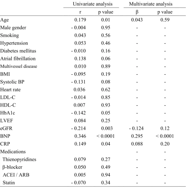
blood sampling did not correlate with SDF-1α levels (r = –0.08, p = 0.28). As shown in Table
2, SDF-1α levels were positively correlated with age, BNP, and CRP but inversely correlated
with eGFR in the univariate linear regression analysis (Table 2). SDF-1α levels did not
9
Prevalence of PCI-related variables was similar between patients with high and low SDF-1α
levels (Supplementary Table 1).
Prospective study
Patients (n = 82) with higher SDF-1α levels (>2162 pg/mL, a cut-off value
determined by ROC analysis) had 26 events during follow-up, whereas patients (n = 110) with
lower SDF-1α levels (<2162 mg/dL) had 4 events (p < 0.01). The cut-off value of SDF-1α
levels as 2162 pg/mL, determined by ROC analysis, provided sensitivity of 86.7%, specificity
of 65.4%, and accuracy of 68.8% for prediction of future events (Fig. 1). Kaplan–Meier
analysis demonstrated a significantly higher probability of a cardiac event in patients with
higher SDF-1α levels compared with those with lower SDF-1α levels (Fig. 2). A univariate
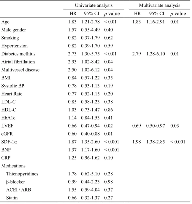
Cox proportional hazards analysis revealed that SDF-1α levels (HR 1.87; 95% CI 1.35–2.60),
age (HR 1.83; 95% CI 1.21–2.78), DM (HR 2.73; 95% CI 1.30–5.75), atrial fibrillation (HR
2.93; 95% CI 1.02–8.42), multivessel disease (HR 2.50; 95% CI 1.02–6.12), LVEF (HR 0.66;
95% CI 0.47–0.94), eGFR (HR 0.60; 95% CI 0.40–0.88), and BNP levels (HR 1.37; 95% CI
1.17–1.60) were significantly associated with future cardiac events, as shown in Table 3. A
forward stepwise multivariate Cox proportional hazards analysis demonstrated that SDF-1α
levels remained significantly associated with future events after adjustment for potential
confounding variables (HR 1.98; 95% CI 1.38–2.85).
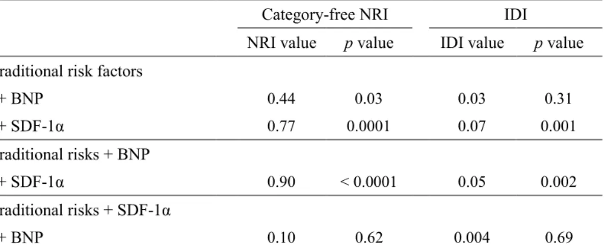
Incremental effects of SDF-1α levels on the predictive value of traditional risk factors Category-free NRI and IDI demonstrated the additive value of SDF-1α to the
baseline conventional risk factors consisting of age, male gender, DM, hypertension, smoking,
10
IDI 0.07, p = 0.001) (Table 4). When BNP was added to the baseline model of risk factors,
SDF-1α had significant additive effect on the predictive value of the model (NRI 0.90, p <
0.0001; IDI 0.05, p = 0.002) (Table 4). In contrast, BNP had no significant additive effect on
the predictive value of a risk factor model with SDF-1α (Table 4).
Discussion
The present study showed that high SDF-1α levels are a predictor of adverse
outcomes in patients with a history of MI. Moreover, high SDF-1α levels had incremental
effects on prognostic value of traditional risk factors in this patient population. Thus, SDF-1α
levels may aid in risk assessment in stable patients with a history of MI. We have previously
shown that SDF-1α is released from the infarcted myocardium into the coronary circulation in
both the early (2 weeks after MI) and chronic phases (6 months after MI) following AMI [7].
The myocardial production and release of SDF-1α in the chronic phase of MI, rather than in
the early phase, was associated with post-MI LV adverse remodeling and progressive
dysfunction [7]. These results support the present finding that SDF-1α levels can predict
secondary cardiac events in patients with a history of MI. Animal experiments have
demonstrated that local infusion or over-expression of SDF-1α attenuates myocardial
ischemic injury following LV dysfunction [18–22]. In contrast, other experimental studies
have shown that SDF-1α/CXCR4 signaling worsens post-MI LV function due to increased
inflammatory injury and apoptotic mechanisms [23–26]. Thus, the effect of SDF-1α on
post-MI LV function in animal models remains unclear. In animal models, the effects of
endogenous SDF-1α on post-MI cardiac tissue may be balanced between angiogenic/
reparative and proinflammatory actions of SDF-1α/CXCR4 signaling [21]. In humans, our
11
myocardium and in the peripheral vein might exert detrimental effects on post-MI cardiac
tissue.
Previous clinical reports have demonstrated that, when SDF-1α levels were measured
in the acute phase of ischemic events, high SDF-1α levels in the peripheral vein correlated
with recurrent cardiac events in patients with AMI and non-ST elevation ACS [8,9]. However,
our previous study showed that myocardial production of SDF-1α in the early phase of AMI
did not reflect post-MI LV adverse remodeling and dysfunction [7]. These results suggest that
SDF-1α in the infarcted myocardium and in the peripheral vein might reflect different tissues
of origin. That is, SDF-1α levels in the peripheral vein during the early phase of cardiac
ischemic events may not originate from the infarcted myocardium. SDF-1α is produced in the
ischemic myocardium through induction of hypoxia-inducible factor [27]. Also, SDF-1α may
be induced by proinflammatory stimuli in various systemic tissues [28,29] as systemic
activation of proinflammatory responses occurs in the chronic phase of MI [30,31]. However,
the precise source of SDF-1α in the peripheral vein remains undefined. A previous report
showed that SDF-1α levels in the peripheral vein increased during the acute phase of MI [9].
In the present stable patients with a history of MI, SDF-1α levels were higher in the patients
with future secondary cardiac events than in control subjects. The mechanism by which high
SDF-1α levels were sustained in the peripheral vein during the chronic phase of MI is unclear.
A systemic inflammatory response occurs during the acute phase of MI, but is resolved in the
chronic phase [1,3]. The mechanisms leading to resolution, including inhibition of
proinflammatory cytokine synthesis [1], may be impaired in the patients who have recurrent
cardiac events. For example, post-MI LV dysfunction might induce systemic activation of the
renin– angiotensin–aldosterone system, leading to prolonged proinflammatory responses in
12
persistent LV adverse remodeling and dysfunction after MI. In support of this, we observed
that SDF-1α levels positively correlated with BNP levels in the present study. In the present
study, BNP did not have a significant predictive value in the multivariate Cox proportional
hazard analysis. This was explained by the strong correlation of BNP with LVEF which was
included in the co-variables of the multivariate Cox hazard analysis. The present study
included a relatively small number of patients evaluated at a single center. The present results
could not be generalized in other cohorts including patients with acute coronary syndrome or
heart failure. A large prospective trial is required to understand the precise role of SDF-1α in
the pathogenesis of chronic phase of MI. In conclusion, high levels of SDF-1α in the
peripheral vein predicted secondary cardiac events in stable patients with a history of MI. The
high SDF-1α levels had incremental effects on the prognostic value of the traditional risk
factors in this patient population. Hence, SDF-1α levels may be a useful risk assessment tool
in patients with a history of MI.
Funding
This study was supported by JSPS KAKENHI Grant Number B2- 19390209 and
B-22390158.
Disclosures
13
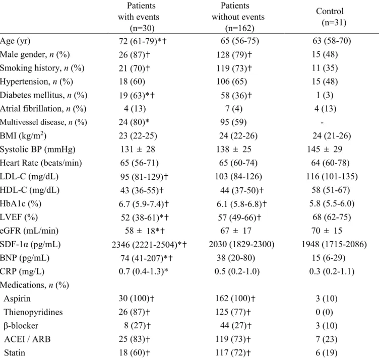
Table 1. Characteristics of study patients with a history of MI and control subjects. Patients with events (n=30) Patients without events (n=162) Control (n=31) Age (yr) 72 (61-79)*✝ 65 (56-75) 63 (58-70) Male gender, n (%) 26 (87)✝ 128 (79)✝ 15 (48) Smoking history, n (%) 21 (70)✝ 119 (73)✝ 11 (35) Hypertension, n (%) 18 (60) 106 (65) 15 (48) Diabetes mellitus, n (%) 19 (63)*✝ 58 (36)✝ 1 (3) Atrial fibrillation, n (%) 4 (13) 7 (4) 4 (13) Multivessel disease, n (%) 24 (80)* 95 (59) - BMI (kg/m2) 23 (22-25) 24 (22-26) 24 (21-26) Systolic BP (mmHg) 131 ± 28 138 ± 25 145 ± 29
Heart Rate (beats/min) 65 (56-71) 65 (60-74) 64 (60-78)
LDL-C (mg/dL) 95 (81-129)✝ 103 (84-126) 116 (101-135) HDL-C (mg/dL) 43 (36-55)✝ 44 (37-50)✝ 58 (51-67) HbA1c (%) 6.7 (5.9-7.4)✝ 6.1 (5.8-6.8)✝ 5.8 (5.5-6.0) LVEF (%) 52 (38-61)*✝ 57 (49-66)✝ 68 (62-75) eGFR (mL/min) 58 ± 18*✝ 67 ± 17 70 ± 15 SDF-1α (pg/mL) 2346 (2221-2504)*✝ 2030 (1829-2300) 1948 (1715-2086) BNP (pg/mL) 74 (41-207)*✝ 38 (20-80) 15 (6-29) CRP (mg/L) 0.7 (0.4-1.3)* 0.5 (0.2-1.0) 0.3 (0.2-1.1) Medications, n (%) Aspirin 30 (100)✝ 162 (100)✝ 3 (10) Thienopyridines 26 (87)✝ 125 (77)✝ 0 (0) β-blocker 8 (27)✝ 44 (27)✝ 3 (10) ACEI / ARB 25 (83)✝ 119 (73)✝ 7 (23) Statin 18 (60)✝ 117 (72)✝ 6 (19)
Data are expressed either as the mean value ± SD, median and range (25th and 75th percentile), or number (%) of patients. MI, myocardial infarction; BMI, body mass index; BP, blood pressure; LDL-C, low-density lipoprotein cholesterol; HDL-C, high-density lipoprotein cholesterol; HbA1c, hemoglobin A1c; LVEF, left ventricular ejection fraction; eGFR, estimated glomerular filtration rate; SDF-1α, stromal cell-derived factor-1α; BNP, brain natriuretic peptide; CRP, C-reactive protein; ACEI, angiotensin-converting enzyme inhibitor; ARB, angiotensin II receptor blocker.
* p < 0.05 vs. patients without events. ✝ p < 0.05 vs. control patients.
14
Table 2. Univariate and multivariate linear regression analysis of the correlations between SDF-1α and baseline clinical parameters in patients with a history of MI.
Univariate analysis Multivariate analysis
r p value β p value Age 0.179 0.01 0.043 0.59 Male gender - 0.004 0.95 - - Smoking 0.043 0.56 - - Hypertension 0.053 0.46 - - Diabetes mellitus - 0.010 0.16 - - Atrial fibrillation 0.138 0.06 - - Multivessel disease 0.010 0.89 - - BMI - 0.095 0.19 - - Systolic BP - 0.131 0.08 - - Heart rate 0.036 0.62 - - LDL-C - 0.014 0.85 - - HDL-C 0.007 0.93 - - HbA1c - 0.142 0.05 - - LVEF 0.084 0.25 - - eGFR - 0.214 0.003 - 0.124 0.12 BNP 0.346 < 0.0001 0.295 < 0.0001 CRP 0.149 0.04 0.088 0.20 Medications - - Thienopyridines 0.079 0.27 - - β-blocker 0.050 0.49 - - ACEI / ARB 0.005 0.94 - - Statin - 0.070 0.34 - -
The multivariate analysis consisted of covariates that demonstrated a significant correlation in the univariate analysis. r = regression coefficient. β = standardized regression coefficient. Aspirin was not included in this analysis because it was used by all patients. SDF-1α, stromal cell-derived factor-1α; MI, myocardial infarction; BMI, body mass index; BP, blood pressure; LDL-C, low-density lipoprotein cholesterol; HDL-C, high-density lipoprotein cholesterol; HbA1c, hemoglobin A1c; LVEF, left ventricular ejection fraction; eGFR, estimated glomerular filtration rate; BNP, brain natriuretic peptide; CRP, C-reactive protein; ACEI, angiotensin-converting enzyme inhibitor; ARB, angio-tensin II receptor blocker.
15
Table 3. Univariate and stepwise multivariate Cox hazard analysis of the risk factors for future cardiac events.
Univariate analysis Multivariate analysis HR 95% CI p value HR 95% CI p value Age 1.83 1.21-2.78 < 0.01 1.83 1.16-2.91 0.01 Male gender 1.57 0.55-4.49 0.40 Smoking 0.82 0.37-1.79 0.62 Hypertension 0.82 0.39-1.70 0.59 Diabetes mellitus 2.73 1.30-5.75 < 0.01 2.79 1.28-6.10 0.01 Atrial fibrillation 2.93 1.02-8.42 0.04 Multivessel disease 2.50 1.02-6.12 0.04 BMI 0.84 0.57-1.22 0.35 Systolic BP 0.78 0.53-1.13 0.19 Heart Rate 0.77 0.52-1.15 0.20 LDL-C 0.85 0.58-1.23 0.38 HDL-C 1.03 0.73-1.47 0.86 HbA1c 1.14 0.84-1.53 0.41 LVEF 0.66 0.47-0.94 0.02 0.69 0.50-0.97 0.03 eGFR 0.60 0.40-0.88 0.01 SDF-1α 1.87 1.35-2.60 < 0.001 1.98 1.38-2.85 < 0.001 BNP 1.37 1.17-1.60 < 0.001 CRP 1.25 0.96-1.62 0.10 Medications Thienopyridines 1.78 0.62-5.10 0.28 β-blocker 0.99 0.44-2.23 0.98 ACEI / ARB 1.55 0.59-4.04 0.37 Statin 0.66 0.32-1.37 0.27
The hazard ratios and 95% CI for continuous variables were estimated by a 1-SD increase. Dichotomous variables were coded as 1 for the presence of, and 0 for the absence of, each factor. BMI, body mass index; BP, blood pressure; LDL-C, low-density lipoprotein
cholesterol; HDL-C, high-density lipoprotein cholesterol; HbA1c, hemoglobin A1c; LVEF, left ventricular ejection fraction; eGFR, estimated glomerular filtration rate; SDF-1α, stromal cell-derived factor-1α; BNP, brain natriuretic peptide; CRP, C-reactive protein; ACEI,
16
Table 4. Incremental effects of SDF-1α levels on the predictive value of traditional risk
factors.
Category-free NRI IDI
NRI value p value IDI value p value
Traditional risk factors
+ BNP 0.44 0.03 0.03 0.31 + SDF-1α 0.77 0.0001 0.07 0.001 Traditional risks + BNP + SDF-1α 0.90 < 0.0001 0.05 0.002 Traditional risks + SDF-1α + BNP 0.10 0.62 0.004 0.69
NRI, net reclassification improvement; IDI, integrated discrimination improve-ment; BNP, brain natriuretic peptide; SDF-1α, stromal cell-derived factor-1α. The traditional risk factors consisted of age, male gender, diabetes mellitus, hypertension, smoking, low-density lipoprotein
cholesterol, high-density lipoprotein cholesterol, C-reactive protein, left ventricular ejection fraction, and multivessel disease.
17
Supplementary Table 1. Comparison of clinical variables between patients with high and low SDF-1α. High SDF-1α ( > 2162 pg/mL) (n=82) Low SDF-1α (< 2162 pg/mL) (n=110) Age (years) 70 (60-77) * 63 (54-73) Male gender, n (%) 65 (79) 89 (81) Smoking history, n (%) 59 (72) 81 (74) Hypertension, n (%) 55 (67) 69 (63) Diabetes mellitus, n (%) 34 (41) 43 (39) Atrial fibrillation, n (%) 8 (10) 3 (3) Multivessel disease, n (%) 51 (62) 68 (62) BMI (kg/m2) 23 (22-25) 24 (22-26) Systolic BP (mmHg) 134 ± 27 139 ± 24
Heart Rate (beats/min) 65 (60-74) 65 (59-73)
LDL-C (mg/dL) 97 (78-123) 106 (87-129) HDL-C (mg/dL) 44 (38-52) 44 (37-50) HbA1c (%) 6.1 (5.8-6.8) 6.2 (5.8-7.0) LVEF (%) 57 (45-64) 56 (49-66) eGFR (mL/min) 60 ± 18 * 69 ± 17 BNP (pg/mL) 66 (33-105) * 32 (17-72) CRP (mg/L) 0.6 (0.3-1.4) 0.5 (0.2-1.0) Medications, n (%) Aspirin 82 (100) 110 (100) Thienopyridines 66 (80) 85 (77) β-Blocker 25 (30) 27 (25) ACEI / ARB 64 (78) 80 (73) Statin 52 (63) 83 (75) PCI-related variables Pre-dilatation, n (%) 37 (45) 49 (45) Post-dilatation, n (%) 12 (15) 15 (14) BMS use, n (%) 42 (51) 51 (46) DES use, n (%) 16 (20) 23 (21)
Total stent length (mm) 20 (16-25) 18 (16-24)
Thrombus aspiration, n (%) 41 (50) 64 (58)
Data are expressed either as the mean value ± SD, median and range (25th and 75th percentile), or number (%) of patients. The cut-off value of SDF-1α levels as 2162 pg/mL
18
was determined by ROC analysis.SDF-1α, stromal cell-derived factor-1α;BMI, body mass index; BP, blood pressure; LDL-C, low-density lipoprotein cholesterol; HDL-C, high-density lipoprotein cholesterol; HbA1c, hemoglobin A1c; LVEF, left ventricular ejection fraction; eGFR, estimated glomerular filtration rate;BNP, brain natriuretic peptide; CRP, C-reactive protein;ACEI, angiotensin-converting enzyme inhibitor; ARB, angiotensin II receptor blocker; PCI, percutaneous coronary intervention that was performed in the culprit coronary lesion at the occurrence of MI; BMS, bare metal stent; DES, drug-eluting stent.
19
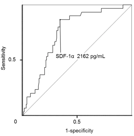
Fig. 1. Receiver operator characteristic curve to obtain optimal cut-off level of stromal cell-derived factor-1α (SDF-1α) for the prediction of cardiac events.
20
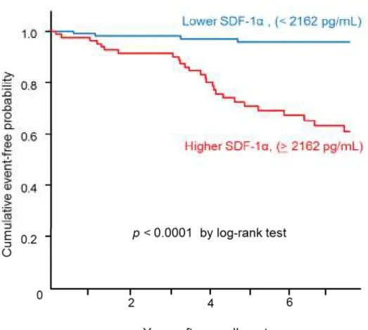
Fig. 2. Kaplan–Meier analysis according to stromal cell-derived factor-1α (SDF-1α) levels. The cut-off value of SDF-1α (2162 pg/mL) was determined by receiver-operating
characteristic analysis in the study patients. High levels of SDF-1α (>2162 pg/mL, n = 82) resulted in higher probability of future cardiac events than the lower one (<2162 pg/mL, n = 110) (p < 0.0001).
21 References
[1] Frangogiannis NG. Regulation of the inflammatory response in cardiac repair. Circ Res
2012;110:159–73.
[2] Nahrendorf M, Pittet MJ, Swirski FK. Monocytes: protagonists of infarct inflammation and
repair after myocardial infarction. Circulation 2010;121: 2437–45.
[3] Nikolaos G, Frangogiannis Dewald O, Ren G, Duerr GD, Zoerlein M, Klemm C, Gersch C,
Tincey S, Michael LH, Entman ML, Frangogiannis NG. Of mice and dogs: species-specific
differences in the inflammatory response following myocardial infarction. Am J Pathol
2004;164:665–77.
[4] Pfeffer MA, Braunwald E. Ventricular remodeling after myocardial infarction. Experimental
observations and clinical implications. Circulation 1990;81: 1161–72.
[5] White HD, Norris RM, Brown MA, Brandt PW, Whitlock RM, Wild CJ. Left ventricular
end-systolic volume as the major determinant of survival after recovery from myocardial
infarction. Circulation 1987;76:44–51.
[6] Peled A, Petit I, Kollet O, Magid M, Ponomaryov T, Byk T, Nagler A, Ben-Hur H, Many A,
Shultz L, Lider O, Alon R, Zipori D, Lapidot T. Dependence of human stem cell engraftment
and repopulation of NOD/SCID mice on CXCR4. Science 1999;283:845–8.
[7] Uematsu M, Yoshizaki T, Shimizu T, Obata JE, Nakamura T, Fujioka D, Watanabe K,
Watanabe Y, Kugiyama K. Sustained myocardial production of stromal cell-derived factor-1α
was associated with left ventricular adverse remodelling in patients with myocardial infarction.
Am J Physiol Heart Circ Physiol 2015;309:H1764–71.
[8] Fortunato O, Spinetti G, Specchia C, Cangiano E, Valgimigli M, Madeddu P. Migratory
activity of circulating progenitor cells and serum SDF-1α predict adverse events in patients with
22
[9] Tong G, Wang N, Zhou Y, Leng J, Gao W, Tong X, Shen Y, Yang J, Ye X, Zhou L, Gao Y.
Role of stromal cell-derived factor-1 in patients with non-ST elevation acute coronary
syndrome. Int Heart J 2014;55:219–27.
[10] Kugiyama K, Motoyama T, Hirashima O, Ohgushi M, Soejima H, Misumi K, Kawano H,
Miyao Y, Yoshimura M, Ogawa H, Matsumura T, Sugiyama S, Yasue H. Vitamin C attenuates
abnormal vasomotor reactivity in spasm coronary arteries in patients with coronary spastic
angina. J Am Coll Cardiol 1998;32: 103–9.
[11] Braunwald E, Antman EM, Beasley JW, Califf RM, Cheitlin MD, Hochman JS, Jones RH,
Kereiakes D, Kupersmith J, Levin TN, Pepine CJ, Schaeffer JW, Smith 3rd EE, Steward DE,
Theroux P, et al. ACC/AHA guidelines for the management of patients with unstable angina
and non-ST-segment elevation myocardial infarction: executive summary and
recommendations. A report of the American College of Cardiology/American Heart
Association task force on practice guidelines (committee on the management of patients with
unstable angina). Circulation 2000;102:1193–209.
[12] Third Report of the National Cholesterol Education Program (NCEP) Expert Panel on
Detection, Evaluation, and Treatment of High Blood Cholesterol in Adults (Adult Treatment
Panel III) final report. Circulation 2002;106:3143–421.
[13] Teichholz LE, Kreulen T, Herman MV, Gorlin R. Problems in echocardiographic volume
determinations: echocardiographic-angiographic correlations in the presence of absence of
asynergy. Am J Cardiol 1976;37:7–11.
[14] Norimatsu K, Miura S, Suematsu Y, Shiga Y, Miyase Y, Nakamura A, Yamada M,
Matsunaga A, Saku K. Associations between glycated albumin or hemo-globin A1c and the
presence of coronary artery disease. Int J Cardiol 2015;65:487–93.
23 Care 2006;29(Suppl. 1):S4–2.
[16] Ikeda N, Hara H, Hiroi Y. 1,5-Anhydro-D-glucitol predicts coronary artery disease
prevalence and complexity. Int J Cardiol 2014;64:297–301.
[17] Makino A, Nakamura T, Hirano M, Kitta Y, Sano K, Kobayashi T, Fujioka D, Saito Y,
Watanabe K, Watanabe Y, Kawabata K, Obata JE, Kugiyama K. High plasma levels of
macrophage migration inhibitory factor are associated with adverse long-term outcome in
patients with stable coronary artery disease and impaired glucose tolerance or type 2 diabetes
mellitus. Atherosclerosis 2010;213:573–8.
[18] Abbott JD, Huang Y, Liu D, Hickey R, Krause DS, Giordano FJ. Stromal cellderived
factor-1α plays a critical role in stem cell recruitment to the heart after myocardial infarction
but is not sufficient to induce homing in the absence of injury. Circulation 2004;110:3300–5.
[19] Askari AT, Unzek S, Popovic ZB, Goldman CK, Forudi F, Kiedrowski M, Rovner A, Ellis
SG, Thomas JD, DiCorleto PE, Topol EJ, Penn MS. Effect of stromal-cellderived factor 1 on
stem-cell homing and tissue regeneration in ischaemic cardiomyopathy. Lancet 2003;362:697–
703.
[20] Elmadbouh I, Haider HKh, Jiang S, Idris NM, Lu G, Ashraf M. Ex vivo delivered stromal
cell-derived factor-1α promotes stem cell homing and induces angiomyogenesis in the infarcted
myocardium. J Mol Cell Cardiol 2007;42: 792–803.
[21] Frangogiannis NG. The stromal cell-derived factor-1/CXCR4 axis in cardiac injury and
repair. J Am Coll Cardiol 2011;58:2424–6.
[22] Saxena A, Fish JE, White MD, Yu S, Smyth JW, Shaw RM, DiMaio JM, Srivastava D.
Stromal cell-derived factor-1α is cardioprotective after myocardial infarction. Circulation
2008;117:2224–31.
24
Tarzami ST. Effects of CXCR4 gene transfer on cardiac function after ischemiareperfusion
injury. Am J Pathol 2010;176:1705–15.
[24] Jujo K, Hamada H, Iwakura A, Thorne T, Sekiguchi H, Clarke T, Ito A, Misener S, Tanaka
T, Klyachko E, Kobayashi K, Tongers J, Roncalli J, Tsurumi Y, Hagiwara N, et al. CXCR4
blockade augments bone marrow progenitor cell recruitment to the neovasculature and reduces
mortality after myocardial infarction. Proc Natl Acad Sci U S A 2010;107:11008–13.
[25] Liehn EA, Tuchscheerer N, Kanzler I, Drechsler M, Fraemohs L, Schuh A, Koenen RR,
Zander S, Soehnlein O, Hristov M, Grigorescu G, Urs AO, Leabu M, Bucur I, Merx MW, et al.
Double-edged role of the CXCL12/CXCR4 axis in experimental myocardial infarction. J Am
Coll Cardiol 2011;58:2415–23.
[26] Proulx C, El-Helou V, Gosselin H, Clement R, Gillis MA, Villeneuve L, Calderone A.
Antagonism of stromal cell-derived factor-1α reduces infarct size and improves ventricular
function after myocardial infarction. Pflugers Arch 2007;455:241–50.
[27] Ceradini DJ, Kulkarni AR, Callaghan MJ, Tepper OM, Bastidas N, Kleinman ME, Capla
JM, Galiano RD, Levine JP, Gurtner GC. Progenitor cell trafficking is regulated by hypoxic
gradients through HIF-1 induction of SDF-1. Nat Med 2004;10:858–64.
[28] Jin C, Fu WX, Xie LP, Qian XP, Chen WF. SDF-1α production is negatively regulated by
mouse estrogen enhanced transcript in a mouse thymus epithelial cell line. Cell Immunol
2003;223:26–34.
[29] Hohensinner PJ, Kaun C, Rychli K, Niessner A, Pfaffenberger S, Rega G, Furnkranz A,
Uhrin P, Zaujec J, Afonyushkin T, Bochkov VN, Maurer G, Huber K, Wojta J. The
inflammatory mediator oncostatin M induces stromal derived factor-1 in human adult cardiac
cells. FASEB J 2009;23:774–82.
25
myocardial infarction. J Geriatr Cardiol 2015;12:305–12.
[31] Libby P, Nahrendorf M, Swirski FK. Leukocytes link local and systemic inflammation in
ischemic cardiovascular disease: an expanded cardiovascular continuum. J Am Coll Cardiol
2016;67:1091–103.
[32] Rouleau JL, de Champlain J, Klein M, Bichet D, Moye´ L, Packer M, Dagenais GR, Sussex
B, Arnold JM, Sestier F, et al. Activation of neurohumoral systems in postinfarction left
ventricular dysfunction. J Am Coll Cardiol 1993;22:390–8.
[33] Sigurdsson A, Held P, Swedberg K. Short- and long-term neurohormonal activation
following acute myocardial infarction. Am Heart J 1993;126: 1068–76.
[34] Nabah YN, Mateo T, Estelle´ s R, Mata M, Zagorski J, Sarau H, Cortijo J, Morcillo EJ,
Jose PJ, Sanz MJ. Angiotensin II induces neutrophil accumulation in vivo through generation
1
High levels of stromal cell-derived factor-1α predict secondary cardiac events in stable patients with a history of myocardial infarction
Satoshi Matsuoka (MD)a, Manabu Uematsu (MD)a,Takamitsu Nakamura (MD, PhD)a, Takuya Shimizu (MD)a, Mika Futamata (MD)a, Jun-ei Obata (MD, PhD)a, Daisuke Fujioka (MD, PhD)a, Kazuto Nakamura (MD, PhD)a, Toru Yoshizaki (MD)a, Kiyotaka Kugiyama
(MD, PhD, FJCC)a
a Department of Internal Medicine II, University of Yamanashi, Faculty of Medicine, Chuo,
Yamanashi, Japan
Key words: stromal cell-derived factor-1α, myocardial infarction, prognosis, cardiac events, inflammatory marker.
This paper was published in the following journal: Journal of Cardiology, 69(1),
2 Abstract
Background: We recently showed that stromal cell-derived factor (SDF)-1α, a
proinflammatory mediator, is produced in infarcted myocardium and is associated with left
ventricular (LV) adverse remodeling and progressive dysfunction following acute myocardial
infarction (AMI). The current study examined whether SDF-1α levels in the peripheral vein
can provide prognostic information of outcomes in stable patients with a history of MI.
Methods: Plasma levels of SDF-1α in the peripheral vein were measured by enzyme-linked
immunosorbent assay in 192 stable patients with a history of MI. All patients were followed
prospectively for a period of 90 months or until occurrence of one of the following cardiac
events: cardiac death, non-fatal myocardial infarction, unstable angina requiring unplanned
coronary revascularization, or worsening heart failure requiring hospital admission.
Results: During the follow-up period (77 ± 26 months), 30 patients had cardiac events.
Multivariate Cox analysis revealed that high levels of SDF-1α ( >2162 pg/mL; a cut-off value
determined by receiver-operating characteristic analysis) were a significant predictor of
cardiac events, independent of traditional risk factors (HR: 1.98; 95% CI: 1.38–2.85; p <
0.001). The addition of high levels of SDF-1α to conventional risk factors including brain
natriuretic peptide improved net reclassification improvement (NRI) and integrated
discrimination improvement (IDI) (NRI 0.90, p < 0.000-1αnd IDI 0.05, p = 0.002).
Conclusions: High levels of SDF-1α predicted secondary cardiac events in stable patients
with a history of MI. SDF-1α levels may be a useful risk assessment tool in patients with a
3 Introduction
Acute myocardial infarction (AMI) induces the production of chemokines and
growth factors that recruit neutrophils and phagocytes to the ischemic cardiac tissue, leading
to clearance of dead cells and matrix debris [1,2]. This acute, localized inflammatory response
is transient, and is followed by resolution of the inflammation [1,3]. However, prolonged
inflammation leads to dilative remodeling and progressive dysfunction of the infarcted
myocardium through matrix degradation and cardiomyocyte apoptosis [1,2]. Left ventricle
(LV) adverse remodeling and dysfunction after MI are predictors of poor outcome following
MI, and are associated with ventricular arrhythmias, heart failure, and increased mortality
[4,5]. Stromal cell-derived factor (SDF)-1α/CXCL12 is a CXC chemokine with chemotactic
effects on CXCR4-expressing progenitor cells [6]. We have recently shown that SDF-1α, a
proinflammatory mediator, is produced in the infarcted myocardial lesion and released into
the circulation in patients with AMI [7]. In addition, we showed that the myocardial
production of SDF-1α in the chronic phase (6 months after MI) but not in the early phase (2
weeks after MI) was associated with LV adverse remodeling and progressive dysfunction in
patients with AMI [7]. Thus, prolonged production of SDF-1α in the infarcted myocardium
may exert detrimental effects on post-MI LV function in the chronic phase following MI. The
prognostic value of SDF-1α levels in the peripheral vein was previously evaluated in patients
with AMI/ acute coronary syndrome (ACS) [8,9]. However, it remains unclear whether
SDF-1α levels can provide prognostic information in patients in the chronic phase of MI. The
current study examined the prognostic value of SDF-1α levels in the peripheral vein in stable
4 Methods Study patients
The study screened 386 patients with a history of MI, who underwent cardiac
catheterization in Yamanashi University Hospital between February 2003 and March 2009.
All patients underwent routine blood testing at the time of discharge. The inclusion criteria
were: (1) stable previous MI; (2) no episode of angina at rest and no changes in the frequency
of angina in response to sublingual nitroglycerin in the previous 2 months. Exclusion criteria
included: (1) ACS, stroke, cardiogenic shock, pulmonary edema, major surgery, trauma or
serious infectious disease within 4 weeks prior to enrollment; (2) neoplasm, significant
hepatic or inflammatory disease; (3) chronic renal failure or serum creatinine >2.5 mg/dL,
congestive heart failure, or left main coronary artery disease; (4) other serious diseases.
Finally, a total of 210 patients were enrolled in the study according to these inclusion and
exclusion criteria. The study also included 31 control patients without echocardiographic
findings of significant valvular disease (moderate or more regurgitation, stenosis),
cardiomyopathy [chamber size dilatation, reduced left ventricular ejection fraction (LVEF),
ventricular asynergy, ventricular hypertrophy, aneurysm], and pulmonary hypertension,
chosen from among the 6-1αngiographically normal patients [without percutaneous coronary
intervention (PCI) history] who were evaluated during the study period. Control patients
served as a reference group for plasma SDF-1α concentrations. Each of the control patients
underwent diagnostic coronary angiography for atypical chest pain at rest at Yamanashi
University Hospital during the study period. Control patients fulfilled all of the following
inclusion criteria: (1) no significant ST segment changes on 12-lead electrocardiogram (ECG)
while having chest pain or on ambulatory ECG; (2) neither chest pain nor ST segment
intra-5
coronary infusion of acetylcholine [10]. Clinical characteristics of patients with a history of
MI and control patients are shown in Table -1αll study participants were ethnic Japanese. All
patients gave written, informed consent at the time of enrollment. The study was approved by
the ethics committee of Yamanashi University Hospital. The investigation conformed to the
principles outlined in the 1975 Declaration of Helsinki.
Prospective study
Patients were followed every month in the hospital or with a clinic visit for 90 months, or
until the occurrence of cardiac death, non-fatal MI, refractory unstable angina pectoris (uAP)
requiring unplanned coronary revascularization, or decompensated heart failure. The time to
the first event was evaluated prospectively. Cardiac death was confirmed by hospital records.
Acute MI and uAP were diagnosed by the presence of acute ischemic symptoms lasting >20
min within 48 h prior to hospital admission, and ECG changes. Acute MI was diagnosed
when creatine kinase-MB levels increased to at least 2 times the upper limit of normal or
when troponin T levels were >0.1 ng/mL [11]. The diagnoses of MI and uAP were confirmed
by coronary angiography. Decompensated heart failure was defined as resting dyspnea with
progressive fluid retention requiring hospitalization and treatment with an intravenous
diuretic. Follow-up data were collected from the patients’ primary physicians every 3 months
by two blinded investigators (T.N., J.O.). During the follow-up period, standard medications
were prescribed to all patients according to the guidelines of the American Heart Association
[12], as shown in Table 1. Diet and lifestyle recommendations were continued throughout the
follow-up period.
6
Venous blood was obtained from all patients on the morning of discharge after a 12-h
overnight fast. The initial volumes of each blood sample, including those forcibly drawn,
were discarded. Serum and EDTA-plasma were aliquoted and stored at –80℃ until time of
analysis. Plasma SDF-1α levels were measured by enzyme-linked immunosorbent assay using
a commercial kit (R & D Systems, Minneapolis, MN, USA). In our laboratory, the intra- and
inter-assay coefficients of variation were 1.9% and 3.5%, respectively. The minimal detection
limit of this assay was 18 pg/mL. Serum C-reactive protein (CRP) levels were assayed by rate
nephelometry (Dade Behring, Tokyo, Japan). Plasma levels of brain natriuretic peptide (BNP)
were measured by immunoradiometric assay (Shionogi Pharmaceutical, Osaka, Japan).
Estimated glomerular filtration rate (eGFR) was calculated by the Modification of Diet in
Renal Disease study equation. Echocardiographic LVEF was calculated by the motion-mode
method using the Teichholz formula [13].
Statistical analysis
All descriptive data were expressed as mean ± SD, median, or frequency (%). The
Shapiro–Wilk test showed that age, body mass index (BMI), heart rate, low-density
lipoprotein cholesterol (LDL-C), high-density lipoprotein cholesterol (HDL-C), hemoglobin
A1c (HbA1c), LVEF, SDF-1α, BNP, and CRP were not normally distributed, hence, these
variables were expressed as the median and inter-quartile ranges (25th and 75th percentiles).
Non-normally distributed variables were log-transformed before the analysis. Continuous
variables were compared between the two groups using the unpaired t-test or the Mann–
Whitney U-test, as appropriate. Frequencies were compared using the chi-square test. For
comparisons of the mean value of >3 groups, one-way analysis of variance (ANOVA)
7
the two groups was examined using univariate and multivariate linear regression analyses.
Kaplan–Meier analysis was performed on the 2 groups and was based on a cut-off SDF-1α
level. The cut-off level of SDF-1α (2162 pg/mL) was determined by receiver-operating
characteristic (ROC) analyses of SDF-1α in the study patients with a history of MI (Fig. 1).
The predictive values were assessed by univariate or forward stepwise multivariate Cox
proportional hazards analysis. For a forward stepwise multivariate Cox hazard analysis, the
significance level was set at 0.1. In the univariate and multivariate Cox proportional hazards
analyses, continuous variables were estimated for 1-SD change. Dichotomous variables were
coded as 1 for the presence of, and 0 for the absence of, each factor. The following factors
were included as dichotomous variables: systemic hypertension (>140/90 mmHg or use of an
antihypertensive medication) [14], diabetes mellitus (DM) (as defined by the American
Diabetes Association or use of an antidiabetic medication) [15,16], smoking history (defined
as smoking >10 cigarettes/day for >10 years), atrial fibrillation (including paroxysmal atrial
fibrillation recorded with an ECG), and multivessel disease. The Cox models included only
variables that showed proportionality in a Schoenfeld residuals test. The category-free net
reclassification improvement (NRI) and the integrated discrimination improvement (IDI)
were used to examine the additive effects of SDF-1α and BNP levels on the predictive value
of the baseline model that consisted of conventional cardiovascular risk factors. Conventional
risk factors included age, male gender, DM, hypertension, smoking, levels of LDL-C,
HDL-C, CRP, LVEF, and multivessel disease. All probability values were presented as 2-tailed with
statistical significance inferred at p < 0.05. All confidence intervals were computed at the 95%
level. Statistical analyses were performed using STATA 10.0 (StataCorp, College Station, TX,
8 Power analysis
Our previous study showed that the composite endpoints occurred in approximately
52% of stable coronary artery disease (CAD) patients with high coronary risk factors and
higher macrophage inhibitory factor (MIF) levels as an inflammatory marker, and in 18% of
patients with lower MIF levels during 5 years of follow-up [17]. On the basis of that study, a
total of 72 patients were required for a two-sided statistical analysis with sufficient statistical
power of 0.90 (b = 0.10 and a = 0.05). This justified the number of patients (n = 220) included
in the current prospective study.
Results
Over the course of the study, 7 patients were lost to follow up, and 11 patients were
excluded due to non-cardiac related death during follow-up. A total of 192 patients completed
the follow-up study (2–90 months, mean = 77 ± 26 months). During the follow-up period, 30
(15.6%) cardiac events occurred, including 5 cardiac deaths, 1 non-fatal MI, 12 uAPs
requiring unplanned coronary revascularization, and 12 episodes of worsening heart failure.
Patient characteristics are shown in Table 1.
Patients and SDF-1α levels
The time between blood sampling for SDF-1α level and the previous MI ranged from
3 to 26 months, with a mean of 7.7 ± 2.1 months. The time between onset of previous MI and
blood sampling did not correlate with SDF-1α levels (r = –0.08, p = 0.28). As shown in Table
2, SDF-1α levels were positively correlated with age, BNP, and CRP but inversely correlated
with eGFR in the univariate linear regression analysis (Table 2). SDF-1α levels did not
9
Prevalence of PCI-related variables was similar between patients with high and low SDF-1α
levels (Supplementary Table 1).
Prospective study
Patients (n = 82) with higher SDF-1α levels (>2162 pg/mL, a cut-off value
determined by ROC analysis) had 26 events during follow-up, whereas patients (n = 110) with
lower SDF-1α levels (<2162 mg/dL) had 4 events (p < 0.01). The cut-off value of SDF-1α
levels as 2162 pg/mL, determined by ROC analysis, provided sensitivity of 86.7%, specificity
of 65.4%, and accuracy of 68.8% for prediction of future events (Fig. 1). Kaplan–Meier
analysis demonstrated a significantly higher probability of a cardiac event in patients with
higher SDF-1α levels compared with those with lower SDF-1α levels (Fig. 2). A univariate
Cox proportional hazards analysis revealed that SDF-1α levels (HR 1.87; 95% CI 1.35–2.60),
age (HR 1.83; 95% CI 1.21–2.78), DM (HR 2.73; 95% CI 1.30–5.75), atrial fibrillation (HR
2.93; 95% CI 1.02–8.42), multivessel disease (HR 2.50; 95% CI 1.02–6.12), LVEF (HR 0.66;
95% CI 0.47–0.94), eGFR (HR 0.60; 95% CI 0.40–0.88), and BNP levels (HR 1.37; 95% CI
1.17–1.60) were significantly associated with future cardiac events, as shown in Table 3. A
forward stepwise multivariate Cox proportional hazards analysis demonstrated that SDF-1α
levels remained significantly associated with future events after adjustment for potential
confounding variables (HR 1.98; 95% CI 1.38–2.85).
Incremental effects of SDF-1α levels on the predictive value of traditional risk factors Category-free NRI and IDI demonstrated the additive value of SDF-1α to the
baseline conventional risk factors consisting of age, male gender, DM, hypertension, smoking,
10
IDI 0.07, p = 0.001) (Table 4). When BNP was added to the baseline model of risk factors,
SDF-1α had significant additive effect on the predictive value of the model (NRI 0.90, p <
0.0001; IDI 0.05, p = 0.002) (Table 4). In contrast, BNP had no significant additive effect on
the predictive value of a risk factor model with SDF-1α (Table 4).
Discussion
The present study showed that high SDF-1α levels are a predictor of adverse
outcomes in patients with a history of MI. Moreover, high SDF-1α levels had incremental
effects on prognostic value of traditional risk factors in this patient population. Thus, SDF-1α
levels may aid in risk assessment in stable patients with a history of MI. We have previously
shown that SDF-1α is released from the infarcted myocardium into the coronary circulation in
both the early (2 weeks after MI) and chronic phases (6 months after MI) following AMI [7].
The myocardial production and release of SDF-1α in the chronic phase of MI, rather than in
the early phase, was associated with post-MI LV adverse remodeling and progressive
dysfunction [7]. These results support the present finding that SDF-1α levels can predict
secondary cardiac events in patients with a history of MI. Animal experiments have
demonstrated that local infusion or over-expression of SDF-1α attenuates myocardial
ischemic injury following LV dysfunction [18–22]. In contrast, other experimental studies
have shown that SDF-1α/CXCR4 signaling worsens post-MI LV function due to increased
inflammatory injury and apoptotic mechanisms [23–26]. Thus, the effect of SDF-1α on
post-MI LV function in animal models remains unclear. In animal models, the effects of
endogenous SDF-1α on post-MI cardiac tissue may be balanced between angiogenic/
reparative and proinflammatory actions of SDF-1α/CXCR4 signaling [21]. In humans, our
11
myocardium and in the peripheral vein might exert detrimental effects on post-MI cardiac
tissue.
Previous clinical reports have demonstrated that, when SDF-1α levels were measured
in the acute phase of ischemic events, high SDF-1α levels in the peripheral vein correlated
with recurrent cardiac events in patients with AMI and non-ST elevation ACS [8,9]. However,
our previous study showed that myocardial production of SDF-1α in the early phase of AMI
did not reflect post-MI LV adverse remodeling and dysfunction [7]. These results suggest that
SDF-1α in the infarcted myocardium and in the peripheral vein might reflect different tissues
of origin. That is, SDF-1α levels in the peripheral vein during the early phase of cardiac
ischemic events may not originate from the infarcted myocardium. SDF-1α is produced in the
ischemic myocardium through induction of hypoxia-inducible factor [27]. Also, SDF-1α may
be induced by proinflammatory stimuli in various systemic tissues [28,29] as systemic
activation of proinflammatory responses occurs in the chronic phase of MI [30,31]. However,
the precise source of SDF-1α in the peripheral vein remains undefined. A previous report
showed that SDF-1α levels in the peripheral vein increased during the acute phase of MI [9].
In the present stable patients with a history of MI, SDF-1α levels were higher in the patients
with future secondary cardiac events than in control subjects. The mechanism by which high
SDF-1α levels were sustained in the peripheral vein during the chronic phase of MI is unclear.
A systemic inflammatory response occurs during the acute phase of MI, but is resolved in the
chronic phase [1,3]. The mechanisms leading to resolution, including inhibition of
proinflammatory cytokine synthesis [1], may be impaired in the patients who have recurrent
cardiac events. For example, post-MI LV dysfunction might induce systemic activation of the
renin– angiotensin–aldosterone system, leading to prolonged proinflammatory responses in
12
persistent LV adverse remodeling and dysfunction after MI. In support of this, we observed
that SDF-1α levels positively correlated with BNP levels in the present study. In the present
study, BNP did not have a significant predictive value in the multivariate Cox proportional
hazard analysis. This was explained by the strong correlation of BNP with LVEF which was
included in the co-variables of the multivariate Cox hazard analysis. The present study
included a relatively small number of patients evaluated at a single center. The present results
could not be generalized in other cohorts including patients with acute coronary syndrome or
heart failure. A large prospective trial is required to understand the precise role of SDF-1α in
the pathogenesis of chronic phase of MI. In conclusion, high levels of SDF-1α in the
peripheral vein predicted secondary cardiac events in stable patients with a history of MI. The
high SDF-1α levels had incremental effects on the prognostic value of the traditional risk
factors in this patient population. Hence, SDF-1α levels may be a useful risk assessment tool
in patients with a history of MI.
Funding
This study was supported by JSPS KAKENHI Grant Number B2- 19390209 and
B-22390158.
Disclosures
13
Table 1. Characteristics of study patients with a history of MI and control subjects. Patients with events (n=30) Patients without events (n=162) Control (n=31) Age (yr) 72 (61-79)*✝ 65 (56-75) 63 (58-70) Male gender, n (%) 26 (87)✝ 128 (79)✝ 15 (48) Smoking history, n (%) 21 (70)✝ 119 (73)✝ 11 (35) Hypertension, n (%) 18 (60) 106 (65) 15 (48) Diabetes mellitus, n (%) 19 (63)*✝ 58 (36)✝ 1 (3) Atrial fibrillation, n (%) 4 (13) 7 (4) 4 (13) Multivessel disease, n (%) 24 (80)* 95 (59) - BMI (kg/m2) 23 (22-25) 24 (22-26) 24 (21-26) Systolic BP (mmHg) 131 ± 28 138 ± 25 145 ± 29
Heart Rate (beats/min) 65 (56-71) 65 (60-74) 64 (60-78)
LDL-C (mg/dL) 95 (81-129)✝ 103 (84-126) 116 (101-135) HDL-C (mg/dL) 43 (36-55)✝ 44 (37-50)✝ 58 (51-67) HbA1c (%) 6.7 (5.9-7.4)✝ 6.1 (5.8-6.8)✝ 5.8 (5.5-6.0) LVEF (%) 52 (38-61)*✝ 57 (49-66)✝ 68 (62-75) eGFR (mL/min) 58 ± 18*✝ 67 ± 17 70 ± 15 SDF-1α (pg/mL) 2346 (2221-2504)*✝ 2030 (1829-2300) 1948 (1715-2086) BNP (pg/mL) 74 (41-207)*✝ 38 (20-80) 15 (6-29) CRP (mg/L) 0.7 (0.4-1.3)* 0.5 (0.2-1.0) 0.3 (0.2-1.1) Medications, n (%) Aspirin 30 (100)✝ 162 (100)✝ 3 (10) Thienopyridines 26 (87)✝ 125 (77)✝ 0 (0) β-blocker 8 (27)✝ 44 (27)✝ 3 (10) ACEI / ARB 25 (83)✝ 119 (73)✝ 7 (23) Statin 18 (60)✝ 117 (72)✝ 6 (19)
Data are expressed either as the mean value ± SD, median and range (25th and 75th percentile), or number (%) of patients. MI, myocardial infarction; BMI, body mass index; BP, blood pressure; LDL-C, low-density lipoprotein cholesterol; HDL-C, high-density lipoprotein cholesterol; HbA1c, hemoglobin A1c; LVEF, left ventricular ejection fraction; eGFR, estimated glomerular filtration rate; SDF-1α, stromal cell-derived factor-1α; BNP, brain natriuretic peptide; CRP, C-reactive protein; ACEI, angiotensin-converting enzyme inhibitor; ARB, angiotensin II receptor blocker.
* p < 0.05 vs. patients without events. ✝ p < 0.05 vs. control patients.
14
Table 2. Univariate and multivariate linear regression analysis of the correlations between SDF-1α and baseline clinical parameters in patients with a history of MI.
Univariate analysis Multivariate analysis
r p value β p value Age 0.179 0.01 0.043 0.59 Male gender - 0.004 0.95 - - Smoking 0.043 0.56 - - Hypertension 0.053 0.46 - - Diabetes mellitus - 0.010 0.16 - - Atrial fibrillation 0.138 0.06 - - Multivessel disease 0.010 0.89 - - BMI - 0.095 0.19 - - Systolic BP - 0.131 0.08 - - Heart rate 0.036 0.62 - - LDL-C - 0.014 0.85 - - HDL-C 0.007 0.93 - - HbA1c - 0.142 0.05 - - LVEF 0.084 0.25 - - eGFR - 0.214 0.003 - 0.124 0.12 BNP 0.346 < 0.0001 0.295 < 0.0001 CRP 0.149 0.04 0.088 0.20 Medications - - Thienopyridines 0.079 0.27 - - β-blocker 0.050 0.49 - - ACEI / ARB 0.005 0.94 - - Statin - 0.070 0.34 - -
The multivariate analysis consisted of covariates that demonstrated a significant correlation in the univariate analysis. r = regression coefficient. β = standardized regression coefficient. Aspirin was not included in this analysis because it was used by all patients. SDF-1α, stromal cell-derived factor-1α; MI, myocardial infarction; BMI, body mass index; BP, blood pressure; LDL-C, low-density lipoprotein cholesterol; HDL-C, high-density lipoprotein cholesterol; HbA1c, hemoglobin A1c; LVEF, left ventricular ejection fraction; eGFR, estimated glomerular filtration rate; BNP, brain natriuretic peptide; CRP, C-reactive protein; ACEI, angiotensin-converting enzyme inhibitor; ARB, angio-tensin II receptor blocker.
15
Table 3. Univariate and stepwise multivariate Cox hazard analysis of the risk factors for future cardiac events.
Univariate analysis Multivariate analysis HR 95% CI p value HR 95% CI p value Age 1.83 1.21-2.78 < 0.01 1.83 1.16-2.91 0.01 Male gender 1.57 0.55-4.49 0.40 Smoking 0.82 0.37-1.79 0.62 Hypertension 0.82 0.39-1.70 0.59 Diabetes mellitus 2.73 1.30-5.75 < 0.01 2.79 1.28-6.10 0.01 Atrial fibrillation 2.93 1.02-8.42 0.04 Multivessel disease 2.50 1.02-6.12 0.04 BMI 0.84 0.57-1.22 0.35 Systolic BP 0.78 0.53-1.13 0.19 Heart Rate 0.77 0.52-1.15 0.20 LDL-C 0.85 0.58-1.23 0.38 HDL-C 1.03 0.73-1.47 0.86 HbA1c 1.14 0.84-1.53 0.41 LVEF 0.66 0.47-0.94 0.02 0.69 0.50-0.97 0.03 eGFR 0.60 0.40-0.88 0.01 SDF-1α 1.87 1.35-2.60 < 0.001 1.98 1.38-2.85 < 0.001 BNP 1.37 1.17-1.60 < 0.001 CRP 1.25 0.96-1.62 0.10 Medications Thienopyridines 1.78 0.62-5.10 0.28 β-blocker 0.99 0.44-2.23 0.98 ACEI / ARB 1.55 0.59-4.04 0.37 Statin 0.66 0.32-1.37 0.27
The hazard ratios and 95% CI for continuous variables were estimated by a 1-SD increase. Dichotomous variables were coded as 1 for the presence of, and 0 for the absence of, each factor. BMI, body mass index; BP, blood pressure; LDL-C, low-density lipoprotein
cholesterol; HDL-C, high-density lipoprotein cholesterol; HbA1c, hemoglobin A1c; LVEF, left ventricular ejection fraction; eGFR, estimated glomerular filtration rate; SDF-1α, stromal cell-derived factor-1α; BNP, brain natriuretic peptide; CRP, C-reactive protein; ACEI,
16
Table 4. Incremental effects of SDF-1α levels on the predictive value of traditional risk
factors.
Category-free NRI IDI
NRI value p value IDI value p value
Traditional risk factors
+ BNP 0.44 0.03 0.03 0.31 + SDF-1α 0.77 0.0001 0.07 0.001 Traditional risks + BNP + SDF-1α 0.90 < 0.0001 0.05 0.002 Traditional risks + SDF-1α + BNP 0.10 0.62 0.004 0.69
NRI, net reclassification improvement; IDI, integrated discrimination improve-ment; BNP, brain natriuretic peptide; SDF-1α, stromal cell-derived factor-1α. The traditional risk factors consisted of age, male gender, diabetes mellitus, hypertension, smoking, low-density lipoprotein
cholesterol, high-density lipoprotein cholesterol, C-reactive protein, left ventricular ejection fraction, and multivessel disease.
17
Supplementary Table 1. Comparison of clinical variables between patients with high and low SDF-1α. High SDF-1α ( > 2162 pg/mL) (n=82) Low SDF-1α (< 2162 pg/mL) (n=110) Age (years) 70 (60-77) * 63 (54-73) Male gender, n (%) 65 (79) 89 (81) Smoking history, n (%) 59 (72) 81 (74) Hypertension, n (%) 55 (67) 69 (63) Diabetes mellitus, n (%) 34 (41) 43 (39) Atrial fibrillation, n (%) 8 (10) 3 (3) Multivessel disease, n (%) 51 (62) 68 (62) BMI (kg/m2) 23 (22-25) 24 (22-26) Systolic BP (mmHg) 134 ± 27 139 ± 24
Heart Rate (beats/min) 65 (60-74) 65 (59-73)
LDL-C (mg/dL) 97 (78-123) 106 (87-129) HDL-C (mg/dL) 44 (38-52) 44 (37-50) HbA1c (%) 6.1 (5.8-6.8) 6.2 (5.8-7.0) LVEF (%) 57 (45-64) 56 (49-66) eGFR (mL/min) 60 ± 18 * 69 ± 17 BNP (pg/mL) 66 (33-105) * 32 (17-72) CRP (mg/L) 0.6 (0.3-1.4) 0.5 (0.2-1.0) Medications, n (%) Aspirin 82 (100) 110 (100) Thienopyridines 66 (80) 85 (77) β-Blocker 25 (30) 27 (25) ACEI / ARB 64 (78) 80 (73) Statin 52 (63) 83 (75) PCI-related variables Pre-dilatation, n (%) 37 (45) 49 (45) Post-dilatation, n (%) 12 (15) 15 (14) BMS use, n (%) 42 (51) 51 (46) DES use, n (%) 16 (20) 23 (21)
Total stent length (mm) 20 (16-25) 18 (16-24)
Thrombus aspiration, n (%) 41 (50) 64 (58)
Data are expressed either as the mean value ± SD, median and range (25th and 75th percentile), or number (%) of patients. The cut-off value of SDF-1α levels as 2162 pg/mL
18
was determined by ROC analysis.SDF-1α, stromal cell-derived factor-1α;BMI, body mass index; BP, blood pressure; LDL-C, low-density lipoprotein cholesterol; HDL-C, high-density lipoprotein cholesterol; HbA1c, hemoglobin A1c; LVEF, left ventricular ejection fraction; eGFR, estimated glomerular filtration rate;BNP, brain natriuretic peptide; CRP, C-reactive protein;ACEI, angiotensin-converting enzyme inhibitor; ARB, angiotensin II receptor blocker; PCI, percutaneous coronary intervention that was performed in the culprit coronary lesion at the occurrence of MI; BMS, bare metal stent; DES, drug-eluting stent.
19
Fig. 1. Receiver operator characteristic curve to obtain optimal cut-off level of stromal cell-derived factor-1α (SDF-1α) for the prediction of cardiac events.
20
Fig. 2. Kaplan–Meier analysis according to stromal cell-derived factor-1α (SDF-1α) levels. The cut-off value of SDF-1α (2162 pg/mL) was determined by receiver-operating
characteristic analysis in the study patients. High levels of SDF-1α (>2162 pg/mL, n = 82) resulted in higher probability of future cardiac events than the lower one (<2162 pg/mL, n = 110) (p < 0.0001).