Abstract. Background: The CD98 expression is high in various human neoplasms. However, the relationship of CD98 expression with the clinicopathological factors of gastric cancer (GC) remains unclear. This study examines the CD98 expression and its clinicopathological impact on GC. Patients and Methods: 331 patients with surgically resected GC were evaluated. Tumor sections were stained and analyzed using immunohistochemistry to assess CD98 expression. Results: CD98 was positively expressed in 19%
(66/331) of our patient cohort. Increased CD98 expression was significantly associated with advanced GC stage, lymph node metastasis, non-signet histology, lymphatic permeation, and vascular invasion. Positive CD98 expression was also a significant prediction marker for unfavorable prognosis postoperatively. However, CD98 was not identified as GC’s independent prognostic predictor. Conclusion: CD98 could be a novel prediction marker for worse prognosis in GC- affected patients. Our data suggests that increased CD98 expression plays an essential role in tumor aggressiveness and metastases.
Gastric cancer (GC) is the most common malignancy of gastrointestinal neoplasms. Recent improvement in survival after treatment is observed; however, patients with advanced
GC still have low survival despite standard chemotherapy.
Moreover, no established biomarker is used to predict survival outcomes and responses after therapeutic modalities, such as surgery or systemic chemotherapy. Thus, finding a viable predictive biomarker in GC-affected patients is crucial. The CD98 family comprises of widely expressed cell surface heterodimer proteins composed of a glycosylated heavy and a non-glycosylated light chain. Particularly, CD98 is a disulfide-linked 125-kDa heterodimeric membrane glycoprotein; it has been described to be associated with cellular proliferation, transformation, adhesion and the L- type amino acid transporter (LAT) system (1, 2). CD98 (4F2hc) expression is elevated in various human neoplasms, such as lung, pancreatic, biliary tract, breast, prostate, and head and neck cancers; hepatocellular carcinoma; and myeloma (3-12). Moreover, it has been reported that increased CD98 expression is significantly associated with shorter survival outcomes, tumor cell proliferation, and metastases (8). CD98 assists in transporting many amino acids in a cooperative function with L-type amino acid transporter 1 (LAT1) and xCT on the membranous surface of cancer cells (9). The LAT1 expression is elevated in various human cancers (2). We have previously reported that enhanced LAT1 expression is a significant prognostic marker correlated with tumor cell proliferation, angiogenesis, and CD98 expression level (3, 4, 8, 9). Recently, Ichinoe et al.
described that LAT1 overexpression is closely associated with worse survival and lymph node metastases. However, since only 87 patients with GC were examined by immunohistochemistry against LAT1, this observation could be biased because of the small sample size. In the same study, LAT1 was also found as an effective predictor for non- scirrhous gastric cancer (13). Inhibition of LAT1 suppresses cancer cell growth via the mTOR signaling pathway (14). As
Correspondence to: Kyoichi Kaira, MD, Ph.D., Department ofOncology Clinical Development, Gunma University Graduate School of Medicine, Showa-machi, Maebashi, Gunma 371-8511, Japan. Tel: +81 27220-8136, Fax: +81 272208136, e-mail:
Key Words: Gastric cancer, CD98, amino acid transporter, immunohistochemistry, prognostic factor.
A
NTICANCERR
ESEARCH 37: xxx-xxx (2017) doi:10.21873/anticanres.11xxxPrognostic Significance of the Expression of CD98 (4F2hc) in Gastric Cancer
TAISUKE SATOH
1, KYOICHI KAIRA
2, KENGO TAKAHASHI
1, NORIHUMI TAKAHASHI
1, YOSHIKATSU KANAI
3, TAKAYUKI ASAO
4, JUN HORIGUCHI
1and TETSUNARI OYAMA
51
Department of Thoracic and Visceral Organ Surgery, Gunma University Graduate School of Medicine, Gunma, Japan;
2
Department of Oncology Clinical Development, Gunma University Graduate School of Medicine, Gunma, Japan;
3
Division of Bio-system Pharmacology, Graduate School of Medicine, Osaka University, Osaka, Japan;
4
Big Data Center for Integrative Analysis, Gunma University Initiative for Advance Research (GIAR), Gunma, Japan;
5
Department of Diagnostic Pathology, Gunma University Graduate School of Medicine, Gunma, Japan section for this paper
■ Experimental
■ Clinical
■ Epidemiological
a novel mechanism, LAT1 inhibition causes cell cycle arrest at G1 phase and several studies reported apoptosis in some cell lines upon LAT1 inhibition (15, 16). Although CD98 expression levels are closely correlated with LAT1 expression, the mechanism of CD98 inhibition and tumor suppression in various human neoplasms remain unclear.
Recent reports demonstrated CD98 as an attractive target for cancer treatment (4, 8). However, it remains unknown whether CD98 is associated with survival outcomes and clinicopathological factors in GC-affected patients. To bridge this gap in knowledge, we investigated the impact of CD98 expression on the pathological dynamics of GC-affected patients.
Patients and Methods
Patients.Under the retrospective study herein, 331 patients were selected who underwent surgery for GC at Gunma University Hospital between January 2000 and December 2009. All clinicopathological data such as age, gender, histology, lymphatic permeation, vascular invasion, lymph node metastasis, and disease staging, were obtained from the institution database. Data were retrospectively analyzed. The Institutional Review Board of Gunma University Hospital (Gunma University Faculty of Medicine’s Ethical Committee for Clinical Studies) approved this study.
Patients’ age ranged from 28 to 90 years, and the median age was 69 years. None of the patients had received neo-adjuvant chemotherapy. All surgical specimens were reviewed and classified according to the WHO classification by an experienced pathologist, blinded to clinical or imaging findings. Pathological tumor–node–
metastasis stages were established using the International System for Staging adopted by the American Joint Committee on Cancer and the Union Internationale Contre le Cancer. In our analysis, the clinicopathological factors such as tumor differentiation, lymphatic permeation and vascular invasion were used. In terms of histology, all patients had adenocarcinoma (AC); 181, 66, 64, and 20 patients had stage I, II, III, and IV tumors, respectively. The day of surgery was considered the starting day for measuring postoperative survival. The follow-up duration ranged from 72 to 5430 days (median, 2047 days). As a comorbid disease in our study, 125 patients were treated with a medication for hypertension. The author’s approach for the evaluation and resection of these tumors has been previously described (17).
Immunohistochemical staining. CD98 expression was determined by immunohistochemical staining using an anti-CD98 antibody. The anti-CD98 antibody is an affinity purified rabbit polyclonal antibody (Santa Cruz Biotechnology, Inc, Dallas, TX, U.S.A. 1:100 dilution) raised against a peptide mapping at the C-terminus of human CD98 (3, 4, 10-12). Immunohistochemical staining was performed on paraffin sections using a polymer peroxidase method (Histofine Simple Stain MAX PO (MULTI) kit; Nichirei Corporation, Tokyo, Japan). Deparaffinized, rehydrated sections were briefly treated with 0.3% hydrogen peroxidase in methanol for 30 min to block endogenous peroxidase activity. After rinsing in phosphate-buffered saline, the sections were incubated with the anti-CD98 antibody (1:100) overnight. Thereafter, sections were incubated with the Histofine Simple Stain MAX PO (MULTI) kit (Nichirei
Corporation). The peroxidase reaction was performed using 0.02%
3,3’-diaminobenzidine tetrahydrochloride and 0.01% hydrogen peroxidase in 0.05 M tris-HCl buffer, pH 7.6. Negative control tissue sections were prepared by omitting the primary antibody.
CD98 expression was considered to be positive only if plasma membrane staining was presen and it was scored by assessing the extent of staining as follows: score=1, ≤10% of tumor area stained;
score=2, 11%-25% stained; score=3, 26%-50% stained; and score=4, ≥51% stained. Tumors with scores ≥2 were defined as CD98 positive. The sections were assessed using light microscopy by at least two authors who were blinded to the results.
Statistical analysis. p-Values of <0.05 indicated statistically significant difference. The significance of difference was determined by Fisher’s exact test. The Kaplan–Meier method was used to estimate survival as a function of time and survival differences were analyzed using the log-rank test. Overall survival (OS) was defined as the time from tumor resection to death from any cause.
Progression-free survival (PFS) was defined as the time between tumor resection and the initial manifestation of disease progression or death. Multivariate analyses were performed using the stepwise Cox proportional hazards model to identify independent prognostic factors. Statistical analyses were performed using GraphPad Prism 4 (Graph Pad Software, San Diego, CA, USA) and JMP 8 (SAS, Institute Inc., Cary, NC, USA) for Windows.
Results
Patient demographics and immunohistochemical analyses.
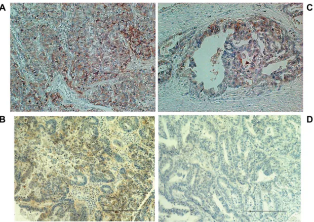
The immunohistochemical analyses were performed on 331 primary GC lesions. CD98 immunostaining was predominantly localized on the plasma membrane (Figure 1).
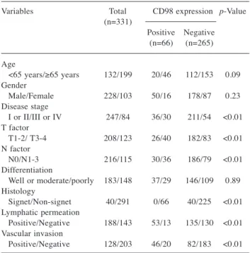
In our patient cohort, the positive expression rate of CD98 was 19% (66/331). Figure 2 shows the CD98 scoring profiles, in which scores of 1, 2, 3, and 4 were observed in 80%, 12%, 7%, and 1% of GC lesions, respectively. Patient demographics according to CD98 expression status are listed in Table I. CD98 expression was significantly associated with disease stage, T factor, lymph node metastases, non- signet cells, lymphatic permeation, and vascular invasion.
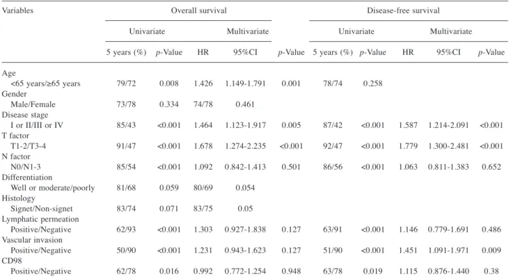
Survival analysis based on CD98 expression. The 5-year OS and PFS rate of all patients was 75% and 74%, respectively.
Out of 331 patients, 102 died after the initial surgery. Using univariate analysis, it was found that age, disease stage, T factor, N factor, lymphatic permeation, vascular invasion and CD98 expression had a significant relationship with OS.
Meanwhile, disease stage, T factor, N factor, lymphatic
permeation and vascular invasion were closely related to
unfavorable PFS (Table II). Multivariable analysis confirmed
that disease stage, age and T factor were independent
prognostic factors for predicting poor OS and that disease
stage, T factor and vascular invasion were significant
predictive factors for PFS in GC-affected patients. Figures
3A and 3B illustrate the Kaplan–Meier survival curve in
patients with high and low CD98 expression, respectively.
The prognostic significance of CD98 expression was analyzed according to disease stage and tumor differentiation. No statistically significant difference in OS and PFS was observed between patients with either positive or negative CD98 expression in early (stage I or II) versus advanced stage disease (III or IV). However, the Kaplan–
Meier analysis of OS demonstrated a significant difference between patients with either positive or negative CD98 expression and with well or moderate differentiation (Figure 3C), but no such significance was observed in patients with poor differentiation (Figure 3D).
Discussion
This study represents the first evaluation of the clinical and pathological significance of CD98 expression in patients with surgically resected GC. We found that increased CD98 expression was significantly associated with tumor aggressiveness and metastasis. CD98 is a potential biomarker that can be used to predict GC patient outcomes after surgical resection. Our study highlighted the prognostic significance of CD98 expression regarding GC-affected patients with well-to-moderate differentiation. These results
suggested that CD98 plays a crucial role in tumor progression and GC invasion. Because the mechanism governing such dynamics is unknown, further studies investigating a GC population without poor differentiation is warranted to verify CD98’s clinical role in GC. While numerous studies have shown that increased CD98
Figure 1.Immunohistochemical staining of CD98 in gastric adenocarcinoma. Positive CD98 staining is localized to the membrane of a malignant melanoma. In the representative images, CD98 immunostaining was scored as grade 4 (A), 3 (B), 2 (C) and grade 1 (D).Figure 2.Scoring of CD98 expression in GC tumor sections. CD98 scores of 1, 2, 3, and 4 were observed in 80%, 12%, 7%, and 1% of GC tumor sections, respectively.
expression is associated with clinical and pathological features in different cancers, the magnitude of positive CD98 expression (19%) in our GC patient cohort seems to be markedly lower in comparison. The positive rates of CD98 expression in lung, pancreatic, biliary tract, breast, and hypopharyngeal cancers are 47%, 56.7%, 36.7%, 31.8%, and 82.3%, respectively (3-5, 8-10). As mentioned above, CD98 expression levels correlate with LAT1 expression levels (3- 5). Our group, has previously noticed that LAT1 expression is higher in patients with squamous cell carcinoma (SQC) than in those with AC (7). Moreover, in patients with lung cancer, LAT1 expression in SQC (with a positive rate of 91%) was significantly higher than in AC (with a positive rate of 29%). The prognostic significance of LAT1 expression has also been shown to be markedly different between patients with SQC and AC. Particularly, LAT1 expression in patients with AC has a direct relationship with worse prognosis, whereas there is a weak association between LAT1 expression and survival in SQC-affected patients. In patients with early stage cancer, LAT1 expression associated more with shorter survival compared to CD98 expression. However, CD98’s prognostic role may be more
Figure 3.Survival outcomes after surgical resection. Kaplan–Meier analysis was conducted to assess the impact of CD98 expression on overall survival (OS) and progression-free survival (PFS) in GC-affected patients. A statistically significant difference in OS (A) and PFS (B) was observed between patients with positive or negative CD98 expression. Additionally, Kaplan–Meier analysis of OS resulted in a significant difference between patients with positive or negative CD98 expression and with either well or moderate differentiation (C), but there was no observed significant difference in patients with poor differentiation (D).
Table I. Patient’s demographics according to CD98 expression.
Variables Total CD98 expression p-Value (n=331)
Positive Negative (n=66) (n=265) Age <65 years/≥65 years 132/199 20/46 112/153 0.09 Gender Male/Female 228/103 50/16 178/87 0.23 Disease stage I or II/III or IV 247/84 36/30 211/54 <0.01 T factor T1-2/ T3-4 208/123 26/40 182/83 <0.01 N factor N0/N1-3 216/115 30/36 186/79 <0.01 Differentiation Well or moderate/poorly 183/148 37/29 146/109 0.89 Histology Signet/Non-signet 40/291 0/66 40/225 <0.01 Lymphatic permeation Positive/Negative 188/143 53/13 135/130 <0.01 Vascular invasion Positive/Negative 128/203 46/20 82/183 <0.01
important than that of LAT1 in advanced stage cancer progression. Additionally, it is uncertain whether the prognostic roles of LAT1 and CD98 are different between patients with early and advanced stages and this warrants rigorous investigation.
Recently, Toyoda et al. investigated the prognostic significance of LAT1 and CD98 in patients with advanced hypopharyngeal SQC and concluded that CD98 and not LAT1, could serve as a prognostic marker to predict survival outcomes postoperatively, although LAT1 and CD98 were positively expressed at 85.7% and 82.8% of the samples studied, respectively. Additionally, this study exhibited a statistically significant correlation between the expression of LAT1 and CD98. Although CD98’s role as a potentially promising and predictive biomarker in advanced human neoplasms (4, 8) is important, there was no significant difference between patients with GC either positive or negative for CD98 expression in terms of advanced stage.
Interestingly, our data demonstrated that a significantly worse prognosis was confirmed in GC-affected patients with positive CD98 expression. As stated earlier, Ichinoe et al.
noted that LAT1 could be an independent prognostic factor in GC-affected patients without scirrhous carcinoma (13). As scirrhous carcinoma is associated with poor differentiation, LAT1 may be a significant prognostic predictor for GC-
affected patients without poor differentiation. Such dynamics are somewhat reflective in our data.
Our study has several limitations. First, we did not investigate the relationship between CD98 expression and the integrin signaling pathway and teasing out this relationship merits future studies. Second, a heterogeneous patient population was evaluated, and such an analysis may bias the results of survival outcomes postoperatively. Next, steps will involve studying survivorship in a homogenous GC patient population. In conclusion, CD98 was positively expressed in 19% of GC-affected patients who underwent surgery and CD98 expression was significantly associated with GC tumor aggression and metastases. CD98 predicted worse prognosis in GC-affected patients, particularly in patients without poor differentiation. However, this result was not confirmed using multivariable analysis. CD98 could be an attractive target for GC therapeutic treatments.
Acknowledgements
The Authors would like to thank Ms. Yuka Matsui for her technical assistance and deeply appreciate Prof. Masahiko Nishiyama of the Department of Molecular Pharmacology and Oncology, Gunma University Graduate School of Medicine for critical reviewing this manuscript. The Authors declare that they have no conflict of interest.
Table II.Univariate and multivariate survival analysis in all patients.
Variables Overall survival Disease-free survival Univariate Multivariate Univariate Multivariate 5 years (%) p-Value HR 95%CI p-Value 5 years (%) p-Value HR 95%CI p-Value Age <65 years/≥65 years 79/72 0.008 1.426 1.149-1.791 0.001 78/74 0.258
Gender
Male/Female 73/78 0.334 74/78 0.461 Disease stage
I or II/III or IV 85/43 <0.001 1.464 1.123-1.917 0.005 87/42 <0.001 1.587 1.214-2.091 <0.001 T factor
T1-2/T3-4 91/47 <0.001 1.678 1.274-2.235 <0.001 92/47 <0.001 1.779 1.300-2.481 <0.001 N factor
N0/N1-3 85/54 <0.001 1.092 0.842-1.413 0.501 86/56 <0.001 1.063 0.811-1.383 0.652 Differentiation
Well or moderate/poorly 81/68 0.059 80/69 0.054 Histology
Signet/Non-signet 83/74 0.071 83/75 0.05 Lymphatic permeation
Positive/Negative 62/93 <0.001 1.303 0.927-1.838 0.127 63/91 <0.001 1.146 0.779-1.691 0.486 Vascular invasion
Positive/Negative 50/90 <0.001 1.231 0.943-1.623 0.127 51/90 <0.001 1.451 1.091-1.971 0.009 CD98
Positive/Negative 62/78 0.016 0.992 0.772-1.254 0.948 63/78 0.019 1.115 0.876-1.440 0.38
References
1 Cantor JM and Ginsberg MH: CD98 at the crossroads of adaptive immunity and cancer. J Cell Sci 125: 1373-1382, 2012.
2 Kanai Y, Segawa H, Miyamoto K, Uchino H, Takeda E and Endou H: Expression cloning and characterization of a transporter for large neutral amino acids activated by the heavy chain of 4F2 antigen (CD98). J Biol Chem 273: 23629-23632, 1998.
3 Kaira K, Sunose Y, Arakawa K, Ogawa T, Sunaga N, Shimizu K, Tominaga H, Oriuchi N, Itoh H, Nagamori S, Kanai Y, Segawa A, Furuya M, Mori M, Oyama T and Takeyoshi I:
Prognostic significance of L-type amino-acid transporter 1 expression in surgically resected pancreatic cancer. Br J Cancer 107: 632-638, 2012.
4 Kaira K, Oriuchi N, Imai H, Shimizu K, Yanagitani N, Sunaga N, Hisada T, Kawashima O, Kamide Y, Ishizuka T, Kanai Y, Nakajima T and Mori M: CD98 expression is associated with poor prognosis in resected non-small-cell lung cancer with lymph node metastases. Ann Surg Oncol 16: 3473-3481, 2009.
5 Furuya M, Horiguchi J, Nakajima H, Kanai Y and Oyama T:
Correlation of L-type amino acid transporter 1 and CD98 expression with triple negative breast cancer prognosis. Cancer Sci 103: 382-389, 2012.
6 Sakata T, Ferdous G, Tsuruta T, Satoh T, Baba S, Muto T, Ueno A, Kanai Y, Endou H and Okayasu I: L-type amino acid transporter 1 as a novel biomarker for high-grade malignancy in prostate cancer. Pathol Int 59: 7-18, 2009.
7 Kaira K, Oriuchi N, Imai H, Shimizu K, Yanagitani N, Sunaga N, Hisada T, Tanaka S, Ishizuka T, Kanai Y, Endou H, Nakajima T and Mori M: Prognostic significance of L-type amino acid transporter 1 expression in resectable stage I-III nonsmall cell lung cancer. Br J Cancer 98: 742-748, 2008.
8 Toyoda M, Kaira K, Shino M, Sakakura K, Takahashi K, Takayasu Y, Tominaga H, Oriuchi N, Nikkuni O, Suzuki M, Iijima M, Tsukamoto N, Nagamori S, Kanai Y, Oyama T and Chikamatsu K: CD98 is a novel prognostic indicator for patients with stage III/IV hypopharyngeal squamous cell carcinoma.
Head Neck 37: 1569-1574, 2015.
9 Toyoda M, Kaira K, Ohshima Y, Ishioka NS, Shino M, Sakakura K, Takayasu Y, Takahashi K, Tominaga H, Oriuchi N, Nagamori S, Kanai Y, Oyama T and Chikamatsu K: Prognostic significance of amino acid transporter expression (LAT1, ASCT2 and xCT) in surgically resected tongue cancer. Br J Cancer 110: 2506- 2513, 2014.
10 Kaira K, Sunose Y, Oriuchi N, Kanai Y and Takeyoshi I: CD98 as a promising biomarker of prognosis in biliary tract cancer.
Hepatobiliary Pancreat Dis Int 13: 654-657, 2014.
11 Isoda A, Kaira K, Iwashina M, Oriuchi N, Tominaga H, Nagamori S, Kanai Y, Oyama T, Asao T, Matsumoto M and Sawamura M: Expression of L-type amino acid transporter 1 (LAT1) as a prognostic and therapeutic indicator in multiple myeloma. Cancer Sci 105: 1496-1502, 2014.
12 Namikawa M, Kakizaki S, Kaira K, Tojima H, Yamazaki Y, Horiguchi N, Sato K, Oriuchi N, Tominaga H, Sunose Y, Nagamori S, Kanai Y, Oyama T, Takeyoshi I and Yamada M:
Expression of amino acid transporters (LAT1, ASCT2 and xCT) as clinical significance in hepatocellular carcinoma. Hepatol Res 45: 1014-1022, 2014.
13 Ichinoe M, Mikami T, Yoshida T, Igawa I, Tsuruta T, Nakada N, Anzai N, Suzuki Y, Endou H and Okayasu I: High expression of L-type amino-acid transporter 1 (LAT1) in gastric carcinomas:
comparison with non-cancerous lesions. Pathol Int 61: 281-289, 2011.
14 Imai H, Kaira K, Oriuchi N, Shimizu K, Tominaga H, Yanagitani N, Sunaga N, Ishizuka T, Nagamori S, Promchan K, Nakajima T, Yamamoto N, Mori M and Kanai Y: Inhibition of L-type amino acid transporter 1 has antitumor activity in non-small cell lung cancer. Anticancer Res 30: 4819-4828, 2010.
15 Kim CS, Cho SH, Chun HS, Lee SY, Endou H, Kanai Y and Kim DK: BCH, an inhibitor of system L amino acid transporters, induces apoptosis in cancer cells. Biol Pharm Bull 31: 1096- 1100, 2008.
16 Kim CS, Moon IS, Park JH, Shin WC, Chun HS, Lee SY, Kook JK, Kim HJ, Park JC, Endou H, Kanai Y, Lee BK and Kim DK:
Inhibition of L-type amino acid transporter modulates the expression of cell cycle regulatory factors in KB oral cancer cells. Biol Pharma Bull 33: 1117-1121, 2010.
17 Takahashi K, Kaira K, Shimizu A, Sato T, Tamahashi N, Ogawa H, Yoshinari D, Yokobori T, Asao T, Takeyoshi I and Oyama T:
Clinical significance of β2–adrenergic receptor expression in patients with surgically resected gastric adenocarcinoma. Tumor Biol: 1-8, 2016.
18 Kaira K, Oriuchi N, Takahashi T, Nakagawa K, Ohde Y, Okumura T, Murakami H, Shukuya T, Kenmotsu H, Naito T, Kanai Y, Endo M, Kondo H, Nakajima T and Yamamoto N:
LAT1 expression is closely associated with hypoxic markers and mTOR in resected non-small cell lung cancer. Am J Transl Res 3: 468-478, 2011.
19 Feral CC, Nishiya N, Fenczik CA, Stuhlmann H, Slepak M and Ginsberg MH: CD98hc (SLC3A2) mediates integrin signaling.
Proc Natl Acad Sci USA 102: 355-360, 2005.
20 Yang Y, Toy W, Choong LY, Hou P, Ashktorab H, Smoot DT, Yeoh KG and Lim YP: Discovery of SLC3A2 Cell Membrane Protein as a Potential Gastric Cancer Biomarker: Implications in Molecular Imaging. J Proteome Res 11: 5736-5747, 2012.