OTHERS
Significance of ERCC1 and Hormonal Receptor Expression
in Ovarian Cancer
Dalia Abd El-moniem Elrawi1, Ahmed Ibrahim El khodary2, Hanan Ramadan Nassar2, Amira Diyaa Darwish2, and Eman Naguib Khorshed3
1Department of Medical Oncology, Ahmed Maher Teaching Hospital, Cairo, Egypt, 2Department of medical oncology, NCI ,Cairo University, Cairo, Egypt, 3Department of Pathology, NCI, Cairo University, Cairo, Egypt
Abstract : Background & Objectives : Ovarian carcinoma usually has a relatively poor prognosis. A rational approach to identify patients, who are likely to benefit from therapy, is urgently needed. Excision repair cross-complemen-tation group 1 enzyme (ERCC1) has been proposed as a molecular predictor of clinical resistance to platinum-based chemotherapy. Steroid hormone receptors are important determinants of prognosis and predictive behavior in tumor tissues of several origins. The present study aimed to investigate the expression profile of ERCC1, ER & AR in patients with Ovarian carcinoma and their association with patient outcome. Methods : This is a prospective study which included 77 patients with ovarian carcinoma who were treated with platinum based chemotherapy at the National Cancer Institute (NCI) in Egypt during the period 7/2016- 7/2018. We evaluated the expression of ER, AR, and Excision repair cross-complementation group 1 enzyme (ERCC1) by immunohistochemistry. Expression profiles were compared to clinical, histologic and prognostic factors, the clinical outcome and survival. All pa-tients received platinum containing chemotherapy regimen. Result : Of the 77 papa-tients with ovarian cancer, 66.2 % (51/77) were ERCC1-positive, 49.4 % (38/77) were AR positive & 75.3 % (58/77) were ER positive. Platinum resistance was found in eight of the tumors with positive ERCC1 protein expression compared with two among the patients with negative tumor staining for ERCC1 (P = 0.643). There was significant association between ER & AR expres-sion and pathological subtypes (p = 0.004, 0.007) respectively. There were no significant association between ER, AR& ERCC1 expression and PFS (P = 0.447,P = 0.162, P = 0.508 respectively) or OS (P = 0.781, P = 0.569, P = 0.381 respectively). Based on Cox proportional hazards regression analysis ERCC1, ER &AR were not independent factors affecting the prognosis of patients with ovarian carcinoma. Conclusion : These results demonstrate that positive ERCC1 expression is not associated with clinical resistance to platinum-based chemotherapy, ERCC1, AR& ER expression are not independent factors affecting the prognosis of patients with epithelial ovarian tumors and not associated with survival benefits. J. Med. Invest. 67 : 391-398, August, 2020
Keywords : Excision repair cross-complementation (ERCC1)expression, ER & AR expression, Ovarian cancer, Platinum-resistance, Survival
INTRODUCTION
Ovarian cancer is the leading cause of gynecologic cancer death in the United States. Ovarian cancer accounting for 3% of cancers among women in the United States, but is the fifth most common cause of cancer-related death. It was estimated that approximately 22,280 women were diagnosed with ovarian can-cer in the United States in 2018, and that approximately 14,070 women died as a result of ovarian cancer in 2018 (1).
According to the National Population-Based Registry Pro-gram of Egypt 2008-2011, ovarian cancer represent 4.12% with crude rate 4.6. It is the 4th most common cancer in females. There is a progressive increase in number of incident ovarian cancer cases from 2288 in 2013 to 5957 in 2050, approximately 260%of 2013 incidence. Proportion of ovarian cancer was highest in upper Egypt (6.1%), and almost similar in middle Egypt (3.8%), and lower Egypt (3.9%) (2).
Platinum-based chemotherapy drugs are first-line treatments for ovarian cancer (3). However, a large number of patients do not respond to platinum-based chemotherapy due to drug resistance.
Previous research shows that the nucleotide excision repair (NER) system plays an important role in platinum resistance to chemotherapy (4). It repairs platinum-induced DNA damage by removing the damaged fragments in the DNA molecule, and then synthesizing DNA by DNA polymerase. ERCC1 (excision repair cross complementation group 1) is a key gene involved in NER.
Endocrine factors play key roles in ovarian cancer develop-ment, with risk reduction related to multiparity and use of oral contraceptives (5,6).
Estrogen regulates growth and differentiation in the normal ovaries and has been demonstrated to have mutagenic effects. Progesterone, on the other hand, induces apoptosis and decreas-es cell membrane permeability, leading to decreased invasive po-tential (7). After menopause, when the estradiol level decreases, androgens are still produced and also seem to influence ovarian cancer development. Androgens promote cell proliferation, and androgen levels are decreased by the use of oral contraceptives (8).
The present study aimed to investigate the following : 1-Prognostic value of immunohistochemical expression of
The Journal of Medical Investigation Vol. 67 2020
Abbreviations
AR : Androgen Receptor ; EOC : Epithelial Ovarian Cancer ; OCSS : Ovarian Cancer Specific Survival ; ERCC1 : Excision repair cross-complementation group 1 ; IHC : Immunohistochem-istry ; ER : Estrogen Receptor ; NER : nucleotide excision repair, PFS : progression-free survival, OS : Overall survival, HR : Hormonal Receptors
Received for publication January 19, 2020 ; accepted March 10, 2020. Address correspondence and reprint requests to Dalia Elrawi, 151 Emtedad Ramsis 2, Nasr City, Cairo, Egypt.
ERCC1 and Sex steroid hormone receptors in the tumor tissue as regards progression free survival and overall survival.
2-Predictive value of ERCC1 expression as regards respose to platinum-based therapy.
MATERIALS AND METHODS
PatientsA total of 77 patients diagnosed with EOC were recruited between July 2016 and July 2018 in National Cancer Institute (Egypt). The median age at diagnosis was 53 years, ranged between 18–74 years. As presented in (Table 1), The tumors
were classified according to the International Federation of Gy-necology and Obstetrics classification system, with 16 (20.8%) samples classified as stage IA, 9 (11.7%) as stage IB, 8 (10.4%) as stage II, 19 (24.7%) as stage III and 25 (32.5%) as stage IV. The pathological types of the tumor samples were as follows : 56 (72.7%) Serous carcinoma samples (9 low G (G1), 7 intermediate (G2), 40 high G (G3)), 5 (6.5%) mucinous carcinoma, 12 (15.6%) endometrium cancer and 2 clear cell carcinoma, 2 transitional carcinoma. 67 (87%). Optimal radical surgery (PAH-BSO) was done in 67 patients (87%), while conservative surgery was done for 9 patients (11.6%) including 7 patients unilateral salpingoo-phrectomy (9%), 2 patients excised ovarian mass. Peritoneal biopsy was performed in 1 patients.
66 patients (85.7%) received systemic platinum-based com-bination chemotherapy, following the surgical procedure, 37 (56.06%) patients received systemic chemotherapy as adju-vant treatment, 14 (21.21%) received as neo-adjuadju-vant and 15 (22.7%) patients received systemic chemotherapy as adjuvant and neoadjuvant.
48 patients (72.2%) received 6 courses of chemotherapy or more.
Chemotherapy regimens consisted of 175 mg/m2 taxol plus car-boplatin calculated at AUC 5–6 every 3 weeks for 6–8 cycles or cycle every week for 18 weeks with AUC 2-3.
Most of patients (n = 37) (57.8%) received carboplatin with AUC 5-6 every 3 weeks and 27 (42.2%) patients received carbo-platin weekly with AUC 2-3.
Ethical approval for the study was granted by the Cairo Uni-versity ethics committee (Egypt), and all patients had given their written informed consent to participate in the study.
Immunohistochemical analysis
Tumor specimens were harvested from 77 patients prior to receiving platinum-based treatment.
1.Paraffin sections were made at 4 microns thickness and mounted on positive charged slides.
2.Immunostaining was done for all cases using Automated BenchMark ULTRA IHC/ISH system, and the following steps occurred automatically :
•Deparaffinization by using the EZ-prep solution.
•Cell conditioning (standard cell conditioning CC1) for 80 minutes.
•Antigen retrieval using reaction buffer (PH 7.4-7.8).
•Application of 100µ of each of the ready-to-use monoclonal antibodies used in the study under specific incubation tem-perature and time for each (Table 2).
•Application of Diaminobenzidine (DAB) as a chromogen (NexES Ultra View DAB Detection Kit).
•Counterstaining with Hematoxylin II for 8 minutes. •Post counter staining with bluing reagent for 4 minutes. 3.Slides were extracted and arranged in racks.
4.Slides were washed in tap water and soap for 5 minutes and then dehydrated in the ascending grades of alcohol for 5 minutes in each container.
5.Slides were cleared in Xylene, and then cover slips were applied.
Chemotherapy outcome
Clinical curative effect was assessed by routine gynecological examination, imaging analysis (color ultrasound, computed tomography, magnetic resonance imaging or positron emission tomography-computed tomography for abdominal or pelvic re-gions) and detection of serum carbohydrate antigen (CA)-125 levels. No recurrence at 6 months post-chemotherapy was re-ferred to as ‘clinically sensitive’ and included normal serum CA-125 levels, no new lesions, or the original residual lesions Table 1. Clinicopathological data of 77 patients with EOC
Characteristics Numbers Percentage Valid percent
Age = < 53 42 54.5 > 53 35 45.5 SA (n = 63) < 1.8 28 36.4 44.4 > = 1.8 35 45.5 55.6 BMI (n = 62) < 30 26 33.8 41.9 > = 30 36 46.8 58.1 CA 125 (n = 67) 0-35 12 15.6 17.9 > 35 55 71.4 82.1 Stage IA 16 20.8 IB 9 11.7 II 8 10.4 III 19 24.7 IV 25 32.5 I, II 33 42.9 III, IV 44 57.1 Malignant ascites present 28 36.4 Peritoneal implants present 24 31.2 Omentum deposits Present 25 32.5 Distant metastasis
Pleural effusin (M1a) 5 6.5 20.0
Liver (HFLs) 10 13.0 40.0
Pulmonary nodules 4 5.2 16.0
Splenic focal lesions 4 5.2 16.0
Anterior abdominal wall 2 2.6 8.0
Non regional LNs 13 16.9 52.0 Histopathology Serous 56 72.7 Endometrioid 12 15.6 mucinous 5 6.5 others 4 5.2 Grades I 12 15.6 II 19 24.7 III 46 59.7 ERCC1 N 26 33.8 P 51 66.2 ER N 19 24.7 P 58 75.3 AR N 39 50.6 P 38 49.4
SA : Surface area, BMI : Body mass index, ER : Estrogen receptor, AR : Androgen receptor, ERCC1 : Excision repair cross-comple-mentation group 1
had decreased in size or disappeared as identified by pelvic and imaging examination. By contrast, disease progression during chemotherapy, a continual increase in serum CA-125 levels or the appearance of new lesions identified by imaging at 6 months post-chemotherapy was recognized as ‘clinical resistance’. Follow-up
The final follow-up occurred on July 2019. The median follow up period was 22.8 months (range, 1.4 – 38.5 months). Disease PFS was described as the time from ovarian cancer surgery or the time from start neoadjuvant chemotherapy to disease re-currence or mortality, whichever came first. The time between surgery or start treatment and mortality or the end of follow-up was described as the overall survival time (OS).
Statistical analysis
Statistical analysis was done using IBM SPSS® Statistics version 22 (IBM® Corp., Armonk, NY, USA). Numerical data were expressed as mean and standard deviation or median and range as appropriate. Qualitative data were expressed as frequency and percentage. Pearson’s Chi-square test or Fisher’s exact test was used to examine the relation between qualitative variables. For not normally distributed quantitative data, com-parison between two groups was done using Mann-Whitney test (non parametric t-test). Survival analysis was done using Ka-plan-Meier method and comparison between two survival curves was done using log-rank test. Multivariate analysis was done using Cox-regression method for the factors affecting survival on univariate analysis. Hazard ratio (HR) with it 95% confidence interval (CI) were used for risk estimation. All tests were two-tailed. A p-value < 0.05 was considered significant.
RESULTS
Expression of ERCC1 and Relation to Outcome
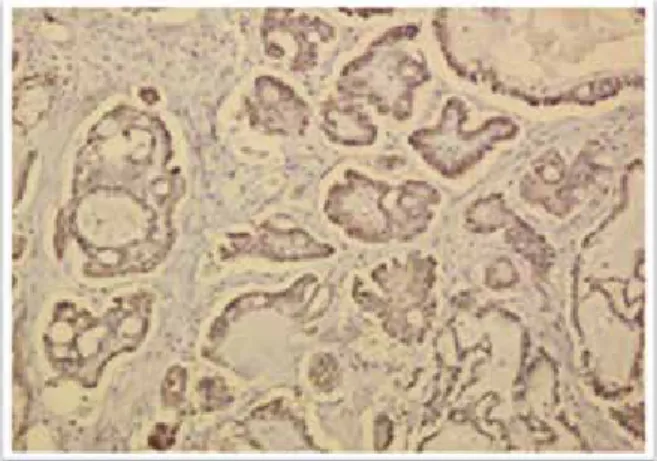
Brown-yellow granules were observed in the majority of tumor cell cytoplasm and nuclei, and corresponded with positive ERCC1 expression (Fig. 1). Immunohistochemistry identified that 51/77 specimens (66.2%) were ERCC1 positive.
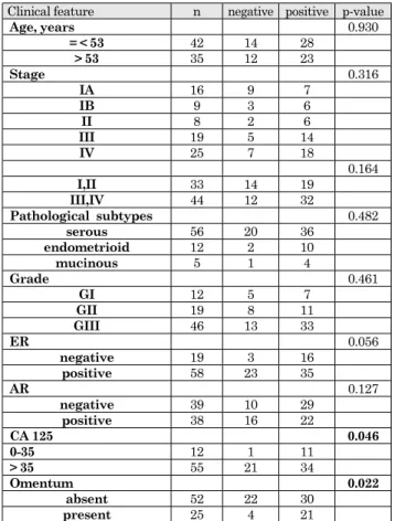
As presented in (Table 3), no significant association was iden-tified between ERCC1 expression and age (P = 0.930), patho-logical type (P = 0.482), cell differentiation (P = 0.461), clinical stage (P = 0.316) and ER expression (P = 0.056) or AR expression (P = 0.127)
While there was significant association between ERCC1 expression and elevated serum level of CA 125 at the time of diagnosis (P = 0.046). Also, presence of omental deposits was significantly correlated with the positive ERCC1 expression in tumor tissue (P = 0.022)
As presented in (Table 4) (Fig.2), the number of resistant cases with positive ERCC1 expression (8/10 ; 80%) was not significantly greater than the number of sensitive cases with positive ERCC1 expression (41/63 ; 65.1%) (P = 0.351). For the 77 EOC cases, there
Table 2. List of used immunohistochemical markers
Antibodies Clone Source Incubation Temp Incubation Time (min) Visualization Positive Control
AR (N-20) Sc-816Rabbit polyclonal Santa Cruz Biotechnology 42°C 32 Nuclear Prostatic carcinoma
ERCC-1 8F1Mouse monoclonal Gene Tex 37°C 32 Nuclear Tonsil
ER SP1Rabbit monoclonal Roche 37°C 36 Nuclear Breast
Figure 1. Positive reaction to ERCC1 in almost all tumor cells, both cytoplasmic and nuclear (X10).
Table 3. Correlation between ERCC1 expression and clinical pathological features.
Clinical feature n negative positive p-value
Age, years 0.930 = < 53 42 14 28 > 53 35 12 23 Stage 0.316 IA 16 9 7 IB 9 3 6 II 8 2 6 III 19 5 14 IV 25 7 18 0.164 I,II 33 14 19 III,IV 44 12 32 Pathological subtypes 0.482 serous 56 20 36 endometrioid 12 2 10 mucinous 5 1 4 Grade 0.461 GI 12 5 7 GII 19 8 11 GIII 46 13 33 ER 0.056 negative 19 3 16 positive 58 23 35 AR 0.127 negative 39 10 29 positive 38 16 22 CA 125 0.046 0-35 12 1 11 > 35 55 21 34 Omentum 0.022 absent 52 22 30 present 25 4 21
ER : Estrogen receptor, AR : Androgen receptor, ERCC1 : Excision repair cross-complementation group 1
was no significant difference in PFS and median OS between patients with positive ERCC1 expression and patients with neg-ative expression (OS, P = 0.381 ; PFS, P = 0.508) (Fig. 3).
Expression of Sex Steroid Hormone Receptors and Relation to Outcome
The expression and the prognostic value were first assessed individually for each marker, with ER positivity detected in 58/77 (75.3%) (Fig 4) and AR positivity detected in 38/77 (49.4%) (Fig 5).
The representation of stage, grade, age at diagnosis, histology and ERCC1 expression in relation to receptor status is outlined in (Table 5). No significant association was identified between AR expression and the clinical pathological features ---) age (P = 0.209), clinicl stage (P = 0.236), pathological subtypes (P = 0.318), pathological grade (P = 0.795), ERCC 1 expression (P = 0.127).
There is a significant association between AR expression and age (P = 0.050), ER expression (P = 0.004) and body surface area > = 1.8 (P = 0.028)
ER positivity was not significant association was identified between ER expression and the clinical pathological features ---) age (P = 0.847), clinicl stage (P = 0.815), pathological grade (P = 0.867), and border significant with ERCC 1 expression (P = 0.056).
There is a significant association between ER expression and pathological subtypes (P = 0.007), AR expression (P = 0.004), HB > = 10 (P = 0.026) and body surface area > = 1.8 (P = 0.049) (Table 6).
Table 4. Correlation between ERCC1 expression and platinum sen-sitivity.
Sensitive Resist P-value
ERCC1 0.351
N (n = 24)(32.9%) 22 (34.9%) 2 (20%)
P (n = 49)(67.1%) 41 (65.1%) 8 (80%) ERCC1 : Excision repair cross-complementation group 1
Figure 2. Correlation between ERCC1 expression and platinum sensitivity.
Figure 3. OS and PFS of patients with positive expression of ERCC1 vs. those with negative.
Figure 4. Moderate diffuse positive nuclear reaction to ER in most of tumor cells (X10).
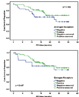
Expression of either ER or AR was not associated with im-proved PFS (P = 0.447, P = 0.162 respectively) (Fig 6) and OS (P = 0.781, P = 0.569 respectively) (Fig 7).
Table 5. Correlation between AR expression and clinical pathologi-cal features.
Clinical feature n negative positive p-value
Age, years 0.050 = < 53 42 17 25 > 53 35 22 13 SA 0.028 < 1.8 28 19 9 > = 1.8 35 14 21 Stage 0.236 IA 16 7 9 IB 9 4 5 II 8 2 6 III 19 9 10 IV 25 17 8 0.087 I-II 33 13 20 II-IV 44 26 18 Pathological subtypes 0.318 serous 56 25 31 endometrioid 12 8 4 mucinous 5 3 2 Grade 0.795 GI 12 5 7 GII 19 10 9 GIII 46 24 22 ER 0.004 negative 19 15 4 positive 58 24 34 ERCC1 0.127 negative 26 10 16 positive 51 29 22
SA : Surface area, ER : Estrogen receptor, AR : Androgen recep-tor, ERCC1 : Excision repair cross-complementation group 1
Table 6. Correlation between ER expression and clinical pathologi-cal features.
Clinical feature n negative positive p-value
Age, years 0.847 = < 53 42 10 32 > 53 35 9 26 SA 0.049 <1.8 28 11 17 >=1.8 35 6 29 Stage 0.815 IA 16 5 11 IB 9 2 7 II 8 1 7 III 19 6 13 IV 25 5 20 I-II 33 8 25 0.939 III-IV 44 11 33 Pathological subtypes 0.007 serous 56 9 47 endometrioid 12 3 9 mucinous 5 4 1 Grade 0.867 GI 12 2 10 GII 19 5 14 GIII 46 12 34 AR 0.004 negative 39 15 24 positive 38 4 34 ERCC1 0.056 negative 26 3 23 positive 51 16 35
SA : Surface area, ER : Estrogen receptor, AR : Androgen recep-tor, ERCC1 : Excision repair cross-complementation group 1
Figure 6. PFS of patients with positive expression of AR or ER vs. those with negative.
Figure 7. OS of patients with positive expression of AR or ER vs. those with negative.
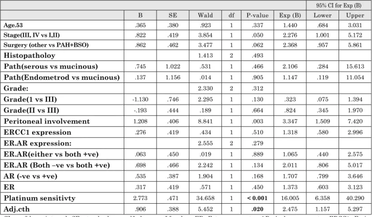
ERCC1 expression, ER expression, AR expression, peritoneal deposits and platinum sensitivity. As presented in (Table 7), age, platinum sensitivity and received adjuvant chemotherapy were identified as independent factors significantly affecting the prognosis of patients (P = 0.048, P < 0.001 and P = 0.020, respec-tively), while other factors did not significantly affect prognosis. Independent risk factors for patient survival time :
Cox proportional hazards regression was used to analyze pos-sible risk factors, including age, histopathological type, degree of cancer cell differentiation, clinical stage, type of surgery and Cox regression analysis demonstrated that ERCC1 expression level, ER or AR expression were not an independent prognostic factor for the survival time of patients with EOC.
DISCUSSION
Chemotherapy drug resistance is a major factor restricting the improvement of patient survival rates, with 20–30% of patients with EOC undergoing primary platinum-resistance ; however, 80% of patients are likely to eventually encounter resistance (9,10). With the rapid development of pharmacogenomics and molecular biology, the mechanism of cisplatin resistance is close-ly associated with NER (11). In DNA repair, ERCC1 is a key gene of the NER pathway due to its binding with DNA repair endonuclease ERCC1-xeroderma pigmentosum group F (XPF) (12,13).
A number of studies have examined the association between ERCC1 expression (14-18) and clinical outcomes including re-sponse to platinum-based therapy, PFS and OS in patients with EOC (19). The outcomes of these studies were discriminative, ranging from increased rate of platinum resistance (22), worse
PFS and overall OS (20-22), similar PFS (23-26), to similar OS (20-23). In our current study, we were unable to confirm any statistically significant association between ERCC1 expression and resistance to platinum-based chemotherapy. PFS and OS did not significantly differ between the positive and negative ERCC1 expression.
A previous meta-analysis evaluated whether response to platinum-based chemotherapy was associated with ERCC1 expression in patients with ovarian cancer (24). It was observed that patients with negative ERCC1 expression had a significant-ly greater response to platinum-based chemotherapy compared with patients with positive ERCC1 expression (24), indicating that ERCC1 protein expression status is correlated with re-sponse to platinum-based chemotherapy in ovarian cancer. Zhao et al. (25) identified a negative correlation between ERCC1 expression and clinical chemosensitivity in EOC.
Stadlmann et al. analyzed 80 samples of ovarian cancer uti-lizing 8F-1 antibody for immunohistochemy and found low overall ERCC1 expression (20.3%) and no association between protein expression and platinum responsiveness (p = 0.21) (16), which correlates with our results. In a study of Steffensen et al., immunohistochemy with the 8F-1 antibody against ERCC1 was used to examine 100 tumor specimens : 45% of specimens were positive, which was associated with a significantly poorer response to platinum-based chemotherapy but not with a worse OS (15). Lin et al. corroborated these findings by demonstrating that low ERCC1 protein expression was significantly associated with drug sensitivity in 63 patients (26).
In a study of Rubatt et al. (27), in which patients who partic-ipated in GOG-172 and GOG-182 trials and provided tumor samples for translational research were included, 27% of tumors were ERCC1-positive. ERCC1 expression was not associated with clinical characteristics or platinum responsiveness. Women
Table 7. Univariate analysis for survival time.
95% CI for Exp (B)
B SE Wald df P-value Exp (B) Lower Upper
Age.53 .365 .380 .923 1 .337 1.440 .684 3.031
Stage(III, IV vs I,II) .822 .419 3.854 1 .050 2.276 1.001 5.172
Surgery (other vs PAH+BSO) .862 .462 3.477 1 .062 2.368 .957 5.861
Histopatholoy 1.413 2 .493 Path(serous vs mucinous) .745 1.022 .531 1 .466 2.106 .284 15.613 Path(Endometrod vs mucinous) .137 1.156 .014 1 .905 1.147 .119 11.054 Grade: 2.330 2 .312 Grade(1 vs III) -1.130 .746 2.295 1 .130 .323 .075 1.394 Grade(II vs III) -.193 .444 .189 1 .664 .824 .345 1.970 Peritoneal involvement 1.208 .406 8.841 1 .003 3.347 1.509 7.420 ERCC1 expression .276 .419 .434 1 .510 1.318 .580 2.996 ER.AR expression: 2.555 2 .279
ER.AR(either vs both +ve) .063 .450 .019 1 .889 1.065 .440 2.575
ER.AR (Both –ve vs both +ve) .698 .466 2.242 1 .134 2.011 .806 5.017
AR (-ve vs +ve) .535 .387 1.904 1 .168 1.707 .799 3.646
ER .317 .419 .571 1 .450 1.373 .603 3.123
Platinum sensitivty 2.773 .471 34.658 1 < 0.001 16.005 6.358 40.290
Adj.cth .906 .388 5.452 1 .020 2.475 1.157 5.297 CI : confidence interval ; SE : standard error, df : degrees of freedom, ER : Estrogen receptor, AR : Androgen receptor, ERCC1 : Exci-sion repair cross-complementation group 1, PAH-BSO : Pan abdominal hysterectomy bilateral salpingoophorectomy
with ERCC1-positive versus negative tumors had similar median PFS (17.9 months versus 17.5 months, respectively, p = 0.59), median OS (52 months versus 47 months, respective-ly, p = 0.30), risk of disease progression [adjusted hazard ratio (HR) = 0.90, 95%CI = 0.71-1.15, p = 0.41), and risk of death (ad-justed HR = 0.81, 95% CI = 0.61-1.07, p = 0.14)
However, Muallem et al. (28) demonstrated that there were no significant differences in the PFS between patients with low, intermediate and high H-scores for ERCC1 expression.
The prognostic value of sex steroid hormone receptor expres-sion in ovarian cancer is not fully defined. In this study, we can however demonstrate that expression of ER and AR not predicts PFS and OS.
In the present study, no information on use of endocrine treatment was available, precluding analyses of possible effects of endocrine treatment on the findings reported. However, endo-crine treatment is not standard in ovarian cancer and is unlikely to have been administered to the study cohort to such an extent that it has influenced the results (29,30).
The finding that aromatase inhibition appears slightly more effective than tamoxifen in ovarian cancer likely reflects the more efficient hormone inhibition of aromatase inhibitors. In support of this notion, epidemiological studies indicate that reduced circulating levels of androgens decrease the risk of developing ovarian cancer, but clinical studies have shown only limited effects of androgen deprivation (32,34,35).
The presence and prognostic value of AR expression in ovari-an covari-ancer vary in different studies, but increased AR expression seems to generally be associated with a favorable prognosis (29,31,33), contrary to the results in the present study.
Zhaojun, et al., (2017) who investigated the correlation between ER expression and epithelial ovarian cancer prognosis in thir-ty-five studies with a total of 5824 patients were included, and demonstrated that the expression of ER, especially ER α, was a positive predictor of overall survival among epithelial ovarian cancer patients (36), contrary to the results in the present study.
In conclusion, the present study demonstrated that high ERCC1 expression in patients with EOC was not associated with resistance to platinum-based chemotherapy or with survival time. In addition, it was also observed that ERCC1 protein ex-pression was not an independent factor affecting the prognosis of patients. We demonstrate a prognostic role of PR and AR ex-pression in ovarian cancer, with independent effects on PFS and OS and the best outcome for patients whose tumors displayed coexpression of ER and AR.
Further studies with larger sample sizes and improved study designs are required to investigate whether or not ERCC1 may function as a predictor for chemotherapy against EOC.
And our data define a basis for further evaluation of the role of sex steroid hormone receptors, and in the future possibly endocrine treatment, in ovarian cancer and support that such studies may be subtype specific to comprehensively evaluate the potential clinical benefit.
STATEMENT CONFLICT OF INTEREST
No conflict of interest.REFERENCES
1. Siegel RL, Miller KD, Jemal A : Cancer statistics, 2018. CA Cancer J Clin 68 : 7-30, 2018
2. Amal SI, Hussein MK, Nabiel NHM, Hoda B, Hossam K : Cancer Incidence in Egypt : Results of the National
Pop-ulation-Based Cancer Registry Program. Journal of Cancer Epidemiology, Volume 2014, Article ID 437971 : 18, https:// doi.org/10.1155/2014/437971
3. Yan L, Pei H, Yu C, Gui-ying W, Na W, Rong-miao Z : Pre-dicting the outcome of platinum-based chemotherapies in epithelial ovarian cancer using the 8092C/a polymorphism of ERCC1 : a meta-analysis. Biomarkers 19(2) : 128-34, 2014
4. Gossage L, Madhusudan S : Current status of excision repair cross complementing-group 1 (ERCC1) in cancer. Cancer Treat Rev 33(6) : 565-77, 2007
5. Collaborative Group on Epidemiological Studies of Ovar-ian Cancer, Beral V, Doll R, Hermon C, Peto R, Reeves G : Ovarian cancer and oral contraceptives : collaborative reanalysis of data from 45 epidemiological studies including 23,257 women with ovarian cancer and 87,303 controls. Lancet 371 : 303-314, 2008
6. Purdie DM, Bain CJ, Siskind V, Webb PM, Green AC : Ovulation and risk of epithelial ovarian cancer. Int J Cancer 104 : 228-232, 2003
7. Ho SM : Estrogen, progesterone and epithelial ovarian can-cer. Reprod Biol Endocrinol 1 : 73, 2003
8. Coenen CM, Thomas CM, Borm GF, Hollanders JM, Rolland R : Changes in androgens during treatment with four low-dose contraceptives. Contraception 53 : 171-176, 1996 9. Lin H, Changchien CC : Management of
relapsed/refracto-ry epithelial ovarian cancer : Current standards and novel approaches. Taiwan J Obstet Gynecol 46 : 379-388, 2007 doi : 10.1016/S1028-4559(08)60007-8.
10. Colombo N, Gore M : Treatment of recurrent ovarian cancer relapsing 6–12 months post platinum-based chemotherapy. Crit Rev Oncol Hematol 64 : 129-138, 2007. doi : 10.1016/j. critrevonc.2007.04.004.
11. Altaha R, Liang X, Yu JJ, Reed E : Excision repair cross complementing-group 1 Gene expression and platinum re-sistance. Int J Mol Med 14 : 959-970, 2004
12. Andrieux LO, Fautrel A, Bessard A, Guillouzo A, Baffet G, Langouët S : GATA-1 is essential in EGF-mediated in-duction of nucleotide excision repair activity and ERCC1 expression through ERK2 in human hepatoma cells. Can-cer Res 67 : 2114-2123, 2007. doi : 10.1158/0008-5472. CAN-06-3821.
13. Niedernhofer LJ, Odijk H, Budzowska M, van Drunen E, Maas A, Theil AF, de Wit J, Jaspers NG, Beverloo HB, Hoeijmakers JH, Kanaar R : The structure-specific endo-nuclease Ercc1-Xpf is required to resolve DNA interstrand cross-link-induced double-strand breaks. Mol Cell Biol 24 : 5776-5787, 2004. doi : 10.1128/MCB.24.13.5776-5787.2004. 14. Steffensen KD, Waldstrøm M, Jakobsen A : The relation-ship of platinum resistance and ERCC1 protein expression in epithelial ovarian cancer. Int J Gynecol Cancer 19 : 820-825, 2009
15. Steffensen KD, Waldstrøm M, Jeppesen U, Brandslund I, Jakobsen A : Prediction of response to chemotherapy by ERCC1 immunohistochemistry and ERCC1 polymorphism in ovarian cancer. Int J Gynecol Cancer 18 : 702-710, 2008 16. Stadlmann S, Dirnhofer S, Guth U, Thies S, Singer G :
ERCC1 immunoexpression does not predict platinum-resis-tance in ovarian cancer. Gynecol Oncol 108 : 252-253, 2008 17. Weberpals J, Garbuio K, O’Brien A, Clark-Knowles K, Doucette S, Antoniouk O, Goss G, Dimitroulakos J : The
DNA repair proteins BRCA1 and ERCC1 as predictive markers in sporadic ovarian cancer. Int J Cancer 124 : 806-815, 2009
18. Dabholkar M, Vionnet J, Bostick-Bruton F, Yu JJ, Reed E : Messenger RNA levels of XPAC and ERCC1 in ovarian
cancer tissue correlate with response to platinumbased che-motherapy. J Clin Invest 94 : 703-708, 1994
19. Krivak TC, Darcy KM, Tian C, Bookman M, Gallion H, Ambrosone CB, Deloia JA : Single nucleotide polypmor-phisms in ERCC1 are associated with disease progression, and survival in patients with advanced-stage ovarian and primary peritoneal carcinoma ; A Gynecologic Oncology Group Study. Gynecol Oncol 122 : 121-126, 2011
20. Krivak TC, Darcy KM, Tian C, Armstrong D, Baysal BE, Gallion H, Ambrosone CB, DeLoia JA : Relationship between ERCC1 polymorphisms, disease progression, and survival in the gynecologic oncology group phase III trial of intraperitoneal versus intravenous cisplatin and pacli-taxel for stage III epithelial ovarian cancer. J Clin Oncol 26 : 3598-3606, 2008
21. Smith S, Su D, Rigault de la Longrais IA, Schwartz P, Puopolo M, Rutherford TJ, Mor G, Yu H, Katsaros D : ERCC1 geno-type and phenogeno-type in epithelial ovarian cancer identify pa-tients likely to benefit from paclitaxel treatment in addition to platinum-based chemotherapy. J Clin Oncol 25 : 5172-5179, 2007.
22. Kim HS, Kim MK, Chung HH, Kim JW, Park NH, Song YS, Kang SB : Genetic polymorphisms affecting clinical outcomes in epithelial ovarian cancer patients treated with taxanes and platinum compounds : A Korean popula-tion-based study. Gynecol Oncol 113 : 264-269, 2009 23. Marsh S, Paul J, King CR, Gifford G, Mcleod HL, Brown
R : Pharmacogenetic assessment of toxicity and outcome after platinum taxane chemotherapy in ovarian cancer : the Scottish randomised trial in ovarian cancer. J Clin Oncol 25 : 4528-4535, 2007
24. Li FY, Ren XB, Xie XY, Zhang J : Meta-analysis of excision repair cross-complementation group 1 (ERCC1) association with response to platinum-based chemotherapy in ovarian cancer. Asian Pac J Cancer Prev 14 : 7203-7206, 2013. doi : 10.7314/APJCP.2013.14.12.7203.
25. Zhao D, Zhang W, Li XG, Wang XB, Li M, Li YF, Tian HM, Song PP, Liu J, Chang QY, Wu LY : The mRNA expression of BRCA1, ERCC1, TUBB3, PRR13 genes and their rela-tionship with clinical chemosensitivity in primary epithelial ovarian cancer. Zhonghua Zhong Liu Za Zhi 34 : 196–200, 2012 (In Chinese)
26. Lin K, Ye D, Xie X : Protein expression levels of excision repair cross-complementation group 1 and xeroderma pig-mentosum D correlate with response to platinum-based che-motherapy in the patients with advanced epithelial ovarian cancer. Int J Gynecol Cancer 18 : 1007-1012, 2008
27. Rubatt JM, Darcy KM, Tian C, Muggia F, Dhir R, Armstrong DK, Bookman MA, Niedernhofer LJ, Deloia J, Birrer M,
Krivak TC : Pre-treatment tumor expression of ERCC1 in women with advanced stage epithelial ovarian cancer is not predictive of clinical outcomes : A gynecologic on-cology group study. Gynecol Oncol 125 : 421-426, 2012. doi : 10.1016/j.ygyno.2012.01.008.
28. Muallem MZ, Marnitz S, Richter R, Köhler C, Sehouli J, Arsenic R : ERCC1 expression as a predictive marker of cervical cancer treated with cisplatin-based
chemoradia-tion. Anticancer Res 34 : 401-406, 2014
29. Lee P, Rosen DG, Zhu C, Silva EG, Liu J : Expression of progesterone receptor is a favorable prognostic marker in ovarian cancer. Gynecol Oncol 96 : 671-677, 2005
30. Sieh W, Köbel M, Longacre TA, Bowtell DD, deFazio A, Goodman MT, Høgdall E, Deen S, Wentzensen N, Moysich KB, Brenton JD, Clarke BA, Menon U, Gilks CB, Kim A, Madore J, Fereday S, George J, Galletta L, Lurie G, Wilkens LR, Carney ME, Thompson PJ, Matsuno RK, Kjær SK, Jensen A, Høgdall C, Kalli KR, Fridley BL, Keeney GL, Vierkant RA, Cunningham JM, Brinton LA, Yang HP, Sherman ME, García-Closas M, Lissowska J, Odunsi K, Morrison C, Lele S, Bshara W, Sucheston L, Jimenez-Linan M, Driver K, Alsop J, Mack M, McGuire V, Rothstein JH, Rosen BP, Bernardini MQ, Mackay H, Oza A, Wozniak EL, Benjamin E, Gentry-Maharaj A, Gayther SA, Tinker AV, Prentice LM, Chow C, Anglesio MS, Johnatty SE, Chenevix-Trench G, Whittemore AS, Pharoah PDP, Goode EL, Huntsman DG, Ramus SJ : Hormone-receptor expres-sion and ovarian cancer survival : an Ovarian Tumor Tis-sue Analysis consortium study. Lancet Oncol 14 : 853-862, 2013
31. Nodin B, Zendehrokh N, Brandstedt J, Nilsson E, Manjer J, Brennan DJ, Jirstrom K : Increased androgen receptor expression in serous carcinoma of the ovary is associated with an improved survival. J Ovarian Res 3 : 14, 2010 32. Elattar A, Warburton KG, Mukhopadhyay A, Freer RM,
Shaheen F, Cross P, Plummer ER, Robson CN, Edmonson RJ : Androgen receptor expression is a biological marker for androgen sensitivity in high grade serous epithelial ovarian cancer. Gynecol Oncol 124 : 142-147, 2012
33. Cardillo MR, Petrangeli E, Aliotta N, Salvatori L, Ravenna L, Chang C, Castagna G : Androgen receptors in ovarian tumors : correlation with oestrogen and progesterone re-ceptors in an immunohistochemical and semiquantitative image analysis study. J Exp Clin Cancer Res 17 : 231-237, 1998
34. Levine D, Park K, Juretzka M, Esch J, Hensley M, Aghajanian C, Lewin S, Konner J, Derosa F, Spriggs D, Iasonos A, Sabbatini P : A phase II evaluation of goserelin and bicalutamide in patients with ovarian cancer in sec-ond or higher complete clinical disease remission. Cancer 110 : 2448-2456, 2007
35. du Bois A, Meier W, Luck HJ, Emon G, Moebus V, Schroed-er W, Costa S, Bauknecht T, Olbricht S, Jackisch C, RichtSchroed-er B, Wagner U : Chemotherapy versus hormonal treatment in platinum- and paclitaxel-refractory ovarian cancer : a ran-domised trial of the German Arbeitsgemeinschaft Gynae-kologische Onkologie (AGO) Study Group Ovarian Cancer. Ann Oncol 13 : 251-257, 2002
36. Shen ZJ, Luo H, Sheng B, Zhao MH, Zhu HY, Zhu XQ : Cor-relation between estrogen receptor expression and prognosis in epithelial ovarian cancer : a meta-analysis. Oncotarget Sep 5 ; 8(37) : 62400-62413, 2017. Published online 2017 May 29.