R E V I E W A R T I C L E
Evidence-based clinical practice guidelines for nonalcoholic fatty liver disease/nonalcoholic steatohepatitis 2020
Katsutoshi Tokushige1,3•Kenichi Ikejima1•Masafumi Ono1•Yuichiro Eguchi1• Yoshihiro Kamada1•Yoshito Itoh1• Norio Akuta1•Masato Yoneda1•
Motoh Iwasa1•Masashi Yoneda1•Motoyuki Otsuka1• Nobuharu Tamaki1• Tomomi Kogiso1• Hiroto Miwa1• Kazuaki Chayama2•Nobuyuki Enomoto1• Tooru Shimosegawa1• Tetsuo Takehara2•Kazuhiko Koike1
Received: 14 May 2021 / Accepted: 14 May 2021 The Author(s) 2021
Abstract
Nonalcoholic fatty liver disease (NAFLD) has become a serious public health issue not only in Western countries but also in Japan. Within the wide spectrum of NAFLD, nonalcoholic steatohepatitis (NASH) is a pro- gressive form of disease that often develops into liver cirrhosis and increases the risk of hepatocellular carcinoma (HCC). While a definite diagnosis of NASH requires liver biopsy to confirm the presence of hepatocyte ballooning,
hepatic fibrosis is the most important prognostic factor in NAFLD. With so many NAFLD patients, it is essential to have an effective screening method for NAFLD with hepatic fibrosis. As HCC with non-viral liver disease has increased markedly in Japan, effective screening and surveillance of HCC are also urgently needed. The most common death etiology in NAFLD patients is cardiovas- cular disease (CVD) event. Gastroenterologists must, therefore, pay close attention to CVD when examining NAFLD patients. In the updated guidelines, we propose screening and follow-up methods for hepatic fibrosis, HCC, and CVD in NAFLD patients. Several drug trials are ongoing for NAFLD/NASH therapy, however, there is currently no specific drug therapy for NAFLD/NASH. In addition to vitamin E and thiazolidinedione derivatives, recent trials have focused on sodium glucose co-transporter 2 (SGLT2) inhibitors and glucagon-like peptide-1 (GLP-1) analogues, and effective therapies are expected to be developed. These practical guidelines for NAFLD/NASH were established by the Japanese Society of Gastroen- terology in conjunction with the Japan Society of Hepa- tology. Clinical evidence reported internationally between 1983 and October 2018 was collected, and each clinical and background question was evaluated using the Grades of Recommendation Assessment, Development and Eval- uation (GRADE) system. This English summary provides the core essentials of these clinical practice guidelines, which include the definition and concept, screening sys- tems for hepatic fibrosis, HCC and CVD, and current therapies for NAFLD/NASH in Japan.
Keywords
NAFLD/NASH guidelines Fibrosis HCC Cardiovascular disease Treatment
The original version of this article appeared in Japanese as ‘‘NAFLD/
NASH Sinryo Guideline 2020’’ from the Japanese Society of Gastroenterologyand the Japan Society of Hepatology, published by Nankodo, Tokyo, in 2020. Please see the article on the standards, methods, and process of developing guidelines.
This article has been co-published with permission in Hepatology Research and Journal of Gastroenterology. This article is published under the Creative Commons CC-BY licence. The articles are identical except for minor stylistic and spelling differences in keeping articles are with each journal’s style. Either citation can be used when citing this article.
The members of the Guidelines Committee are listed in the Appendix.
& Katsutoshi Tokushige
1 Guidelines Committee for Creating and Evaluating the
‘‘Evidence-Based Clinical Practice Guidelines for Nonalcoholic Fatty Liver Disease/Nonalcoholic
Steatohepatitis’’, The Japanese Society of Gastroenterology / The Japan Society of Hepatology, 6F Shimbashi i-MARK Building, 2-6-2 Shimbashi, Minato-ku, Tokyo 105-0004, Japan
2 The Japan Society of Hepatology, Kashiwaya 2 Building 5F, 3-28-10 Hongo, Bunkyo-ku, Tokyo 113-0033, Japan
3 Institute of Gastroenterology, Department of Internal Medicine, Tokyo Women’s Medical University, 8-1 Kawada- cho, Shinjuku-ku, Tokyo, Japan
https://doi.org/10.1007/s00535-021-01796-x
Introduction
Nonalcoholic fatty liver disease (NAFLD) has become the most prevalent cause of chronic liver disease worldwide [1]
and is now the fastest-growing indication for liver trans- plantation among waitlist registrants [2]. There are more than 20 million NAFLD patients in Japan, and it is feared that this number will increase in the future [3]. In recent years, hepatocellular carcinoma (HCC) based on non-viral liver disease has increased, and the need for a screening method has become urgent.
The Japanese NAFLD/NASH guidelines were estab- lished in 2014 [4]. These clinical guidelines have received considerable attention and been widely used. However, new knowledge has since been reported relating to their concepts, diagnostic imaging methods, and treatment methods. Therefore, a joint committee of the Japanese Society of Gastroenterology and the Japanese Society of Hepatology has reviewed and revised these guidelines.
The current guidelines summarize reports from 1983 to the end of October 2018, focusing on the following: (1) concept and clinical significance of liver fibrosis, (2) screening and follow-up systems for liver fibrosis, (3) surveillance of HCC, (4) a system for consultation with specialists regarding cardiovascular risk in NAFLD, and (5) new therapeutics. We hope that these guidelines will be used widely in clinical practice, and also hope to discuss and improve any problems that may arise in the clinical field so that further revisions may be made as necessary.
The concept and definition of NAFLD
In the 2014 guidelines [4], NAFLD was characterized by evidence of hepatic steatosis by either imaging or histology and the appropriate exclusion of other liver diseases.
NAFLD is histologically characterized by macrovesicular steatosis and further categorized into nonalcoholic fatty liver (NAFL) and nonalcoholic steatohepatitis (NASH). In the revised guidelines, the concept and diagnosis of NAFLD are essentially the same (Table
1).NAFLD is characterized by evidence of hepatic steatosis as determined by either imaging or histology, associated with any metabolic factor. Other liver diseases, such as alcoholic liver disease, viral liver disease, and drug-in- duced liver disorder, are excluded.
The European Association for the Study of the Liver (EASL) defines NAFLD as being characterized by ‘‘ex- cessive hepatic fat accumulation associated with insulin resistance (IR)’’ [5]. In Japan, 60–70% of NAFLD cases are complicated with diabetes mellitus or IR, while others may be complicated with obesity, hypertension, or dys- lipidemia without IR [6,
7]. Therefore, we defined NAFLDas ‘‘hepatic steatosis associated with any metabolic factor.’’
Another issue is NAFL progression. McPherson et al. [8]
report that of 27 NAFL patients, 12 (44%) had progressed to NASH at the second biopsy. NAFL and NASH are not completely different diseases; there is a certain amount of overlap. Therefore, we specifically state in the guidelines that ‘‘NAFL and NASH are not completely different dis- eases. Some NAFL patients show slow progression of hepatic fibrosis.’’ In addition, the American Association for the Study of Liver Diseases (AASLD) [9] and EASL management guidelines [5] state that hepatic steatosis induced by drugs should be excluded from NAFLD. We,
Table 1 New definition and concept of NAFLD
Nonalcoholic fatty liver disease (NAFLD) is characterized by evidence of hepatic steatosis as determined by either imaging or histology, associated with metabolic factors. Other liver diseases, such as alcoholic liver disease, viral liver disease, and drug-induced liver disorder are excluded. NAFLD is categorized into nonalcoholic fatty liver (NAFL) and nonalcoholic steatohepatitis (NASH). NAFL is a mostly benign, nonprogressive clinical entity, while NASH can progress to cirrhosis or even hepatocellular carcinoma (HCC)
1. Fat deposition in the liver is histologically significant at 5% or more
2. NASH is histologically characterized by hepatic steatosis associated with evidence of liver cell injury (ballooning degeneration) and inflammation
3. NAFL and NASH are not completely different diseases. Some NAFL patients show slow progression of hepatic fibrosis 4. The upper limit of alcohol drinking is 30 g/day in males and 20 g/day in females
5. Hepatic steatosis induced by drugs is treated as a drug-induced liver disorder
6. Reye’s syndrome and acute fatty liver of pregnancy, which show microvesicular steatosis, are excluded from NAFLD
7. In NASH cirrhosis, certain histological characteristics of NASH, such as hepatic steatosis and ballooning degeneration, are sometimes lost and this is known as ‘‘burned-out NASH.’’
*The most important factor in the prognosis is hepatic fibrosis, and follow-up and treatment methods should be selected depending on the degree of hepatic fibrosis
therefore, specify that ‘‘hepatic steatosis induced by drugs is treated as a drug-induced liver disorder, not as NAFLD.’’
After the publication of the 2014 guidelines, many cohort studies demonstrated that hepatic fibrosis, but no other histological features of NAFLD, was associated independently with long-term overall mortality, liver transplantation, and liver-related events in NAFLD patients [10–12]. Therefore, we added the following statement to the revised guidelines: ‘‘the most important factor in the prognosis is hepatic fibrosis, and follow-up and treatment methods should be selected depending on the degree of hepatic fibrosis.’’
We also added new clinical questions (CQs).
CQ. Which histological factor is most important in assessing survival?
•
The stage of hepatic fibrosis is strongly associated with both total and liver-related mortalities. It is important to evaluate the grade of hepatic fibrosis in NAFLD patients. (Evidence Level A, Strength 1).
Flowchart for the follow-up and screening of hepatic fibrosis, HCC, and cardiovascular disease
There is currently no overall flowchart for the screening and follow-up of hepatic fibrosis, HCC, or cardiovascular disease (CVD). As noted, the most important factor in the prognosis is hepatic fibrosis. We, therefore, propose that the first step on the flowchart be a screening system for NAFLD by a general physician (Fig.
1). Castera et al. [13]report that simple inexpensive and widely available serum biomarkers, such as the FIB-4 index or the NAFLD fibrosis score (NFS), which have a high negative predictive value for ruling out advanced fibrosis, should be used as the first line. Patients with low risk of advanced fibrosis (FIB- 4
\1.3 or NFS
\-1.455) do not need further assess- ment, while liver stiffness should be measured by vibra- tion-controlled transient elastography (VCTE) in those with intermediate risk (FIB-4 = 1.3–3.25 or NFS =
-1.455–0.672) or high risk (FIB-4
[3.25 or NFS
[0.672).
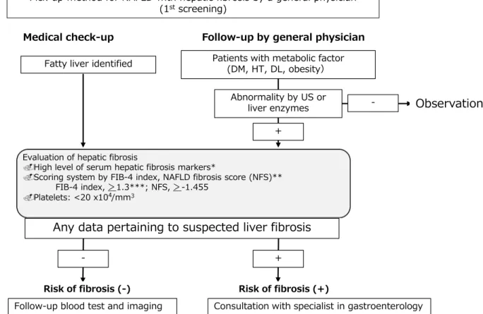
Following Castera et al. [13], we propose a screening method for NAFLD with hepatic fibrosis by a general physician (Fig.
1). At the first screening for NAFLD,physicians are recommended to measure serum hepatic fibrosis markers, such as hyaluronic acid, Type IV collagen 7S, Mac-binding protein glycosylation isomer, and to use a fibrosis scoring system such as the FIB-4 index, NFS, or platelet count. If any data indicate liver fibrosis,
consultation with a gastroenterology specialist should be considered.
Figure
2is a flowchart of the next screening, to be conducted by a gastroenterology specialist. First, the FIB-4 index or NFS is calculated. If the FIB-4 index is under 1.3 or the NFS is under
-1.455, the patient is at low risk of having fibrosis and should repeat the evaluation every 1 year. If moderate hepatic fibrosis (FIB-4 index of 1.3–2.66 or NFS of
-1.455–0.674) is suspected, liver biopsy or VCTE or magnetic resonance elastography (MRE) should be considered. If severe hepatic fibrosis (FIB-4 index:
C2.67 or NFS:
C0.675) is suspected, liver biopsy or elastography is recommended and, depending on fibrosis stage or liver stiffness, surveillance and therapy may be considered. The most important point is to evaluate hepatic fibrosis stage by liver biopsy, elastography, or some other method.
Regarding HCC surveillance, we recommend that in NAFLD with F0-1, the evaluation for fibrosis stage should be performed after 1 year without HCC screening. In male NAFLD with F2 or F3 and female NAFLD with F3, patients have a mild risk of HCC and abdominal ultrasound (US) should be considered every 6–12 months.
HCC screening should be performed by US and tumor markers every 6 months in NAFLD with liver cirrhosis (LC; F4). However, the best screening method in terms of cost and manpower remains to be determined. Loomba et al. [14] recently reported on the American Gastroen- terological Association (AGA) Clinical Practice Update on screening and surveillance for HCC in patients with NAFLD. Screening for HCC should be considered in patients with LC or advanced fibrosis. In the revised guidelines, we have added new CQs regarding HCC fol- low-up and screening in NAFLD/NASH patients.
CQ. How should NAFLD/NASH patients be followed up?
•
Depending on the grade of hepatic fibrosis, it is important to follow-up not only liver-related events, but also CVD and extrahepatic malignancies. (Evidence Level A, Strength 1).
CQ. How should NAFLD/NASH patients be screened for HCC?
•
HCC screening should be performed depending on the stage of hepatic fibrosis and risk factors for HCC.
However, the best screening method in terms of cost
and manpower remains to be determined. (Evidence Level C, Strength 2).
In terms of HCC risk factors, old age, male, advanced fibrosis, diabetes, and moderate alcohol intake have been reported to be risk factors for NAFLD-HCC [15–17].
CVD, the most common death etiology in NAFLD patients, is another issue. Francque et al. [18] reported the screening method for CVD in NAFLD patients. It is important to note that NASH and NAFLD with hepatic fibrosis increase the risk of cardiovascular event [19,
20].In cases with hepatic fibrosis, CVD screening is recommended.
Therefore, we propose the flowchart for cardiovascular event screening in NAFLD patients shown in Fig.
3. Wefirst check for CVD complications and/or a past history of CVD, and perform an electrocardiogram (ECG). If any
abnormality is found, we then consult a specialist in car- diology or neurology. In NAFLD with a reduced platelet count or increased FIB-4 index, we should evaluate risk based on cardiovascular examination, such as loaded ECG and/or US of the carotid artery. CQ5-3 concerns cardio- vascular events in NAFLD patients.
CQ. Do NAFLD/NASH patients have an increased rate of cardiovascular events?
•
In NAFLD/NASH, cardiovascular events are increased, especially in the advanced hepatic fibrosis group.
Examination for and evaluation of CVD should be considered in NAFLD patients with advanced fibrosis.
(Evidence Level A, Strength 1).
Fig. 1. 1st Screening system for NAFLD with hepatic fibrosis:
flowchart. First screening system for NAFLD is performed by a general physician or medical checkup. If any data indicate liver fibrosis, consultation with a gastroenterology specialist should be
considered.FIB-4 indexFibrosis-4 index;NFSNAFLD fibrosis score;
DMdiabetes mellitus;HT hypertension,DLdyslipidemia;M2BPGi Mac-2 Binding Protein Glycosylation isomer;BMIbody mass index;
IFGimpaired fasting glucose;USultrasonography
Epidemiology
The reported prevalence of NAFLD [6,
21–23] varieswidely depending on the population studied and the defi- nition or method used. Age, gender, and ethnic differences have an effect on both the prevalence and severity of NAFLD/NASH [6,
21–24]. These differences are also seenin the prevalence of obesity and metabolic syndrome. The prevalence of NAFLD is reported to be 20–40% in Western countries and 12–30% in Asian countries [1,
25]. Annualhealth checkup data show that 9–30% of Japanese adults have US-diagnosed NAFLD, and NASH is diagnosed in 10–20% or more of NAFLD cases [6,
25, 26]. The esti-mated prevalence of NASH is approximately 3–5%
worldwide [27,
28]. NAFLD/NASH is predominantlydiagnosed in middle-aged men and postmenopausal women [28]. A Markov model has estimated there to be approxi- mately 22,600,000 NAFLD patients in Japan and 3,700,000 NASH patients, and by 2030, the prevalence of NASH and advanced fibrosis will have increased [3].
Regarding the rate of HCC development from NAFLD, Younossi et al. [1] report global data showing that the incidence of HCC among NAFLD and NASH patients is 0.44 per 1000 person-years (range 0.29–0.66) and 5.29 per 1000 person-years (range 0.75–37.56), respectively. It is higher in LC due to NAFLD, at 2–3% per year [16,
17].Pathogenesis and genomic background
Given the important role of insulin resistance and oxidative stress in the pathophysiology of NAFLD/NASH [29–31], several studies have attempted to investigate their effects.
The I148M variant of patatin-like phospholipase domain-containing 3 (PNPLA3) is widely known to be associated with the occurrence and progression of NAFLD/
NASH worldwide [32–34]. The mechanism remains unknown, however, it has been suggested that the 148M variant disrupts ubiquitylation and proteasomal degradation of
PNPLA3, resulting in an accumulation of PNPLA3- Fig. 2. 2nd screening system for NAFLD with hepatic fibrosis andHCC screening: flowchart. Second screening is conducted by a gastroenterology specialist. First, the FIB-4 index or NFS is calculated. If the FIB-4 index is increased, liver biopsy or vibration-controlled transient elastography (VCTE) or magnetic
resonance elastography (MRE) should be considered or recom- mended.FIB-4 indexFibrosis-4 index;NFSNAFLD fibrosis score;
DMdiabetes mellitus;BMIbody mass index;IFGimpaired fasting glucose;USultrasonography;HCChepatocellular carcinoma
148M and impaired mobilization of triglycerides (TGs) from lipid droplets (LDs) [35–37]. There is known to be a significant association between HCC and the 148M variant [38,
39].TM6SF2, GCKR, GATAD2Aand
DYSFhave been reported as genomic background candidates [39–43]. In addition, there has been a recent focus on the relationship between NAFLD and gut dysbiosis [44,
45]. Henao-Mejiaet al. [46] found that inflammasome-mediated dysbiosis regulates the progression of NAFLD and obesity, however, a more detailed analysis using human samples is needed.
Sarcopenia has also been examined [47].
Diagnosis and imaging
The diagnosis of NAFLD is described as part of the con- cept and definition of NAFLD (Table
1). ‘‘Nonalcoholic’’is defined as an upper limit of alcohol drinking of 30 g/day in males and 20 g/day in females. NASH is diagnosed from liver biopsy on the basis of the presence of steatohepatitis, however, liver biopsy has several drawbacks. It is an expensive and invasive procedure and there is potential for sampling error and variability in interpretation by pathol- ogists [48,
49]. However, liver biopsies remain the goldstandard for the diagnosis of NASH and are, therefore, often recommended, particularly in NAFLD with suspected advanced fibrosis and in suspected coexisting chronic liver
disease, where there is a need to distinguish NASH from other chronic liver diseases [9].
Regarding the noninvasive assessment of NASH and advanced fibrosis, there are no practically useful surrogate markers for diagnosing NASH. Platelet count [50] and scoring systems such as the NFS [51] and the FIB-4 index [52,
53] have proven to be useful for predicting fibrosis in theJapanese population and worldwide. However, care must be taken, because age is one of the factors in the FIB-4 index, and higher FIB-4 index titers in older patients do not nec- essarily reflect actual liver stiffness or fibrosis grade [54,
55].A number of imaging modalities can detect liver fat and liver stiffness [56–58], including VCTE, which measures liver stiffness noninvasively, has shown promising results for assessing the severity of liver fibrosis. Recently, MRE was found to have statistically significantly higher diag- nostic accuracy than VCTE in the detection of each stage of fibrosis [59]. MRE and VCTE each have a role to play in the detection of fibrosis in patients with NAFLD, depend- ing on the level of accuracy desired [60].
Therapy
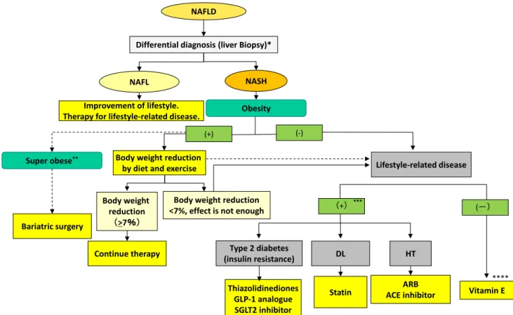
Figure
4is a flowchart of therapy for NAFLD patients. It is similar to that in the previous guidelines [4]. NAFLD is usually associated with metabolic disturbance such as
Fig. 3 Flowchart for cardiovascular event screening in NAFLDpatients. We have to check for cardiovascular disease (CVD) complications and/or a past history of CVD, and perform an electrocardiogram (ECG). If any abnormality is found, we consult a specialist in cardiology or neurology. In NAFLD with a reduced
platelet count or increased FIB-4 index, we should evaluate risk based on cardiovascular examination, such as loaded ECG and/or US of the carotid artery. FIB-4 index Fibrosis-4 index; PLT platelet, DM diabetes mellitus;HThypertension,DLdyslipidemia;USultrasonog- raphy;ECGelectrocardiogram
visceral obesity, insulin resistance, type 2 diabetes mellitus, or dyslipidemia, and these underlying conditions play a crucial role in its pathogenesis. Therefore, it makes sense to treat not only the liver disease itself but also these asso- ciated metabolic morbidities, and it is likewise essential to prevent stimulation or pathogenesis of these diseases, so- called ‘‘2nd hits,’’ in the management of NAFLD/NASH [61]. Treatments for these associated conditions include lifestyle modification, weight loss, and increased physical activity, all of which have been shown to be effective and represent the cornerstone of treatment [61].
Lifestyle-related interventions such as diet and exercise therapies have been reported to improve serum transami- nase levels as well as liver fat as measured by US and magnetic resonance imaging (MRI) in NAFLD patients [62,
63]. Musso et al. [64] evaluated the effects of weightloss in NAFLD in eight randomized controlled trials, four of which included posttreatment histology. Their meta- analysis showed that a 5% or greater weight loss improved hepatic steatosis, and a 7% or greater weight loss correlated with improvement in the NAFLD Activity Score (NAS).
Unfortunately, only 50% of subjects were able to attain a weight loss of 7% or greater even with significant inter- vention. Vilar-Gomez et al. [65] evaluated the effects of weight loss through lifestyle modifications in 261 patients with paired liver biopsies, finding that the degree of weight loss was associated independently with improvements in all NASH-related histology. Furthermore, for those indi- viduals who lost 10% or more of their body weight, 45%
experienced regression of fibrosis, 90% had resolution of steatohepatitis, and 100% demonstrated improvements in NAS. These findings indicate the effectiveness of body weight reduction for patients with obesity-related NAFLD/
NASH. In almost all reports of dietary interventions for patients with obesity-related NAFLD, a low-calorie diet is prescribed and, in terms of dietary contents, the proportions of energy intake from carbohydrates and lipids are often restricted to 50–60% and 20–25%, respectively. Recent attention has focused on low-carbohydrate and Mediter- ranean diets [66,
67], and coffee intake has been reported toinhibit hepatic fibrosis and hepatocarcinogenesis [68,
69].Fig. 4 Flowchart of therapy for NAFLD/NASH. BMI body mass index; DM diabetes mellitus; HT hypertension, DL dyslipidemia;
GLP-1 glucagon-like peptide-1; SGLT2 sodium glucose co-
transporter;ARBangiotensin II receptor antagonist;ACEangiotensin II converting enzyme inhibitor
Exercise therapy is another useful lifestyle-related intervention for NAFLD/NASH. Consistent aerobic exer- cise in 30- to 60-min sessions held 3–4 times weekly for 4–12 weeks in patients with NAFLD complicated by obesity has been shown to improve liver fat content, even without accompanying body weight reduction [70,
71]. Ohet al. [72] report that physical activity of moderate to vigorous intensity for
C250 min/week as part of lifestyle management improves NAFLD pathophysiology in obese men in Japan. The benefits seem to be acquired through reducing inflammation and oxidative stress levels and altering the fatty acid metabolism. While these reports do not examine histological changes, liver fat are thought to improve even with exercise therapy alone.
CQ. What kind of diet is recommended to improve NAFLD/NASH?
•
Body weight reduction through a low-calorie diet improves liver function and fatty changes in the liver in patients with NAFLD. To improve NAFLD/NASH, we recommend prioritizing the optimization of energy intake and restricting lipids or carbohydrates in terms of the proportions of nutrient intake. (Evidence Level C, Strength 2).
CQ. Is exercise beneficial for improving NAFLD/
NASH?
•
Although the effects of exercise on liver histology have not been fully clarified, we recommend implementing exercise therapy because, even alone, it improves liver function and liver fat in the liver in patients with NAFLD. (Evidence Level B, Strength 2).
Pharmacological treatments
Vitamin E and pioglitazone have been shown to improve liver function and liver histological findings [73–76].
However, their safety over the long term remains to be evaluated.
CQ. Is vitamin E effective for patients with NAFLD/NASH?
•
Vitamin E improves hepatic biological and histological parameters in patients with NASH, and is recom- mended. However, its safety over the long term in
patients with CVD, congestive heart failure, or bladder cancer has not yet been fully assessed.
CQ. Are thiazolidinediones effective for patients with NAFLD/NASH?
•
Pioglitazone is recommended for NASH patients with insulin resistance. (Evidence Level A, Strength 2).
Other studies have focused on other diabetic drugs such as incretin-related drugs like glucagon-like peptide-1 (GLP-1) receptor analogue [77–80] dipeptidyl peptidase 4 (DPP-4) inhibitor [81,
82], and sodium glucose co-transporter 2(SGLT2) inhibitor [83–85]. Both GLP-1 analogue and SGLT2 inhibitor not only decrease body weight but also improve the glucose metabolism. As SGLT2 inhibitor is known to be effective to heart failure [86], additional effects can reasonably be expected in NASH patients.
Some reports have demonstrated that GLP-1 analogue and SGLT2 improve liver function and liver histological find- ings [79,
80, 85, 87, 88]. In the revised guidelines, twodrugs have been added.
CQ. Is SGLT2 inhibitor effective for NAFLD/
NASH?
•
In NAFLD/NASH patients with type 2 diabetes, SGLT2 inhibitor improves liver enzymes and histo- logical findings, and its administration is, therefore, suggested. SGLT2 inhibitor is not adaptive for insur- ance of NAFLD/NASH. (Evidence Level C, Strength 2).
CQ. Are incretin-related drugs such as GLP-1 analogue and/or DPP-4 inhibitor effective for NAFLD/NASH?
•
In NAFLD with type 2 diabetes, GLP-1 analogue has been found to improve liver function and liver histo- logical findings. SGLT2 inhibitor is not adaptive for insurance of NAFLD/NASH. The effect of DPP-4 inhibitor is not constant. (Evidence Level C, Strength 2).
Regarding to both drugs, randomized trials with greater numbers of patients are needed.
Drugs for dyslipidemia and hypertension are reported to
improve liver enzymes [89–93], as described in previous
guideline.
CQ. Are drugs for dyslipidemia effective for patients with NAFLD/NASH?
•
HMG-CoA reductase inhibitors (statins) are recom- mended for NAFLD/NASH patients with hyperc- holesterolemia. However, the effect of ezetimibe is not constant. (Evidence Level C, Strength 2).
CQ. Is an angiotensin II receptor antagonist (ARB) or angiotensin II converting enzyme inhibitor (ACE) effective for hypertensive patients with NAFLD/NASH?
•
We recommend an ARB or ACE for NASH patients with hypertension. (Evidence Level C, Strength 2).
Ursodeoxycholic acid (UDCA) and biguanides have no significant effect on liver histology and we do not recom- mend them as a specific treatment for liver disease in patients with NASH [94–96], as described in previous guideline.
BQ. Are conventional doses of ursodeoxycholic acid effective for patients with NAFLD/NASH?
•
We do not recommend UDCA at conventional dose levels for the treatment of NAFLD/NASH.
CQ. Is biguanides effective for patients with NAFLD/NASH?
•
We do not recommend biguanides for the treatment of NAFLD/NASH, because there is no evidence, sug- gesting improvement of liver enzyme and liver histol- ogy in NAFLD/NASH patients. (Evidence Level B, Strength 2).
Other candidate drugs
FRQ. Are there any other effective drugs for treatment of patients with NAFLD/NASH in the future?
There are several ongoing drug trials for NAFLD/NASH therapy, including trials of obeticholic acid (OCA), elafi- branor, selonsertib (SEL), apoptosis signal-regulating kinase 1 (ASK1), cenicriviroc (CVC), fibroblast growth
factor (FGF)-21, Aramchol, acetyl-CoA carboxylase (ACC) inhibitor (GS-0976), FGF19 (NGM-282), pemafi- brate, emricasan, toll-like receptor 4 (TLR4) inhibitor (JKB-21), solithromycin, SSAO/VAP-1 inhibitor (BI 1467335), IMM-124E, galectin-3 inhibitor (GR-MD-02), and heat shock protein (HSP) 47 [97–108] to be addressed in future research questions.
Nevertheless, the trials for NAFLD/NASH had several problems. First, even in placebo groups, about 20% of patients improved due to lifestyle improvements. Second, to confirm improvement of liver fibrosis, which associated with prognosis, an observation period of 5 years or more is necessary. In addition, histological evaluation differs among pathologists. It is also problematic that NAFLD is a syndrome, and therefore, includes many pathogeneses. The effects of drugs are, therefore, not always constant. Finally, the prevalence of the
PNPLA3variant, which is a risk factor of progression of NASH, is different among races. It is expected that these problems will be resolved, and that new drugs will be approved for NASH therapy.
Elucidating the pathogenesis of NAFLD/NASH and developing therapies are now worldwide issues, and it is important that Japanese medical studies of NAFLD/NASH advance. For that purpose, these guidelines were prepared by searching for relevant evidence worldwide without regard to ethnic characteristics, and the obtained evidence was then summarized from a Japanese perspective. Of course, ethnic differences correlate with susceptibility to metabolic syndrome-related diseases including NAFLD/
NASH [109], possibly as a result of genomic polymor- phism. Therefore, it is also important to develop Japanese- based clinical research work and to interpret other coun- tries’ evidence in light of ethnic considerations. The revised guidelines not only summarize the current clinical state of NAFLD/NASH but also point to future directions for study.
Acknowledgements The authors thank to Dr. Kento Imajo, Dr. Yuji Ogawa (Yokohama City University), Dr. Ryuta Shigefuku, Dr.
Kazushi Sugimoto (Mie University), Dr. Yuya Seko (Kyoto Prefec- tural University of Medicine), Dr. Hirokazu Takahashi (Saga University), Prof. Atsushi Nakajima, Dr. Yasushi Honda (Yokohama City University). We also special appreciate for Prof. Masahiro Yoshida (International University of Health and Welfare) for advice for writing and Mr. Naohiko Yamaguchi (Library of Seirei Sakura citizen hospital) for searching the references.
Declarations
Conflict of interest Any financial relationship with enterprises, businesses, or academic institutions in the subject matter or materials discussed in the manuscript are listed as follows: (1) those from which the authors, the spouse, partner or immediate relatives of authors, have received individually any income, honoraria, or any other types of remuneration—MSD, Otsuka Pharmaceutical, Mitsubishi Tanabe Pharma, Bristol-Myers Squibb, Lundbeck Japan, AbbVie, Gilead
Sciences, Sumitomo Dainippon Pharma, Novo Nordisk, EA Pharma, Astellas Pharma, Mylan, Mochida Pharmaceutical, Kowa Company, Tsumura & CO., and (2) those from which the academic institutions of the authors received support (commercial/academic cooperation)—
EA Pharma, Astellas Pharma, Eisai, MSD, Otsuka Pharmaceutical, Suntory Global Innovation Center, Shionogi & Co., Daiichi Sankyo, Sumitomo Dainippon Pharma, Taiho Pharmaceutical, Takeda Phar- maceutical, Mitsubishi Tanabe Pharma, Chugai Pharmaceutical, Bayer Yakuhin, Bristol-Myers Squibb, Mochida Pharmaceutical, Yakult Honsha, AbbVie, Gilead Sciences, Gadelius Medical, Novartis Pharma, Eli Lilly Japan, Mylan N.V., Century Medical, Sogo Rinsho Medefi, Nissan Chemical, Fujirebio. Kowa, Kyokuto Pharmaceutical Industrial. Biofermin Pharmaceutical, and (3) those from which the authors have received individually endowed chair—NichiNichi Pharmaceutical.
Open Access This article is licensed under a Creative Commons Attribution 4.0 International License, which permits use, sharing, adaptation, distribution and reproduction in any medium or format, as long as you give appropriate credit to the original author(s) and the source, provide a link to the Creative Commons licence, and indicate if changes were made. The images or other third party material in this article are included in the article’s Creative Commons licence, unless indicated otherwise in a credit line to the material. If material is not included in the article’s Creative Commons licence and your intended use is not permitted by statutory regulation or exceeds the permitted use, you will need to obtain permission directly from the copyright holder. To view a copy of this licence, visithttp://creativecommons.
org/licenses/by/4.0/.
Appendix
The members of the Guidelines Committee who created and evaluated the Japanese Society of Gastroenterology
‘‘Evidence-based clinical practice guidelines for nonalco- holic fatty liver disease/nonalcoholic steatohepatitis2020’’
are listed below.
Creation Committee
Chair: Katsutoshi Tokushige (Institute of Gastroenterology, Department of Internal Medicine, Tokyo Women’s Medi- cal University). Vice-Chair: Kenichi Ikejima (Department of Gastroenterology, Juntendo University Graduate School of Medicine). Members: Masafumi Ono (Division of Gastroenterology and Hepatology, Department of Internal Medicine, Tokyo Women’s Medical University Medical Center East), Yuichiro Eguchi, (Loco Medical General Institute), Yoshihiro Kamada, (Department of Gastroen- terology and Hepatology, Osaka University Graduate School of Medicine), Yoshito Itoh (Department of Gas- troenterology and Hepatology, Graduate School of Medi- cine, Kyoto Prefectural University of Medicine), Norio Akuta (Department of Hepatology, Toranomon Hospital), Masato Yoneda (Department of Gastroenterology and Hepatology, Yokohama City University Graduate School of Medicine), Motoh Iwasa (Department of
Gastroenterology and Hepatology, Mie University Hospi- tal), Masashi Yoneda (Division of Hepatology and Pan- creatology, Department of Internal Medicine, Aichi Medical University), Motoyuki Otsuka (Department of Gastroenterology, Graduate School of Medicine, The University of Tokyo), Nobuharu Tamaki (Department of Gastroenterology and Hepatology, Musashino Red Cross Hospital), Tomomi Kogiso (Institute of Gastroenterology, Department of Internal Medicine, Tokyo Women’s Medi- cal University).
Evaluation Committee
Chair: Yoshiyuki Takei (Department of Gastroenterology and Hepatology, Mie University Graduate School of Medicine). Vice-Chair: Hitoshi Yoshiji (Third Department of Internal Medicine, Nara Medical University). Members:
Masataka Seike (Department of Gastroenterology, Oita Cardiovascular Hospital), Sumiko Nagoshi (Department of Gastroenterology and Hepatology, Saitama Medical Cen- ter, Saitama Medical University).
The Japanese Society of Gastroenterology
President: Kazuhiko Koike (Kanto Central Hospital).
Past President: Tooru Shimosegawa (South Miyagi Medical Center).
Director Responsible: Hiroto Miwa (Division of Gas- troenterology, Department of Internal Medicine, Hyogo College of Medicine), Nobuyuki Enomoto (First Depart- ment of Internal Medicine, Faculty of Medicine, University of Yamanashi).
The Japan Society of Hepatology
President: Tetsuo Takehara (Department of Gastroenterol- ogy and Hepatology, Osaka University Graduate School of Medicine).
Past President: Kazuhiko Koike (Kanto Central Hospital).
Director Responsible: Kazuaki Chayama (Department of Gastroenterology and Metabolism, Graduate School of Biomedical Sciences, Hiroshima University).
References
1. Younossi ZM, Koenig AB, Abdelatif D, Fazel Y, Henry L, Wymer M. Global epidemiology of nonalcoholic fatty liver
disease-Meta-analytic assessment of prevalence, incidence, and outcomes. Hepatology. 2016;64:73–84.
2. Wong RJ, Aguilar M, Cheung R, et al. Nonalcoholic steato- hepatitis is the second leading etiology of liver disease among adults awaiting liver transplantation in the United States. Gas- troenterology. 2015;148:547–55.
3. Estes C, Anstee QM, Arias-Loste MT, et al. Modeling NAFLD disease burden in China, France, Germany, Italy, Japan, Spain, United Kingdom, and United States for the period 2016–2030.
J Hepatol. 2018;69:896–904.
4. Watanabe S, Hashimoto E, Ikejima K, et al. Evidence-based clinical practice guidelines for nonalcoholic fatty liver disease/
nonalcoholic steatohepatitis. J Gastroenterol. 2015;50:364–77.
5. EASL-EASD-EASO. Clinical Practice Guidelines for the man- agement of non- alcoholic fatty liver disease. J Hepatol.
2016;64:1388–1402.
6. Eguchi Y, Hyogo H, Ono M, et al. Prevalence and associated metabolic factors of nonalcoholic fatty liver disease in the general population from 2009 to 2010 in Japan: a multicenter large retrospective study. J Gastroenterol. 2012;47:586–95.
7. Hashiba M, Ono M, Hyogo H, et al. Glycemic variability is an independent predictive factor for development of hepatic fibrosis in nonalcoholic fatty liver disease. PLoS ONE. 2013;8:e76161.
8. McPherson S, Hardy T, Henderson E, Burt AD, Day CP, Anstee QM. Evidence of NAFLD progression from steatosis to fibros- ing-steatohepatitis using paired biopsies: implications for prog- nosis and clinical management. J Hepatol. 2015;62:1148–55.
9. Chalasani N, Younossi Z, Lavine JE, et al. The diagnosis and management of nonalcoholic fatty liver disease: practice guid- ance from the American Association for the Study of Liver Diseases. Hepatology. 2018;67:328–57.
10. Angulo P, Kleiner DE, Dam-Larsen S, et al. Liver fibrosis, but no other histologic features, associates with long-term outcomes of patients with nonalcoholic fatty liver disease. Gastroenterol- ogy. 2015;149(2):389–97.e10.
11. Dulai PS, Singh S, Patel J, et al. Increased risk of mortality by fibrosis stage in nonalcoholic fatty liver disease: systematic review and meta-analysis. Hepatology. 2017;65:1557–65.
12. Kogiso T, Sagawa T, Kodama K, Taniai M, Hashimoto E, Tokushige K. Long-term outcomes of non-alcoholic fatty liver disease and the risk factors for mortality and hepatocellular carcinoma in a Japanese population. J Gastroenterol Hepatol. In press. 2020.
13. Castera L, Friedrich-Rust M, Loomba R. Noninvasive assess- ment of liver disease in patients with nonalcoholic fatty liver disease. Gastroenterology. 2019;156:1264-1281.e4.
14. Loomba R, Lim JK, Patton H, El-Serag HB. aga clinical practice update on screening and surveillance for hepatocellular carci- noma in patients with nonalcoholic fatty liver disease: expert review. Gastroenterology. 2020;158:1822–30.
15. Kawamura Y, Arase Y, Ikeda K, et al. Large-scale long-term follow-up study of Japanese patients with non-alcoholic Fatty liver disease for the onset of hepatocellular carcinoma. Am J Gastroenterol. 2012;107:253–61.
16. Ascha MS, Hanouneh IA, Lopez R, Tamimi TA, Feldstein AF, Zein NN. The incidence and risk factors of hepatocellular car- cinoma in patients with nonalcoholic steatohepatitis. Hepatol- ogy. 2010;51:1972–8.
17. Tokushige K, Hashimoto E, Kodama K. Hepatocarcinogenesis in non-alcoholic fatty liver disease in Japan. J Gastroenterol Hepatol. 2013;28(Suppl 4):88–92.
18. Francque SM, van der Graaff D, Kwanten WJ. Non-alcoholic fatty liver disease and cardiovascular risk: Pathophysiological mechanisms and implications. J Hepatol. 2016;65:425–43.
19. Sinn DH, Cho SJ, Gu S, et al. Persistent nonalcoholic fatty liver disease increases risk for carotid atherosclerosis. Gastroen- terology. 2016;151:481-488.e1.
20. Targher G, Byrne CD, Lonardo A, Zoppini G, Barbui C. Non- alcoholic fatty liver disease and risk of incident cardiovascular disease: a meta-analysis. J Hepatol. 2016;65:589–600.
21. Summart U, Thinkhamrop B, Chamadol N, Khuntikeo N, Songthamwat M, Kim CS. Gender differences in the prevalence of nonalcoholic fatty liver disease in the Northeast of Thailand:
a population-based cross-sectional study. F1000Res.
2017;6:1630.
22. Younossi Z, Anstee QM, Marietti M, et al. Global burden of NAFLD and NASH: trends, predictions, risk factors and pre- vention. Nat Rev Gastroenterol Hepatol. 2018;15:11–20.
23. Cotter TG, Rinella M. Nonalcoholic fatty liver disease 2020: the state of the disease. Gastroenterology. 2020;158:1851–64.
24. Sheka AC, Adeyi O, Thompson J, Hameed B, Crawford PA, Ikramuddin S. Nonalcoholic steatohepatitis: a review. JAMA.
2020;323:1175–83.
25. Chitturi S, Farrell GC, Hashimoto E, et al. Non-alcoholic fatty liver disease in the Asia-Pacific region: definitions and overview of proposed guidelines. J Gastroenterol Hepatol.
2007;22:778–87.
26. Hamaguchi M, Takeda N, Kojima T, et al. Identification of individuals with non- alcoholic fatty liver disease by the diag- nostic criteria for the metabolic syndrome. World J Gastroen- terol. 2012;18:1508–16.
27. Vernon G, Baranova A, Younossi ZM. Systematic review: the epidemiology and natural history of non-alcoholic fatty liver disease and non-alcoholic steatohepatitis in adults. Aliment Pharmacol Ther. 2011;34:274–85.
28. Hashimoto E, Tokushige K. Prevalence, gender, ethnic varia- tions, and prognosis of NASH. J Gastroenterol. 2011;46(Suppl 1):63–9.
29. Houstis N, Rosen ED, Lander ES. Reactive oxygen species have a causal role in multiple forms of insulin resistance. Nature.
2006;440:944–8.
30. Maher JJ, Leon P, Ryan JC. Beyond insulin resistance: Innate immunity in nonalcoholic steatohepatitis. Hepatology.
2008;48:670–8.
31. Rolo AP, Teodoro JS, Palmeira CM. Role of oxidative stress in the pathogenesis of nonalcoholic steatohepatitis. Free Radic Biol Med. 2012;52:59–69.
32. Romeo S, Kozlitina J, Xing C, et al. Genetic variation in PNPLA3 confers susceptibility to nonalcoholic fatty liver dis- ease. Nat Genet. 2008;40:1461–5.
33. Valenti L, Alisi A, Galmozzi E, et al. I148M patatin-like phospholipase domain- containing 3 gene variant and severity of pediatric nonalcoholic fatty liver disease. Hepatology.
2010;52:1274–80.
34. Kawaguchi T, Sumida Y, Umemura A, et al. Genetic poly- morphisms of the human PNPLA3 gene are strongly associated with severity of non-alcoholic fatty liver disease in Japanese.
PLoS ONE. 2012;7:e38322.
35. He S, McPhaul C, Li JZ, et al. A sequence variation (I148M) in PNPLA3 associated with nonalcoholic fatty liver disease dis- rupts triglyceride hydrolysis. J Biol Chem. 2010;285:6706–15.
36. Pingitore P, Pirazzi C, Mancina RM, et al. Recombinant PNPLA3 protein shows triglyceride hydrolase activity and its I148M mutation results in loss of function. Biochim Biophys Acta. 2014;1841:574–80.
37. BasuRay S, Smagris E, Cohen JC, Hobbs HH. The PNPLA3 variant associated with fatty liver disease (I148M) accumulates on lipid droplets by evading ubiquitylation. Hepatology.
2017;66:1111–24.
38. Seko Y, Sumida Y, Tanaka S, et al. Development of hepato- cellular carcinoma in Japanese patients with biopsy-proven non- alcoholic fatty liver disease: association between PNPLA3 genotype and hepatocarcinogenesis/fibrosis progression. Hepa- tol Res. 2017;47:1083–92.
39. Kawaguchi T, Shima T, Mizuno M, et al. Risk estimation model for nonalcoholic fatty liver disease in the Japanese using mul- tiple genetic markers. PLoS ONE. 2018;13:e0185490.
40. Kozlitina J, Smagris E, Stender S, et al. Exome-wide association study identifies a TM6SF2 variant that confers susceptibility to nonalcoholic fatty liver disease. Nat Genet. 2014;46:352–6.
41. Dongiovanni P, Petta S, Maglio C, et al. Transmembrane 6 superfamily member 2 gene variant disentangles nonalcoholic steatohepatitis from cardiovascular disease. Hepatology.
2015;61:506–14.
42. Mancina RM, Dongiovanni P, Petta S, et al. The MBOAT7- TMC4 variant rs641738 increases risk of nonalcoholic fatty liver disease in individuals of European descent. Gastroenterology.
2016;05(150):1219-30.e6.
43. Donati B, Dongiovanni P, Romeo S, et al. MBOAT7 rs641738 variant and hepatocellular carcinoma in non-cirrhotic individu- als. Sci Rep. 2017;7:4492.
44. Boursier J, Mueller O, Barret M, et al. The severity of nonal- coholic fatty liver disease is associated with gut dysbiosis and shift in the metabolic function of the gut microbiota. Hepatol- ogy. 2016;63:764–75.
45. Loomba R, Seguritan V, Li W, et al. Gut microbiome-based metagenomic signature for non-invasive detection of advanced fibrosis in human nonalcoholic fatty liver disease. Cell Metab.
2017;25:1054-62.e5.
46. Henao-Mejia J, Elinav E, Jin C, et al. Inflammasome-mediated dysbiosis regulates progression of NAFLD and obesity. Nature.
2012;482:179–85.
47. Koo BK, Kim D, Joo SK, et al. Sarcopenia is an independent risk factor for non- alcoholic steatohepatitis and significant fibrosis. J Hepatol. 2017;66:123–31.
48. Ratziu V, Charlotte F, Heurtier A, et al. Sampling variability of liver biopsy in nonalcoholic fatty liver disease. Gastroenterol- ogy. 2005;128:1898–906.
49. Merriman RB, Ferrell LD, Patti MG, et al. Correlation of paired liver biopsies in morbidly obese patients with suspected nonal- coholic fatty liver disease. Hepatology. 2006;44:874–80.
50. Yoneda M, Fujii H, Sumida Y, et al. Platelet count for predicting fibrosis in nonalcoholic fatty liver disease. J Gastroenterol.
2011;46:1300–6.
51. Ito T, Ishigami M, Ishizu Y, et al. Utility and limitations of noninvasive fibrosis markers for predicting prognosis in biopsy- proven Japanese non-alcoholic fatty liver disease patients.
J Gastroenterol Hepatol. 2019;34:207–14.
52. Sumida Y, Yoneda M, Hyogo H, et al. Validation of the FIB4 index in a Japanese nonalcoholic fatty liver disease population.
BMC Gastroenterol. 2012;12:2.
53. Kawamura Y, Ikeda K, Arase Y, et al. New discriminant score to predict the fibrotic stage of non-alcoholic steatohepatitis in Japan. Hepatol Int. 2015;9:269–77.
54. McPherson S, Hardy T, Dufour JF, et al. Age as a confounding factor for the accurate non-invasive diagnosis of advanced NAFLD fibrosis. Am J Gastroenterol. 2017;112:740–51.
55. Ishiba H, Sumida Y, Tanaka S, et al. The novel cutoff points for the FIB4 index categorized by age increase the diagnostic accuracy in NAFLD: a multi-center study. J Gastroenterol.
2018;53:1216–24.
56. Ochi H, Hirooka M, Koizumi Y, et al. Real-time tissue elas- tography for evaluation of hepatic fibrosis and portal hyperten- sion in nonalcoholic fatty liver diseases. Hepatology.
2012;56:1271–8.
57. Cassinotto C, Boursier J, de Le´dinghen V, et al. Liver stiffness in nonalcoholic fatty liver disease: a comparison of supersonic shear imaging, FibroScan, and ARFI with liver biopsy. Hepa- tology. 2016;63:1817–27.
58. Yoneda M, Fujita K, Inamori M, et al. Transient elastography in patients with non- alcoholic fatty liver disease (NAFLD). Gut.
2007;56:1330–1.
59. Imajo K, Kessoku T, Honda Y, et al. Magnetic resonance imaging more accurately classifies steatosis and fibrosis in patients with nonalcoholic fatty liver disease than transient elastography. Gastroenterology. 2016;150:626-37.e7.
60. Xiao G, Zhu S, Xiao X, Yan L, Yang J, Wu G. Comparison of laboratory tests, ultrasound, or magnetic resonance elastography to detect fibrosis in patients with nonalcoholic fatty liver dis- ease: a meta-analysis. Hepatology. 2017;66:1486–501.
61. Thoma C, Day CP, Trenell MI. Lifestyle interventions for the treatment of non- alcoholic fatty liver disease in adults: a sys- tematic review. J Hepatol. 2012;56:255–66.
62. Promrat K, Kleiner DE, Niemeier HM, et al. Randomized controlled trial testing the effects of weight loss on nonalcoholic steatohepatitis. Hepatology. 2010;51:121–9.
63. Lazo M, Solga SF, Horska A, et al. Effect of a 12-month intensive lifestyle intervention on hepatic steatosis in adults with type 2 diabetes. Diabetes Care. 2010;33:2156–63.
64. Musso G, Cassader M, Rosina F, Gambino R. Impact of current treatments on liver disease, glucose metabolism and cardiovas- cular risk in non-alcoholic fatty liver disease (NAFLD): a sys- tematic review and meta-analysis of randomised trials.
Diabetologia. 2012;55:885–904.
65. Vilar-Gomez E, Martinez-Perez Y, Calzadilla-Bertot L, et al.
Weight loss through lifestyle modification significantly reduces features of nonalcoholic steatohepatitis. Gastroenterology.
2015;149:367-378.e5.
66. Ryan MC, Itsiopoulos C, Thodis T, et al. The Mediterranean diet improves hepatic steatosis and insulin sensitivity in individuals with non-alcoholic fatty liver disease. J Hepatol. 2013;59:138–43.
67. Romero-Go´mez M, Zelber-Sagi S, Trenell M. Treatment of NAFLD with diet, physical activity and exercise. J Hepatol.
2017;67:829–46.
68. Wijarnpreecha K, Thongprayoon C, Ungprasert P. Coffee con- sumption and risk of nonalcoholic fatty liver disease: a sys- tematic review and meta-analysis. Eur J Gastroenterol Hepatol.
2017;29:e8–12.
69. Bravi F, Bosetti C, Tavani A, Gallus S, La Vecchia C. Coffee reduces risk for hepatocellular carcinoma: an updated meta- analysis. Clin Gastroenterol Hepatol. 2013;11:1413-21.e1.
70. Johnson NA, Sachinwalla T, Walton DW, et al. Aerobic exercise training reduces hepatic and visceral lipids in obese individuals without weight loss. Hepatology. 2009;50:1105–12.
71. van der Heijden GJ, Wang ZJ, Chu ZD, et al. A 12-week aerobic exercise program reduces hepatic fat accumulation and insulin resistance in obese, Hispanic adolescents. Obesity (Silver Spring). 2010;18:384–90.
72. Oh S, Shida T, Yamagishi K, et al. Moderate to vigorous physical activity volume is an important factor for managing nonalcoholic fatty liver disease: a retrospective study. Hepa- tology. 2015;61:1205–15.
73. Sanyal AJ, Mofrad PS, Contos MJ, et al. A pilot study of vitamin E versus vitamin E and pioglitazone for the treatment of non- alcoholic steatohepatitis. Clin Gastroenterol Hepatol.
2004;2:1107–15.
74. Sanyal AJ, Chalasani N, Kowdley KV, et al. Pioglitazone, vitamin E, or placebo for nonalcoholic steatohepatitis. N Engl J Med. 2010;362:1675–85.
75. Sawangjit R, Chongmelaxme B, Phisalprapa P, et al. Compar- ative efficacy of interventions on nonalcoholic fatty liver disease
(NAFLD): a PRISMA-compliant systematic review and network meta-analysis. Medicine (Baltimore). 2016;95:e4529.
76. Musso G, Cassader M, Paschetta E, Gambino R. Thiazolidine- diones and advanced liver fibrosis in nonalcoholic steatohep- atitis: a meta-analysis. JAMA Intern Med. 2017;177:633–40.
77. Svegliati-Baroni G, Saccomanno S, Rychlicki C, et al. Gluca- gon-like peptide-1 receptor activation stimulates hepatic lipid oxidation and restores hepatic signalling alteration induced by a high-fat diet in nonalcoholic steatohepatitis. Liver Int.
2011;31:1285–97.
78. Armstrong MJ, Gaunt P, Aithal GP, et al. Liraglutide safety and efficacy in patients with non-alcoholic steatohepatitis (LEAN): a multicentre, double-blind, randomised, placebo-controlled phase 2 study. Lancet. 2016;387:679–90.
79. Armstrong MJ, Hull D, Guo K, et al. Glucagon-like peptide 1 decreases lipotoxicity in non-alcoholic steatohepatitis. J Hepa- tol. 2016;64:399–408.
80. Eguchi Y, Kitajima Y, Hyogo H, et al. Pilot study of liraglutide effects in non-alcoholic steatohepatitis and non-alcoholic fatty liver disease with glucose intolerance in Japanese patients (LEAN-J). Hepatol Res. 2015;45:269–78.
81. Cui J, Philo L, Nguyen P, et al. Sitagliptin vs placebo for non- alcoholic fatty liver disease: a randomized controlled trial.
J Hepatol. 2016;65:369–76.
82. Kawakubo M, Tanaka M, Ochi K, et al. Dipeptidyl peptidase-4 inhibition prevents nonalcoholic steatohepatitis-associated liver fibrosis and tumor development in mice independently of its anti-diabetic effects. Sci Rep. 2020;10:983.
83. Seko Y, Sumida Y, Sasaki K, et al. Effects of canagliflozin, an SGLT2 inhibitor, on hepatic function in Japanese patients with type 2 diabetes mellitus: pooled and subgroup analyses of clinical trials. J Gastroenterol. 2018;53:140–51.
84. Bajaj HS, Brown RE, Bhullar L, Sohi N, Kalra S, Aronson R.
SGLT2 inhibitors and incretin agents: associations with alanine aminotransferase activity in type 2 diabetes. Diabetes Metab.
2018;44:493–9.
85. Akuta N, Kawamura Y, Fujiyama S, et al. SGLT2 inhibitor treatment outcome in nonalcoholic fatty liver disease compli- cated with diabetes mellitus: the long-term effects on clinical features and liver histopathology. Intern Med. 2020;59:1931–7.
86. Lam CSP, Chandramouli C, Ahooja V, Verma S. SGLT-2 inhibitors in heart failure: current management, unmet needs, and therapeutic prospects. J Am Heart Assoc. 2019;8:e013389.
87. Lv X, Dong Y, Hu L, Lu F, Zhou C, Qin S. Glucagon-like peptide-1 receptor agonists (GLP-1 RAs) for the management of nonalcoholic fatty liver disease (NAFLD): a systematic review.
Endocrinol Diabetes Metab. 2020;3:e00163.
88. Akuta N, Watanabe C, Kawamura Y, et al. Effects of a sodium- glucose cotransporter 2 inhibitor in nonalcoholic fatty liver disease complicated by diabetes mellitus: preliminary prospec- tive study based on serial liver biopsies. Hepatol Commun.
2017;1:46–52.
89. Dongiovanni P, Petta S, Mannisto V, et al. Statin use and non- alcoholic steatohepatitis in at risk individuals. J Hepatol.
2015;63:705–12.
90. Kargiotis K, Athyros VG, Giouleme O, et al. Resolution of non- alcoholic steatohepatitis by rosuvastatin monotherapy in patients with metabolic syndrome. World J Gastroenterol.
2015;21:7860–8.
91. Torres DM, Jones FJ, Shaw JC, Williams CD, Ward JA, Har- rison SA. Rosiglitazone versus rosiglitazone and metformin versus rosiglitazone and losartan in the treatment of nonalco- holic steatohepatitis in humans: a 12-month randomized, prospective, open- label trial. Hepatology. 2011;54:1631–9.
92. Goh GB, Pagadala MR, Dasarathy J, et al. Renin-angiotensin system and fibrosis in non-alcoholic fatty liver disease. Liver Int. 2015;35:979–85.
93. Hyogo H, Ikegami T, Tokushige K, et al. Efficacy of pitavastatin for the treatment of non-alcoholic steatohepatitis with dyslipi- demia: an open-label, pilot study. Hepatol Res. 2011;41:1057–65.
94. Lindor KD, Kowdley KV, Heathcote EJ, et al. Ursodeoxycholic acid for treatment of nonalcoholic steatohepatitis: results of a randomized trial. Hepatology. 2004;39:770–8.
95. Leuschner UF, Lindenthal B, Herrmann G, et al. High-dose ursodeoxycholic acid therapy for nonalcoholic steatohepatitis: a double-blind, randomized, placebo-controlled trial. Hepatology.
2010;52:472–9.
96. Musso G, Gambino R, Cassader M, Pagano G. A meta-analysis of randomized trials for the treatment of nonalcoholic fatty liver disease. Hepatology. 2010;52:79–104.
97. Neuschwander-Tetri BA, Loomba R, Sanyal AJ, et al. Farnesoid X nuclear receptor ligand obeticholic acid for non-cirrhotic, non-alcoholic steatohepatitis (FLINT): a multicentre, ran- domised, placebo-controlled trial. Lancet. 2015;385:956–65.
98. Ratziu V, Harrison SA, Francque S, et al. Elafibranor, an agonist of the peroxisome proliferator-activated receptor-a and -d, induces resolution of nonalcoholic steatohepatitis without fibrosis worsening. Gastroenterology. 2016;150:1147-1159.e5.
99. Younossi ZM, Loomba R, Rinella ME, et al. Current and future therapeutic regimens for nonalcoholic fatty liver disease and nonalcoholic steatohepatitis. Hepatology. 2018;68:361–71.
100. Loomba R, Lawitz E, Mantry PS, et al. The ASK1 inhibitor selonsertib in patients with nonalcoholic steatohepatitis: a ran- domized, phase 2 trial. Hepatology. 2018;67:549–59.
101. Friedman SL, Ratziu V, Harrison SA, et al. A randomized, pla- cebo-controlled trial of cenicriviroc for treatment of nonalcoholic steatohepatitis with fibrosis. Hepatology. 2018;67:1754–67.
102. Wong VWS, Adams LA. Fibroblast growth factor 21 for non- alcoholic steatohepatitis. Lancet. 2019;392:2658–60.
103. Safadi R, Konikoff FM, Mahamid M, et al. The fatty acid-bile acid conjugate Aramchol reduces liver fat content in patients with nonalcoholic fatty liver disease. Clin Gastroenterol Hepa- tol. 2014;12:2085-91.e1.
104. Ishibashi S, Yamashita S, Arai H, et al. Effects of K-877, a novel selective PPARa modulator (SPPARMa), in dyslipidaemic patients: a randomized, double blind, active- and placebo-con- trolled, phase 2 trial. Atherosclerosis. 2016;249:36–43.
105. Harrison SA, Marri SR, Chalasani N, et al. Randomised clinical study: GR-MD-02, a galectin-3 inhibitor, vs. placebo in patients having non-alcoholic steatohepatitis with advanced fibrosis.
Aliment Pharmacol Ther. 2016;44:1183–98.
106. Stiede K, Miao W, Blanchette HS, et al. Acetyl-coenzyme A carboxylase inhibition reduces de novo lipogenesis in over- weight male subjects: a randomized, double-blind, crossover study. Hepatology. 2017;66:324–34.
107. Loomba R, Noureddin M, Kowdley KV, et al. Combination therapies including cilofexor and firsocostat for bridging fibrosis and cirrhosis due to NASH. Hepatology. 2020;73(2):625–43.
108. Sumida Y, Okanoue T, Nakajima A, (JSG-NAFLD) JSGoN.
Phase 3 drug pipelines in the treatment of non-alcoholic steatohepatitis. Hepatol Res. 2019;49:1256–62.
109. Kodama K, Tojjar D, Yamada S, Toda K, Patel CJ, Butte AJ.
Ethnic differences in the relationship between insulin sensitivity and insulin response: a systematic review and meta-analysis.
Diabetes Care. 2013;36:1789–96.
Publisher’s Note Springer Nature remains neutral with regard to jurisdictional claims in published maps and institutional affiliations.
![Figure 4 is a flowchart of therapy for NAFLD patients. It is similar to that in the previous guidelines [4]](https://thumb-ap.123doks.com/thumbv2/123deta/7560085.2523038/6.892.124.765.80.447/figure-flowchart-therapy-nafld-patients-similar-previous-guidelines.webp)