Acta Med. Nagasaki 29: 40-52
Experimental Hypertension by Long-Term Emotional Stress and Changes of Adrenal Cortex
Morphological and Morphometrical Study
Kazuhiro SHIMIZU
Department of Pathology, Atomic Disease Institute, Nagasaki University School of Medicine
Nagasaki, Japan
Received for publication, March 1, 1984
A part of this study was presented at the 72nd Annual Meeting of the Japanese Pathological Society, Apr. 6, 1983.
Emotional stress in rats was induced by "stick-poking harassment" carried out over lengthy periods of 3 months and 1 year, and experimental hypertension was produced.
Electron microscopic examination of the adrenal zona fasciculata cells in the experimental group revealed a significant enlargement of the mitochondrion and an increase in the number of lipid droplets in the 3-month group, and a significant enlargement of the mitochondrion in the 1-year group. The plasma corticosterone levels, which were meas- ured at the same time, were significantly higher in the 3-month experimental group, but there was no significant difference noticed in the 1-year group. Corticosterone is a hy- pertensinogenic steroid, and its involvement in the hypertension was conjectured.
INTRODUCTION
There have been various reports concerning the production of hypertension in ani- mals by the application of stress. A "stick-poking harassment" method was employed, which involves numerous emotional factors, and findings were presented with regard to experimental hypertension caused by stress. The adrenal gland is an organ which re- sponds in a sensitive way to stress from the outside. An investigation was focused chiefly on the adrenal cortex and a morphological and morphometrical approach to the two experimental groups was taken.
清 水 和 宏
MATERIAL AND METHOD
About three-month old male Wistar rats with an average weight of 280g were used for experimental animals, and "stick-poking harassment" was selected as the method to induce emotional stress. Bamboo chopsticks were used, but care was taken not to inflict trauma. The harassment was carried out everyday for 20 minutes over periods of 3 months and 1 year. 10 rats were used in the 3-month "harassed" group and 9 in the 1- year group, while 5 each were used in the control groups. The systolic pressure was measured by the tail-cuff method (RAT automatic blood pressure recorder USM-105-R type, Ueda Manufactory) biweekly, as well as the weight of the rats. Starting one week
before completion of the experiments handling was carried out for 5 minutes everyday, and then the rats were sacrificed by decapitation. Stress application was not conducted on the final day. The corticosterone was measured after dissociating plasma from the collected blood, and immediately the adrenal glands were trimmed of adherent fat before weighing. The left adrenal gland was set aside for examination of the medulla. The right adrenal gland was divided into two equal parts, one of which was fixed in em- bedding medium for frozen tissue specimens and immediately frozen at a temperature of
-20°C . It was cut into thin slices 10 p in thickness with a cryostat, and used as a specimen for Glucose-6-phosphate dehydrogenase (G6PD), 3g-Hydroxy steroid dehydro- genase (3IHSD) and Oil-red-O (ORO), while the remainder was fixed in 10% aqueous
formaldehyde solution. As staining for light microscopy, Hemotoxylin-Eosin staining, MALLORY-AZAN's method, WATANABE's method for reticulin fiber, Periodic Acid SCHIFF method, and GOMORI's methenamine silver method were employed. The other half of the right adrenal gland was cut in slender pieces and fixed in 1.5% glutar- aldehyde. After fixation in osmium tetroxide it was dehydrated in alcohol series and embedded in Luveak-812. The semithin sections were stained with toluidine blue, to identify three zones, and then the observational part was decided by random sampling.
The ultrathin sections were stained with uranyl acetate and lead citrate, and then observed using JEOL 100B electron microscopy. From 12 to 24 photographs were taken of the zona glomerulosa and zona reticularis at 2000 magnifications, and the zona fasciculata at 1700 magnifications. These were enlarged to 5000 and 3400 magnifications respectively.
Cells with sufficiently visible nuclei were selected, and the areas of cell, nucleus, mi- tochondrion, and lipid droplet were measured and the number of mitochondria and lipid droplets was measured. The percent area of the mitochondria and lipid droplets with respect to cytoplasm were also calculated. For measurement the modular system for semiautomatic quantitative evaluation images of A.S.M. LEITZ, West Germany, and for statistical procedure the Student's t-test was employed.
RESULT
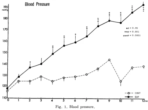
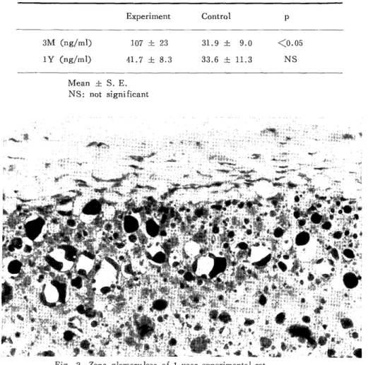
There was already a significant difference in blood pressure after 3 months, and the difference widened even more with the continuing passage of time (Fig. 1). After one year the average blood pressure was a high 191 mmHg, but there was no significant difference in body weight and in relative adrenal weight (Table 1) . Plasma corticosterone was significantly higher in the 3-month experimental group, although no significant dif- ference was encountered between the two groups in the 1-year group (Table 2). G6PD and 3(3HSD enzyme activity was noticed, but there was no difference in intensity or in staining behavior between the two groups in the 3-month and 1-year groups. The ORO revealed a tendency toward concentration of numerous lipids in the zona glomerulosa of both the 3-month and 1-year experimental groups (Fig. 2). A so-called PN-like lesion
Fig. 1. Blood pressure.
Table 1. Comparison of blood pressure, body weight and relative adrenal weight
3Months lYear
Experiment Control P Experiment Control P
Blood pressure (mmHg) 142.8± 2.4 131.6±15.6 X0.05 191± 3 137± 2 <0.001
Body weight (g) 461.0±13.1 461.4± 2.9 NS 490±18 529±14 NS
Relative adrenal weight (mg/100gBW) 12.2± 0.7 12.2± 0.2 NS 11.4±0.4 9.7±0.7 NS
Mean ± S. E.
NS: not significant
Table 2. Comparison of corticosterone
Experiment Control p
3M (ng/ml) 107 ~. 23 31.9 ± 9.0 X0.05
1Y (ng/ml) 41.7 f 8.3 33.6 ± 11.3 NS
Mean ± S. E.
NS: not significant
Fig. 2. Zona glomerulosa of 1-year experimental rat.
Concentration of lipid droplets is seen. O.R.O. X550
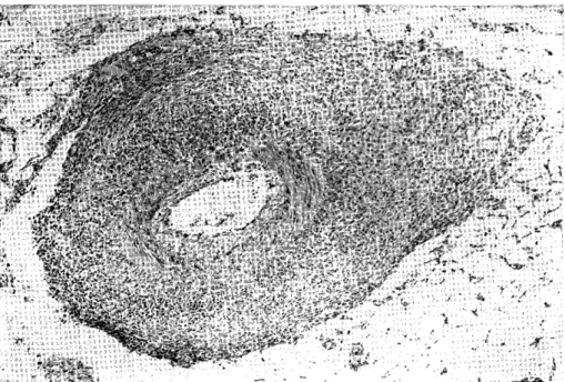
(comparatively extensive fibrinoid necrosis of the arteries in the mesentery and heart, etc. , infiltration of neutrophils and lymphocytes, and the appearance of fibroblast) was encountered light microscopically in one rat of the 1-year experimental group (Fig. 3), but aside from that there were no findings of special interest in either the 3-month or 1-year group. Morphometrical findings on the basis of electron microscopy were as fol-
lows :
a. Zona Glomerulosa
In the experimental group, the number of lipid droplets was significantly higher, and the percent area of lipid droplets was significantly greater in the experimental group.
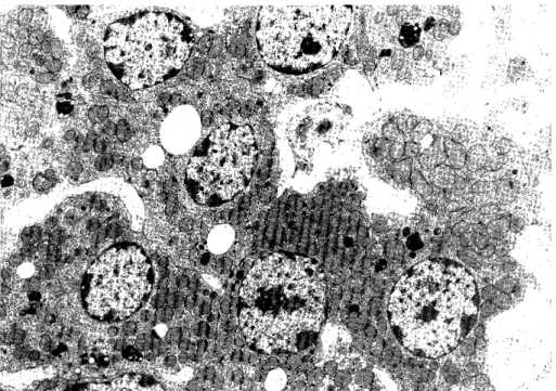
In the 1-year group, the areas of the cell, cytoplasm, lipid droplet, and mitochondrion were all significantly larger in the experimental group. The number of lipid droplets was significantly higher, and the percent area of lipid droplets was significantly greater in the experimental group, but the percent area of mitochondria was significantly greater in the control group (Fig. 4) (Table 3)
Fig. 3. A mesenteric artery of 1-year experimental rat.
Panarteritis nodosa like lesion is seen. H.E . X95
Fig. 4. Zona glomerulosa cells of 1-year experimental rat.
Numerous lipid droplets are seen. X4200
Table 3. Synopsis of morphometric parameters of the zona glomerulosa
3Months lYear
Experiment Control p Experiment Control p
Area of cell (pm2) 61.51 ±1.26 67.23±1.85 <0.05 52.87±1.20 48.49±1.22 <0.05 Area of cytoplasm (pmt) 39.22 ±1.14 42.48±1.53 NS 38.39±1.12 33.11±1.03 X0.001 Area of nucleus (pmt) 22.22±0.34 24.75±0.74 <0.01 14.43±0.45 15.39±0.46 NS Area of lipid droplet (um2) 0.67±0.02 0.73 ±0.03 NS 0.48±0.03 0.37±0.03 <0.01
Number of lipid droplets 4.23±0.43 2.44±0.54 <0.05 10.59±0.65 4.55±0.52 X0.001 Percent area of lipid roplets (%) 6.50±0.74 3.16±0.87 d 00.01 13.44 0.94 5.83±0.86 X0.001 Area of mitochondrion (um2) 0.49±0.01 0.50±0.01 N S 0.34±0.01 0.31±0.01 <0.05 Number of mitochondria 27.07±0.95 27.48±1.19 NS 35.56±1.12 37.75±1.39 NS Percent area of
mitochondria (%) 33.12±0.63 32.52±0.89 NS 29.46±0.53 33.80±0.78 <0.001 Mean ± S. E.
NS: not significant
b. Zona Fasciculata
In the 3-month group, the areas of the cell, cytoplasm, and mitochondrion were all significantly larger in the experimental group, whereas the areas of the nucleus and lipid droplet were significantly larger in the control group. The number of lipid droplets was significantly greater in the experimental group, but there was no significant differen- ce in percent areas of lipid droplets between the two groups. There was no significant difference in total area of lipid droplets either : experimental group (20.10±1.67) pmt, and control group (18.11±2.06) pmt (Fig. 5). In the 1-year group, the area of the mi- tochondrion was significantly higher in the experimental group, but no significant dif-
ference was encountered in the other parameters (Table 4).
c. Zona Reticularis
In the 3-month group, the areas of the cell and cytoplasm were significantly larger in the control group, as were the number of mitochondria, but the percent area of mi- tochondria was significantly greater in the experimental group. In the 1-year group, the areas of the cell, cytoplasm, nucleus, lipid droplet and mitochondrion were all signifi- cantly larger in the control group (Fig. 6) (Table 5).
Fig. 5. Zona fasciculata cells of 3-month experimental rat.
A crystal-shaped body (arrow) is seen. X4200
Table 4. Synopsis of morphometric parameters of the zona fasciculata
3Months lYear
Experiment Control p Experiment Control p
Area of cell (pm2) 153.82±5.55 137.90±5.11 <0.05 143.21±4.02 134.68±4.26 NS Area of cytoplasm (jam') 129.25±5.10 110.62±4.60 <0.01 118.80±3.84 112.01±4.08 NS Area of nucleus (um2) 24.57±0.74 27.28±0.92 <0.05 24.41±0.56 22.67±0.61 NS Area of lipid droplet (um2) 2.31±0.11 3.42±0.36 X0.01 1.47±0.07 1.75±0.19 NS Number of lipid droplets 8.32±0.68 5.36±0.43 <0.001 12.37±1.30 8.86±0.91 NS Percent area of lipid oplets (%) 14.71±0.95 14.87±1.49 NS 13.67±0.87 13.42±2.44 NS dr Area of mitochondrion (pmt) 0.52±0.01 0.46±0.01 <0.001 0.52±0.01 0.41±0.01 <0.001 Number of mitochondria 83.49±2.63 76.89±2.69 NS 69.92±2.22 70.49±2.90 NS
Percent area of 33.15±0.82 31.46±0.78 NS 29.86±0.56 29.42±0.89 NS mitochondria (/)
Mean ± S. E.
NS: not significant
Fig. 6. Zona reticularis cells of 1-year experimental rat.
Numerous lipofuscins are seen. X4200
Table 5. Synopsis of morphometric parameters of the zona reticularis
3Months lYear
Experiment Control p Experiment Control p
Area of cell (pmt) 104.30±1.85 119.82±3.74 X0.001 58.82±1.11 71.73±2.26 <0.001 Area of cytoplasm (pmt) 75.78±1.74 90.92±3.62 X0.001 43.31±1.00 52.55±2.04 X0.001 Area of nucleus (pIm2) 28.52±0.38 28.90±0.67 NS 15.51±0.26 19.18±0.69 <0.001 Area of lipid droplet (pmt) 3.19±0.39 4.36±0.57 NS 1.23±0.09 1.61±0.15 <0.M05 Number of lipid droplets 1.93±0.19 1.62±0.16 NS 2.38±0.23 2.04±0.25 NS Percent area of lipid oplets (/) 7.27±0.73 7.13±0.92 NS 5.77±0.63 5.60±0.81 NS dr
Area of mitochondrion (pmt) 0.54±0.01 0.56±0.01 NS 0.31±0.01 0.40±0.01 <0.001 Number of mitochondria 44.65±1.17 51.11±1.79 <0.01 44.37±1.12 44.23±1.78 NS Percent area of
mitochondria (%) 31.66±0.57 27.76±2.27 <0.05 32.65±0.55 30.54; 0.92 NS Mean ± S. E.
NS: not significant
DISCUSSION
There have been reports by numerous scholars such as HUDAK10) and MARWOOD12) concerning the production of hypertension by repeated stress application. In the present experiment a "stick-poking harassment" method was employed in which tormented rats were continually poked with bamboo chopsticks, therby involving emotional factors to a large extent. Because the tall-cuff method entails the measurement o f blood pressure during the awake state as well as the restrictional stately, the possibility, in the present experiment, that the blood pressure levels were higher than actual levels cannot be de- nied. However, the control group did not show high levels using the same method, and so the difference between the two groups could be assessed . FRIEDMAN8) emphasized the significance of a "recovery period", or period when the animals are not harassed, in emotional experimental hypertension. Because such a recovery period was not estab- lished in the present experiment, it is not possible to conclude that all of the experi- mental group suffered from fixed hypertension. Although it was only encountered in one rat, however, the 1-year experimental group displayed PN-like lesion thought to be a hypertensive change. Even if a recovery period had been established, therefore, it cannot be presumed that the blood pressure of all the experimental animals would have returned to the normal range. Moreover, intense hypertensive vascular lesions were en- countered in a preliminary experiment which, pathologically, were histological indications of malignant hypertension.
DOMOTO et alY and FISHER et al.') reported laminated crista of the mitochondria as one characteristic of a hyperfunctional state in the zona glomerulosa, and KAWAI et al.") reported an increase in number o f mitochondria. In the present experiment, neither an increase in number of mitochondria nor the presence of laminated crista was encountered. A concentration of lipid droplets was noticed in the zona glomerulosa and considered to be a relevant indication of hypo, function").
BUUCK et al.') focused their attention on the mitochondria and lipid droplets in changes of the zona fasciculata during chronic exercise, and made observations over a period ranging from 2 to 8 weeks. As a result, they encountered a continuous enlarge- ment of the mitochondrion and an early increase in number of lipid droplets, and de- scribed the relationship between the number of lipid droplets and plasma corticosterone.3) On the other hand, SHARAWY et al .21> reported an increase in plasma corticosterone and free cholesterol in the zona fasciculata after stress application. The present experi- ment as well, showed a significant increase in both the number of lipid droplets in the zona fasciculata and plasma corticosterone, but after one year the difference had disap- peared. Moreover, the mitochondrion became larger in both the 3-month and 1-year groups. NUSSDORFER et al.") said that ACTH acts to enlarge the mitochondrion') 17 , and that corticosterone feeds back, directly or indirectly, to the adrenal glands."") It is known generally that when stress is applied, an invigoration of the CRF-ACTH system deviating from the feedback mechanism takes place. It can be said that the continually
large size of the mitochondrion in the zona fasciculata during the present experiment was due to the action of an excess of ACTH caused by stress, and that . the gradual decrease in the number of lipid droplets and plasma corticosterone was due to the feedback of corticosterone to the adrenal glands. Also, one step in the process of steroid synthesis takes place in the mitochondira, and the zona fasciculata is thought to be in a hyper- functional state. RHODINI9) produced a lipid decrease in cells of the zona fasciculata during a stress experiment by injecting ACTH, and ROHR et al.20) obtained the same result in stress experiments using hypothermia and catabolism. In the zona fasciculata during the present experiment there was no significant difference encountered in total area of lipid droplets-which means a different result from the above was obtained-but the disparity is probably due to the fact that immediate effect played an influential role in the experiment by RHODIN et al., as did hypothermia and catabolism in the experiment by ROHR et al.. The pattern of results in the experiment by BUUCK et al.
is similar to that of the present experiment because stress was applied at regular intervals in both, and a space of time was left between the last application and sacrifice.
On the other hand, the time lag between the results of BUUCK's experiment and the present experiment can be accounted for by the genetic differences in the experimental animals and kinds of stress.
Corticosterone, one of the glucocorticoids, is a hypertensinogenic steroid. In the 3-month group, the corticosterone levels were significantly higher in the experimental group than in the control group, as were the blood pressure levels. The 1-year group displayed significantly high blood pressure, but no significant difference in corticosterone was noticed though the levels were still high. It can be conjectured therefore that corticosterone is involved more in the establishment than in the development of hyperten- sion.
In the present experiment using long-term harassment, a pronounced manifestation o f hypertension was confirmed. As far as the mechanism o f occurence o f hypertension and that of maintenance of hypertension are concerned, however, investigation into the relationship between hypertension and hypothalamus neurosecretion, as well as the in- volvement of the sympathetic nervous system, is still in progress. Colleagues MATSUO et al.") confirmed by measurement of catecholamine in the urine that from 4 to 7 months after commencement of the experiment there was a significant increase in epinephrine volume, and that dopamine displayed high levels after 5 months and 8-9 months. There- fore, involvement of the sympathetic nerve-adrenal medulla system too cannot be denied.
HANIU9) stated that experimental glucocorticoid hypertension is brought about by the overall action of various vasopressive agents such as the accumulation of sodium and wa- ter in the body, an increase in reactivity of the vascular wall toward norepinephrine, and invigoration of the renin-angiotensin system. Here too, it is necessary to make further investigations into the mutual relationship with the medulla and the other vasopressive agents, as well as the adrenal cortex, but in the present experiment the author concentrat- ed for the time being on the adrenal cortex and achieved morphological and morphometri- cal results in particular.
NICKERSON et al. 14) compared the zona reticularis of aging SHR with that of aging WKY. The results of their investigation showed that the cell volume, total volume of lipid droplets, total volume of mitochondria, and weight of the SHR were significantly less than that of the WKY, and blood pressure was higher in the SHR. Similar results were obtained in the present investigation of the zona reticularis. NICKERSON et al.
emphasized the genetic difference in the SHR and WKY, but the possibility that it is an effect of hypertension cannot be denied.
CONCLUSION
By long-term stress application, namely "stick-poking harassment", pronounced hy- pertension and hypertensive vascular lesion were produced in rats, though the occurence
frequency of the latter was low.
Investigations were also carried out into the relationship between the neurosecretion system and the adrenal medulla system etc., and the manifestation and maintenance o f hypertension, but the present thesis examines chiefly morphological changes in the adre- nal cortex.
The results were as follows :
1 . Hypertension o f an average 191 mmHg was produced in the 1-year group.
2. A significant rise in plasma corticosterone was encountered in the 3-month experi- mental group, but there was no significant difference in the 1-year group.
3. A PN-like lesion was noticed in the 1-year experimental group, although only in one rat.
4. A concentration o f lipid droplets was encountered in the zona g lomeru losa o f the ex- perimental groups, and considered to be a relevant indication of hypofunction.
5. A significant enlargement of the mitochondrion and proliferation of lipid droplets were noticed in the zona fasciculata of the 3-month experimental group, and a
significant enlargement o f the mitochondrion was noticed in the 1-year experimental
group and thought to be a hyperfunctional state.
6. The necessity for further investigation into the zona reticularis was perceived.
ACKNOWLEDGEMENT
The author acknowledge Professor I. NISHIMORI, Associate Professor I. SEKINE and Associate Professor M. K ISHIKA WA for their valuable advices and encouragements.
And the author would like to acknowledge Professor H. TSUCHIYAMA and Associate Professor K. KAWAI, Ilnd Department of Pathology, Nagasaki University School of Medicine, for their suggestive and valuable criticism and comments. The cooperative research students and skillful technical assistants in the Department of Pathology, Atomic Disease Institute, Nagasaki University School of Medicine are acknowledged.
REFERENCE
1) BUNAG, R. D.: Pressor effects of the tail-cuff method in awake normotensive and hypertensive rats. J. Lab. Clin. Med. 78: 675-682, 1971.
2) BURROW, G. N.: A steroid inhibitory effect on adrenal mitochondria. Endocrinolo-
gy 84: 979-985, 1969.
3) BUUCK, R. J. and THARP, G. D.: Effect of chronic exercise on adrenocortical
function and structure in rat. J. Appl. Physiol. 31: 880-883, 1971.
4) BUUCK, R. J., THAPP, G. D. and BRUMBAUGH, J. A.: Effects of chronic exercise on the ultrastructure of the adrenocortical cells in the rat. Cell. Tiss. Res. 168:
261-270, 1976.
5) CANICK, J. A. and PURVIS, J. L.: The maintenance of mitochondrial size in the rat adrenal cortex zona fasciculata by ACTH. Exp. Mol. Pathol. 16: 79-93, 1972.
6) DOMOTO, D. T., BOYD, J. E., MULROW, P. J. and KASHGARIAN, M.: The ultrastructure of the adrenal zona glomerulosa of rats on potassium-supplemented or
sodium-depleted diets. Am. J. Pathol. 72: 433-446, 1973.
7) FISHER, E. R. and HORVAT, B.: Ultrastructural features of aldosterone production.
Arch. Path. 92: 172-179, 1971.
8) FRIEDMAN, R.: Experimental psychogenic hypertension. In: Stress and the Heart, ed. Wheathey D., 2nd edition, Raven press, New York, pp. 209-228, 1981.
9) HANIU, K.: Glucocorticoid and hypertension. In: HORMONE AND HYPERTEN-
SION, ed. Yoshinaga K., CHUGAI IGAKU Co., Tokyo, pp. 57-86, 1979 (Japa- nese).
10) HUDAK, W. J. and BUCKLEY, J. P.: Production of hypertensive rats by experi- mental stress. J. Pharm. Sci. 50: 263-264, 1961.
11) KAWAI, K,, SUGIHARA, H. and TSUCHIYAMA, H.: The effect caused by the double condition of potassium loading and simultaneous sodium restriction on the glome-
rular zone of rats. Histochemical and ultrastructural study. Acta. Path. Jap. 28:
265-278, 1978.
12) MARWOOD, J. F. and LOCKETT, M. F.: Stress-induced hypertension in rats. In:
Stress and the Heart, ed. Wheathey D., 2nd edition, Raven press, New York, pp.
229-243, 1981.
13) MATSUO, K . , SEKINE, I., FUJINO, K . , NISHIMORI, I., KOIWA, T., NIWA, M. and OZAKI, M.: Experimental hypertension induced by chronic stress and urinary
catechoamines. Medicine and Biology 106: 123-127, 1983 (Japanese).
14) NICKERSON, P. A., FELD, L. G. and VANLIEW, J. B.: Zona reticularis in aging spontaneously hypertensive rats. Am. J. Pathol. 97: 433-448, 1979.
15) NUSSDORFER, G. G. and MAZZOCCHI, G.: Correlated morphometric and autoradi- ographic studies of the effects of corticosterone on adrenocortical cells of intact and
hypophysectomized ACTH-treated rats. Z. Zellforsch. 111: 90-105, 1970.
16) NUSSDORFER, G., MAZZOCCHI, G. and REBONATO, L.: Long-term trophic effect of ACTH on rat adrenocortical cells. An ultrastructural, morphometric and autora-
diographic study. Z. Zellforsch. 115: 30-45, 1971.
17) NUSSDORFER, G. G. and MAZZOCCHI, G.: A stereologic study of the effects of ACTH and cyclic 3',5'-AMP on adrenocortical cells of intact and hypophysectomized
rats. Lab. Invest. 26: 45-52, 1972.
18) NUSSDORFER, G. G., MAZZOCCHI, G. and REBUFFAT, P.: An ultrastructural
stereologic study of the effects of ACTH and adenosine 3',5'-cyclic monophosphate
on the zona glomerulosa of rat adrenal cortex. Endocrinology 92: 141-151, 1973.
19) RHODIN, J. A. G.: The ultrastructure of the adrenal cortex of the rat under normal and experimental conditions. J. Ultrastruct. Res. 34: 23-71, 1971.
20) ROHR, H. P., BARTSCH, G., PELTENBURG, M., GYsIN, C. and TRIPPEL, J.:
Stereological study of the zona fasciculata of the adrenal cortex in stress situations
(hypothermia, catabolism). Path. Res. Pract. 162: 380-397, 1978.
21) SHARAWY, M., DIRKSEN, T. and CHAFFIN, J.: Increase in free cholesterol content of the adrenal cortex after stress: Radioautographic and biochemical study. Am. J.
Anat. 156: 567-576, 1979.