• 検索結果がありません。

福島県立医科大学 学術機関リポジトリ

N/A
N/A
Protected

Academic year: 2021

シェア "福島県立医科大学 学術機関リポジトリ"

Copied!
8
0
0

読み込み中.... (全文を見る)

全文

(1)

Fukushima Medical University

福島県立医科大学 学術機関リポジトリ

This document is downloaded at: 2021-11-07T23:31:08Z

Title Contrast-enhanced harmonic endoscopic ultrasonography in gallbladder cancer and pancreatic cancer

Author(s)

Sugimoto, Mitsuru; Takagi, Tadayuki; Suzuki, Rei; Konno, Naoki; Asama, Hiroyuki; Watanabe, Ko; Nakamura, Jun;

Kikuchi, Hitomi; Waragai, Yuichi; Takasumi, Mika; Sato, Yuki; Hikichi, Takuto; Ohira, Hiromasa

Citation Fukushima Journal of Medical Science. 63(2): 39-45

Issue Date 2017

URL http://ir.fmu.ac.jp/dspace/handle/123456789/639

Rights © 2017 The Fukushima Society of Medical Science

DOI 10.5387/fms.2017-04

Text Version publisher

(2)

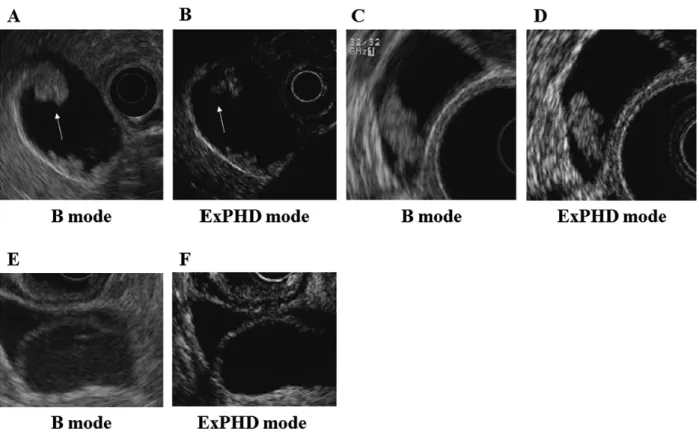
Vol. 63, No. 2, 2017

[Review]

Contrast

-

enhanced harmonic endoscopic ultrasonography in gallbladder cancer and pancreatic cancer

Mitsuru Sugimoto

1)

, Tadayuki Takagi

1)

, Rei Suzuki

1)

, Naoki Konno

1)

, Hiroyuki Asama

1)

, Ko Watanabe

1,2)

, Jun Nakamura

1,2)

, Hitomi Kikuchi

1,2)

, Yuichi Waragai

1)

, Mika Takasumi

1)

,

Yuki Sato

1)

, Takuto Hikichi

2)

and Hiromasa Ohira

1)

1)

Department of Gastroenterology, Fukushima Medical University, School of Medicine, Fukushima Ja- pan,

2)

Department of Endoscopy, Fukushima Medical University Hospital, Fukushima, Japan

(Received February 22, 2017, accepted February 22, 2017)

Abstract

Endoscopic ultrasonography (EUS) plays a major role in diagnosing gallbladder (GB) cancer and pan- creatic cancer (PC). In cases of GB cancer, EUS allows for precise observations of morphology and wall layers. However, proficiency is required for the morphologic diagnosis of GB tumors. There- fore, contrast

-

enhanced harmonic EUS (CH

-

EUS) began to be performed to diagnose GB lesions.

CH

-

EUS enables real

-

time observation of the hemodynamics of GB tumors. The enhanced pat- terns generated by CH

-

EUS improve precision in the diagnosis of such tumors.

PC appears as a hypoechoic mass on EUS. However, distinguishing between PC and mass

-

forming pancreatitis or focal autoimmune pancreatitis (AIP) is difficult via conventional EUS. CH

-

EUS al- lows for differentiating among these diseases (PC is hypoenhanced and heterogeneously enhanced, pancreatitis is isoenhanced, and a pancreatic neuroendocrine tumor is hyperenhanced). EUS

-

guid- ed fine needle aspiration (EUS

-

FNA) also contributes to pathological diagnoses of pancreatic le- sions. However, certain PC patients cannot be diagnosed via EUS

-

FNA. PC is heterogeneously enhanced on CH

-

EUS, and unenhanced regions have been reported to be areas of fibrosis or necro- sis. CH

-

EUS

-

guided fine needle aspiration (CH

-

EUS

-

FNA) permits puncturing of the enhanced area while avoiding necrotic and fibrotic regions. Moreover, as CH

-

EUS findings have been quanti- tatively analyzed, a time

-

intensity curve (TIC) has become usable for diagnosing solid pancreatic le- sions. CH

-

EUS

-

related techniques have been developed and increasingly utilized in the pancreati- cobiliary area.

Introduction

Biliary tract cancer and pancreatic cancer (PC) have poor prognoses. More precise methods for diagnosing these diseases were desired. Endo- scopic ultrasonography (EUS) has played an impor- tant role in diagnosing gallbladder (GB) cancer and PC

1-5)

. However, when EUS is used, GB cancer must be diagnosed morphologically, and it is difficult to differentiate between pancreatic inflammatory tu- mor

-

like lesions and PC

6,7)

.

Recently, contrast

-

enhanced harmonic EUS (CH

-

EUS) was reported to be useful in diagnosing

pancreaticobiliary diseases

8-27)

. In this report, we describe the efficacy of CH

-

EUS in diagnosing GB cancer and PC, including in experiments at our insti- tution.

Contrast agent for ultrasound

Ultrasound contrast agents have been classified based on their ability to cross the pulmonary arterial bed and their motion under an ultrasound beam with a low mechanical index (MI) (Table 1)

28)

.

Historically, Levovist (Schering, Berlin, Ger- many) was widely used for contrast

-

enhanced ab- Corresponding author : Tadayuki Takagi E

-

mail : [email protected]

https://www.jstage.jst.go.jp/browse/fms http://www.fmu.ac.jp/home/lib/F

-

igaku/

39

(3)

40 M. Sugimoto et al.

dominal ultrasonography. Levovist features air surrounded by a galactose shell. When Levovist is used, enhanced images are created from harmonic signals received when microbubbles are destroyed by high MI ultrasound. Therefore, for continued observation, contrast agents must be repeatedly in- jected and destroyed.

However, second

-

generation contrast agents are used today. In particular, SonoVue (Bracco, Mi- lan, Italy) and Sonazoid (GE Healthcare, Little Chal- font, United Kingdom) have been used for pancreati- cobiliary CH

-

EUS. These microbubbles of contrast agents resonate under low MI ultrasound without being destroyed. Thus, second

-

generation contrast agents have allowed for the prolonged observation of enhancement effects.

Methods of CH

-

EUS

In our hospital, the endoscopes and ultrasonic equipment used for CH

-

EUS are GF

-

UCT260 and GF

-

UE260 ultrasound gastrovideoscopes (Olympus Medical Systems, Tokyo, Japan), the ProSound α

-

10 ultrasound system (Aloka, Tokyo, Japan), and the EU

-

ME2 ultrasound processor (Olympus Medical Systems, Tokyo, Japan). Ideally, patients are ade- quately sedated with midazolam or another agent prior to endoscope insertion. After target lesions are visualized on the monitor in the B and extended pure harmonic detection (ExPHD) modes, 0.015 mL/kg of the contrast medium (16 µg of Sonazoid in 2 mL of distilled water) is injected. Subsequently, the target lesions are evaluated in the arterial and early venous phases that occur approximately 90

seconds after these lesions are enhanced

15,29)

.

CH

-

EUS using first

-

generation enhancing agents for GB cancer

Several investigators have previously described the superiority of EUS relative to abdominal ultraso- nography (US) for diagnosing neoplastic GB lesions (adenomas and adenocarcinomas)

1,2,30,31)

. However, these reports did not intend to imply that EUS can only be used to diagnose GB adenocarcinomas. The efficacy of CH

-

EUS for diagnosing GB malignant le- sions has also been reported. In 1997 and 1998, Hirooka et al.

8,10)

observed that the visualization of GB adenocarcinoma was enhanced by the contrast medium Albunex, a first

-

generation enhancing agent (Table 1). In those studies, 11 GB adenocarcinoma patients (91.7%) exhibited enhancement effects, but such effects were not observed for patients with ad- enosquamous carcinoma or cholesterol polyps.

CH

-

EUS using second

-

generation enhancing agents for GB cancer

Recently, the differentiation of GB lesions via CH

-

EUS with second

-

generation enhancing agents (SonoVue or Sonazoid) has been reported. In these reports, GB polyps were diagnosed based on en- hanced patterns.

Choi et al.

13)

and Park et al.

14)

described the effi- cacy of CH

-

EUS with the contrast medium SonoVue.

Choi et al.

13)

reported that irregular vessels observed via CH

-

EUS were useful in the diagnosis of malig- nant polyps, with a sensitivity and a specificity of Table 1. Ultrasound contrast agents

Passes through the pulmonary

arterial bed

Response MI

Diameter of microbubbles

(µm) Gas Shell Developer

Echovist

®

× 99% < 12.0

95% < 8.0 Air Galactose Schering

Albunex

®

○ High 4.3 Air Albumin MBI

Mallinckrodt

Levovist

®

○ High 2.0

-

4.0 Air Galactose Schering

Echogen

®

○ Low 3.0

-

5.0 Perfluoropentane Surfactant Sonus

Abbott

Optison

®

○ Low 3.0

-

4.5 Perfluoropentane Albumin MBI

Amersham Health Definity

®

○ Low 1.1

-

3.3 Perfluoropentane Phospholipids ImaRx, Bristol

-

Myers

ImavistTM ○ Low 5.0 Perfluoropentane Phospholipids Alliance

SonoVue

®

○ Low 2.5 Sulfur hexafluoride Phospholipids Bracco

SonazoidTM ○ Low 3.0 Perflubutane Lipids GE Healthcare

MI, Mechanical index

(4)

90.3% and 96.6%, respectively. Furthermore, per- fusion defects observed via CH

-

EUS could be used to efficiently diagnose malignant polyps, with a sen- sitivity and a specificity of 90.3% and 94.9%, respec- tively. In addition, when using SonoVue and the same procedures, Park et al.

14)

found that 80% (8/10) of GB adenocarcinomas were heterogeneously en- hanced and that 75% of GB adenomas (6/8) were ho- mogeneously enhanced.

In Japan, Sonazoid is used as the second

-

gener- ation contrast agent for EUS for pancreaticobiliary or other abdominal diseases

11,15,24-26,32-35)

. Imazu et al. performed CH

-

EUS with Sonazoid and reported finding inhomogeneously enhanced patterns that in- dicated malignant GB wall thickening. In that re- port, the sensitivity, specificity and accuracy for con- ventional EUS vs CH

-

EUS for diagnosing malignant GB wall thickening were 83.3% vs 89.6%, 65.0% vs 98.0% (p<0.001) and 73.1% vs 94.4% (p<0.001), re- spectively. Specificity and accuracy for GB wall thickening were significantly higher for CH

-

EUS

than for conventional EUS. We retrospectively re- viewed the efficacy of CH

-

EUS for diagnosing large (>10 mm) malignant and benign GB

-

protruding le- sions

17)

. In that review, the sensitivity, specificity, and malignant accuracy of CH

-

EUS were 100%

(7/7), 94.1%, (16/17) and 95.8% (23/24), respectively.

Cases treated at our hospital are presented in Figure 1.

CH

-

EUS for PC

CH

-

EUS using SonoVue or Sonazoid has been used to diagnose solid pancreatic lesions (SPLs)

18,19,21,24,26,27,36)

. CH

-

EUS can be utilized to clearly visualize SPL microvasculature and blood flow of the pancreatic parenchyma. PC has been observed as a hypoenhanced heterogeneous tumor on CH

-

EUS

18,19,24,26,27)

(Figure 2).

In 2008, Kitano et al. reported that on CH

-

EUS with SonoVue, hypovascular and heterogeneous im- ages were observed for 80% (4/5) of malignant pan- creatic lesions and isovascular images were ob-

Fig. 1. Enhanced pattern of contrast

-

enhanced harmonic endoscopic ultrasonography (CH

-

EUS) for gallbladder (GB) lesions.

A. GB cancer was detected in a B

-

mode image.

B. After contrast agent (Sonazoid) injection, GB cancer was enhanced heterogeneously in an extended pure harmonic detection (ExPHD)

-

mode image.

C. GB adenoma was detected in a B

-

mode image.

D. After contrast agent (Sonazoid) injection, GB adenoma was enhanced homogeneously in an ExPHD mode image.

E. GB cyst was detected in a B

-

mode image.

F. After contrast agent (Sonazoid) injection, a GB cyst was not enhanced in an ExPHD

-

mode image.

(5)

42 M. Sugimoto et al.

served for 100% (3/3) of pancreatitis lesions

18)

. In 2010, Fusaroli et al. stated that hypoenhanced and inhomogeneous images on CH

-

EUS with Sono

-

Vue were a good identifier for diagnosing pancreatic ade- nocarcinoma. In 2014, Park et al. found that 57 out of 62 pancreatic adenocarcinomas produced hypoen- hanced images on CH

-

EUS with Sono

-

Vue, which had a sensitivity of 92%, a specificity of 68% and an accuracy of 82%

27)

.

In 2012, Kitano et al. described findings ob- tained when using CH

-

EUS with Sonazoid to assess small (≤2 cm in diameter) SPLs

26)

. In this report, small pancreatic ductal carcinomas (n=67) appeared as hypoenhanced lesions, and the sensitivity and specificity of the tested imaging approach were 91.2% and 94.4%, respectively. This approach was superior to multidetector

-

row computed tomogra-

phy (which had a sensitivity of 70.6% and a specifici- ty of 91.9%). Furthermore, these researchers re- ported sensitivities of 90.6% and 92.2% for CH

-

EUS and EUS

-

guided fine needle aspiration (EUS

-

FNA), respectively, in patients who underwent surgical re- section of tumors (n=91), although sensitivity in- creased to 100% if findings for both procedures were considered.

CH

-

EUS

-

guided fine needle aspiration (CH

-

EUS

-

FNA) for SPLs

EUS

-

FNA is used to collect biopsy samples from many organs throughout the digestive tract and is useful for diagnosing SPLs

37-40)

. The reported di- agnostic accuracy, sensitivity, and specificity of EUS

-

FNA for SPLs are 85

-

89.4%, 82

-

94.7%, and 100%, Fig. 2. Enhanced pattern of pancreatic cancer (PC) on contrast

-

enhanced harmonic EUS (CH

-

EUS) and CH

-

EUS

-

guided fine needle aspiration (CH

-

EUS

-

FNA).

A. PC was imaged using B mode.

B. (Left : B mode ; right : extended pure harmonic detection (ExPHD) mode) CH

-

EUS was performed and vi- sualized using ExPHD mode. PC was hypoenhanced and enhanced heterogeneously on CH

-

EUS.

C. (Left : B mode ; right : ExPHD mode) Fine needle aspiration was performed in the enhanced region of PC.

(6)

respectively

41-43)

. However, certain SPL patients could not be diagnosed via EUS

-

FNA. In 2015, Hou et al. described findings obtained using CH

-

EUS

-

FNA with Sono

-

Vue

44)

. They punctured SPLs after they had confirmed these lesions’ enhanced patterns and retrospectively analyzed 58 cases in- volving CH

-

EUS

-

FNA and 105 cases involving EUS

-

FNA. Sufficient biopsy specimens were more frequently obtained in the CH

-

EUS group (96.6%) than in the EUS

-

FNA group (86.7%).

As mentioned above, PC appears as hypoen- hanced heterogeneous tumors on CH

-

EUS. Unen- hanced areas reportedly reflect necrosis and fibro- sis

45)

. Therefore, we performed CH

-

EUS

-

FNA by puncturing the enhanced region of SPLs

33)

(Figure 2). In our report, sufficient biopsy samples were obtained with a single needle pass for 60% (12/20) of the CH

-

EUS

-

FNA group compared with 25% (5/20) of the conventional EUS

-

FNA group (P=0.027). In many cases, when EUS

-

FNA is performed, four needle passes are needed to obtain sufficient biopsy samples

46)

. In all reports about pancreatic tumor seeding associated with EUS

-

FNA, multiple needle punctures were performed

47-51)

. CH

-

EUS

-

FNA is expected to require a minimal number of needle passes.

Quantitative evaluation of CH

-

EUS for SPLs In the reports described above, enhanced pat- terns were judged subjectively. However, as CH

-

EUS findings for SPLs have been subjected to objec- tive quantitative analyses, a time

-

intensity curve (TIC) has been determined.

In 2011, Matsubara et al. observed that the rate of echo intensity reduction from the peak at one minute was greatest for PC, followed by mass

-

form- ing pancreatitis, autoimmune pancreatitis (AIP), and pancreatic neuroendocrine tumors

24)

. The reported sensitivity, specificity, and accuracy of EUS with a TIC were 95.8%, 92.6%, and 94.7%, respectively. In 2012, Imazu et al. reported that peak intensity and maximum intensity gain were significantly higher for AIP than for PC

25)

.

In 2015, Saftoiu et al. described TIC analysis with an artificial neural network classification model for diagnosing PC and chronic pancreatitis

52)

. The sensitivity, specificity, positive predictive value, and negative predictive value for this approach were 94.64%, 94.44%, 97.24%, and 89.4%, respectively.

Conclusion

CH

-

EUS has been reported to be extremely ef- fective in the diagnosis of pancreaticobiliary diseas- es. The combination of CH

-

EUS with EUS

-

FNA recently began being utilized ; furthermore, tech- niques to quantitatively evaluate enhancement ef- fects, such as determination of a TIC, have been performed. Procedures related to CH

-

EUS play a major role in the pancreaticobiliary field.

References

1. Sugiyama M, Atomi Y & Yamato T. Endoscopic ultrasonography for differential diagnosis of polyp- oid gall bladder lesions : analysis in surgical and follow up series. Gut, 46 : 250

-

254, 2000.

2. Kimura K, Fujita N, Noda Y, Kobayashi G & Ito K. Differential diagnosis of large

-

sized peduncu- lated polypoid lesions of the gallbladder by endo- scopic ultrasonography : a prospective study.

J Gastroenterol, 36 : 619

-

622, 2001.

3. DeWitt J, Devereaux B, Chriswell M, et al. Com- parison of endoscopic ultrasonography and multi- detector computed tomography for detecting and staging pancreatic cancer. Ann Intern Med, 141 : 753

-

763, 2004.

4. Tamm EP, Loyer EM, Faria SC, Evans DB, Wolff RA & Charnsangavej C. Retrospective analysis of dual

-

phase MDCT and follow

-

up EUS/EUS

-

FNA in the diagnosis of pancreatic cancer. Abdom Im- aging, 32 : 660

-

667, 2007.

5. Azuma T, Yoshikawa T, Araida T & Takasaki K.

Differential diagnosis of polypoid lesions of the gallbladder by endoscopic ultrasonography. Am J Surg, 181 : 65

-

70, 2001.

6. Abraham SC, Wilentz RE, Yeo CJ, et al. Pancreati- coduodenectomy (Whipple resections) in patients without malignancy : are they all ‘chronic pancre- atitis’? Am J Surg Pathol, 27 : 110

-

120, 2003.

7. Weber SM, Cubukcu

-

Dimopulo O, Palesty JA, et al. Lymphoplasmacytic sclerosing pancreatitis : inflammatory mimic of pancreatic carcinoma. J Gastrointest Surg, 7 : 129

-

137 ; discussion 137

-

129, 2003.

8. Hirooka Y, Naitoh Y, Goto H, Ito A, Taki T & Haya- kawa T. Usefulness of contrast

-

enhanced endo- scopic ultrasonography with intravenous injection of sonicated serum albumin. Gastrointest En- dosc, 46 : 166

-

169, 1997.

9. Mizuguchi M, Kudo S, Fukahori T, et al. Endo-

scopic ultrasonography for demonstrating loss of

multiple

-

layer pattern of the thickened gallbladder

wall in the preoperative diagnosis of gallbladder

cancer. Eur Radiol, 7 : 1323

-

1327, 1997.

(7)

44 M. Sugimoto et al.

10. Hirooka Y, Naitoh Y, Goto H, et al. Contrast

-

en- hanced endoscopic ultrasonography in gallbladder diseases. Gastrointest Endosc, 48 : 406

-

410, 1998.

11. Imazu H, Uchiyama Y, Matsunaga K, et al. Con- trast

-

enhanced harmonic EUS with novel ultraso- nographic contrast (Sonazoid) in the preoperative T

-

staging for pancreaticobiliary malignancies.

Scand J Gastroenterol, 45 : 732

-

738, 2010.

12. Inui K, Yoshino J & Miyoshi H. Diagnosis of gall- bladder tumors. Intern Med, 50 : 1133

-

1136, 2011.

13. Choi JH, Seo DW, Choi JH, et al. Utility of con- trast

-

enhanced harmonic EUS in the diagnosis of malignant gallbladder polyps (with videos).

Gastrointest Endosc, 78 : 484

-

493, 2013.

14. Park CH, Chung MJ, Oh TG, et al. Differential di- agnosis between gallbladder adenomas and choles- terol polyps on contrast

-

enhanced harmonic endo- scopic ultrasonography. Surg Endosc, 27 : 1414

-

1421, 2013.

15. Imazu H, Mori N, Kanazawa K, et al. Contrast

-

enhanced harmonic endoscopic ultrasonography in the differential diagnosis of gallbladder wall thick- ening. Dig Dis Sci, 59 : 1909

-

1916, 2014.

16. Xu JM, Guo LH, Xu HX, et al. Differential diagno- sis of gallbladder wall thickening : the usefulness of contrast

-

enhanced ultrasound. Ultrasound Med Biol, 40 : 2794

-

2804, 2014.

17. Sugimoto M, Takagi T, Konno N, et al. The effica- cy of contrast

-

enhanced harmonic endoscopic ul- trasonography in diagnosing gallbladder cancer.

Sci Rep, 6 : 25848, 2016.

18. Kitano M, Sakamoto H, Matsui U, et al. A novel perfusion imaging technique of the pancreas : con- trast

-

enhanced harmonic EUS (with video).

Gastrointest Endosc, 67 : 141

-

150, 2008.

19. Fusaroli P, Spada A, Mancino MG & Caletti G.

Contrast harmonic echo

-

endoscopic ultrasound improves accuracy in diagnosis of solid pancreatic masses. Clin Gastroenterol Hepatol, 8 : 629

-

634 e621

-

622, 2010.

20. Ishikawa T, Itoh A, Kawashima H, et al. Useful- ness of EUS combined with contrast

-

enhancement in the differential diagnosis of malignant versus be- nign and preoperative localization of pancreatic en- docrine tumors. Gastrointest Endosc, 71 : 951

-

959, 2010.

21. Napoleon B, Alvarez

-

Sanchez MV, Gincoul R, et al. Contrast

-

enhanced harmonic endoscopic ul- trasound in solid lesions of the pancreas : results of a pilot study. Endoscopy, 42 : 564

-

570, 2010.

22. Seicean A, Badea R, Stan

-

Iuga R, Mocan T, Gulei I

& Pascu O. Quantitative contrast

-

enhanced har- monic endoscopic ultrasonography for the discrimi- nation of solid pancreatic masses. Ultraschall

Med, 31 : 571

-

576, 2010.

23. Ang TL, Teo EK, Ang D, Kwek AB & Fock KM.

A pilot study of contrast harmonic endosonography using DEFINITY in the evaluation of suspected pancreatic and peri

-

ampullary malignancies. J In- terv Gastroenterol, 1 : 160

-

165, 2011.

24. Matsubara H, Itoh A, Kawashima H, et al. Dy- namic quantitative evaluation of contrast

-

enhanced endoscopic ultrasonography in the diagnosis of pancreatic diseases. Pancreas, 40 : 1073

-

1079, 2011.

25. Imazu H, Kanazawa K, Mori N, et al. Novel quan- titative perfusion analysis with contrast

-

enhanced harmonic EUS for differentiation of autoimmune pancreatitis from pancreatic carcinoma. Scand J Gastroenterol, 47 : 853

-

860, 2012.

26. Kitano M, Kudo M, Yamao K, et al. Characteriza- tion of small solid tumors in the pancreas : the val- ue of contrast

-

enhanced harmonic endoscopic ul- trasonography. Am J Gastroenterol, 107 : 303

-

310, 2012.

27. Park JS, Kim HK, Bang BW, Kim SG, Jeong S &

Lee DH. Effectiveness of contrast

-

enhanced har- monic endoscopic ultrasound for the evaluation of solid pancreatic masses. World J Gastroenterol, 20 : 518

-

524, 2014.

28. Sanchez MV, Varadarajulu S & Napoleon B.

EUS contrast agents : what is available, how do they work, and are they effective ? Gastrointest Endosc, 69 : S71

-

77, 2009.

29. Piscaglia F, Nolsoe C, Dietrich CF, et al. The EF- SUMB Guidelines and Recommendations on the Clinical Practice of Contrast Enhanced Ultrasound (CEUS) : update 2011 on non

-

hepatic applica- tions. Ultraschall Med, 33 : 33

-

59, 2012.

30. Sugiyama M, Xie XY, Atomi Y & Saito M.

Differential diagnosis of small polypoid lesions of the gallbladder : the value of endoscopic ultraso- nography. Ann Surg, 229 : 498

-

504, 1999.

31. Sadamoto Y, Oda S, Tanaka M, et al. A useful ap- proach to the differential diagnosis of small polyp- oid lesions of the gallbladder, utilizing an endoscop- ic ultrasound scoring system. Endoscopy, 34 : 959

-

965, 2002.

32. Kitano M, Kamata K, Imai H, et al. Contrast

-

en- hanced harmonic endoscopic ultrasonography for pancreatobiliary diseases. Dig Endosc, 27 Suppl 1 : 60

-

67, 2015.

33. Sugimoto M, Takagi T, Hikichi T, et al. Conven- tional versus contrast

-

enhanced harmonic endo- scopic ultrasonography

-

guided fine

-

needle aspira- tion for diagnosis of solid pancreatic lesions : A prospective randomized trial. Pancreatology, 15 : 538

-

541, 2015.

34. Xia Y, Kitano M, Kudo M, et al. Characterization

of intra

-

abdominal lesions of undetermined origin

(8)

by contrast

-

enhanced harmonic EUS (with vid- eos). Gastrointest Endosc, 72 : 637

-

642, 2010.

35. Sakamoto H, Kitano M, Matsui S, et al. Estima- tion of malignant potential of GI stromal tumors by contrast

-

enhanced harmonic EUS (with videos).

Gastrointest Endosc, 73 : 227

-

237, 2011.

36. Michiko Y, Yoshimitsu Y, Yasuyuki S & Hideyuki H.

Education and Imaging. Gastrointestinal : con- trast

-

enhanced harmonic EUS for pancreatic can- cer. J Gastroenterol Hepatol, 24 : 1698, 2009.

37. Vilmann P, Jacobsen GK, Henriksen FW & Hancke S. Endoscopic ultrasonography with guided fine needle aspiration biopsy in pancreatic disease.

Gastrointest Endosc, 38 : 172

-

173, 1992.

38. Bhutani MS, Hawes RH, Baron PL, et al. Endo- scopic ultrasound guided fine needle aspiration of malignant pancreatic lesions. Endoscopy, 29 : 854

-

858, 1997.

39. Voss M, Hammel P, Molas G, et al. Value of endo- scopic ultrasound guided fine needle aspiration bi- opsy in the diagnosis of solid pancreatic masses.

Gut, 46 : 244

-

249, 2000.

40. Chang KJ, Katz KD, Durbin TE, et al. Endoscopic ultrasound

-

guided fine

-

needle aspiration. Gas- trointest Endosc, 40 : 694

-

699, 1994.

41. Williams DB, Sahai AV, Aabakken L, et al. Endo- scopic ultrasound guided fine needle aspiration bi- opsy : a large single centre experience. Gut, 44 : 720

-

726, 1999.

42. Eloubeidi MA, Jhala D, Chhieng DC, et al. Yield of endoscopic ultrasound

-

guided fine

-

needle aspi- ration biopsy in patients with suspected pancreatic carcinoma. Cancer, 99 : 285

-

292, 2003.

43. Ryozawa S, Kitoh H, Gondo T, et al. Usefulness of endoscopic ultrasound

-

guided fine

-

needle aspi- ration biopsy for the diagnosis of pancreatic can- cer. J Gastroenterol, 40 : 907

-

911, 2005.

44. Hou X, Jin Z, Xu C, et al. Contrast

-

enhanced har- monic endoscopic ultrasound

-

guided fine

-

needle aspiration in the diagnosis of solid pancreatic le-

sions : a retrospective study. PLoS One, 10 : e0121236, 2015.

45. D’Onofrio M, Zamboni G, Tognolini A, et al.

Mass

-

forming pancreatitis : value of contrast

-

en- hanced ultrasonography. World J Gastroenterol, 12 : 4181

-

4184, 2006.

46. Suzuki R, Irisawa A, Bhutani MS, et al. Prospec- tive evaluation of the optimal number of 25

-

gauge needle passes for endoscopic ultrasound

-

guided fine

-

needle aspiration biopsy of solid pancreatic le- sions in the absence of an onsite cytopathologist.

Dig Endosc, 24 : 452

-

456, 2012.

47. Micames C, Jowell PS, White R, et al. Lower fre- quency of peritoneal carcinomatosis in patients with pancreatic cancer diagnosed by EUS

-

guided FNA vs. percutaneous FNA. Gastrointest En- dosc, 58 : 690

-

695, 2003.

48. Paquin SC, Gariepy G, Lepanto L, Bourdages R, Raymond G & Sahai AV. A first report of tumor seeding because of EUS

-

guided FNA of a pancre- atic adenocarcinoma. Gastrointest Endosc, 61 : 610

-

611, 2005.

49. Ahmed K, Sussman JJ, Wang J & Schmulewitz N.

A case of EUS

-

guided FNA

-

related pancreatic can- cer metastasis to the stomach. Gastrointest En- dosc, 74 : 231

-

233, 2011.

50. Chong A, Venugopal K, Segarajasingam D &

Lisewski D. Tumor seeding after EUS

-

guided FNA of pancreatic tail neoplasia. Gastrointest Endosc, 74 : 933

-

935, 2011.

51. Katanuma A, Maguchi H, Hashigo S, et al. Tumor seeding after endoscopic ultrasound

-

guided fine

-

needle aspiration of cancer in the body of the pan- creas. Endoscopy, 44 Suppl 2 UCTN : E160

-

161, 2012.

52. Saftoiu A, Vilmann P, Dietrich CF, et al. Quantita-

tive contrast

-

enhanced harmonic EUS in differen-

tial diagnosis of focal pancreatic masses (with vid-

eos). Gastrointest Endosc, 82 : 59

-

69, 2015.

Fig. 1.  Enhanced pattern of contrast - enhanced harmonic endoscopic ultrasonography (CH - EUS) for gallbladder  (GB) lesions.

参照

関連したドキュメント

[r]

静岡大学 静岡キャンパス 静岡大学 浜松キャンパス 静岡県立大学 静岡県立大学短期大学部 東海大学 清水キャンパス

[r]