− 1 −
9
1. Introduction
To visualize the inside of biomedical objects, we usually use X-ray computed tomography (CT) scanners corresponding to the objectives, and we have developed several photon-counting energy-dispersive CT scanners [1-3]. Using iodine and gadolinium contrast media, enhanced K-edge CT has been performed to image blood vessels with high contrasts.
Recently, we have constructed near-infrared-ray (NIR) CT scanners [4-6] with wavelengths of 850 and Abstract
To perform computed tomography (CT) using visible-ray (VR) and near-infrared-ray (NIR) photons, we measured penetrating-photon spectra using a white power light-emitting diode (LED). Penetrating- photon spectra through the object were measured using a spectrometer through a 1.5-m-length light fiber, and the wavelength was calibrated using two LEDs with wavelengths of 567 and 630 nm. The white power LED produced VR and NIR photons with wavelengths ranging from 400 to 900 nm, and the spectra had two peaks of 444 and 565 nm. The penetrating peak wavelengths of 3-mm-thick chicken breast, human finger and glass vial filled with 10-mg/ml indigo carmine solutions were 570, 616 and 787 nm, respectively.
(Accepted December 6, 2019)
Keywords:penetrating-photon spectra, white power LED, human-body window, living-body window, visible rays, near-infrared rays
a
Department of Physics, Iwate Medical University, 1-1-1 Idaidori, Yahaba, Iwate 028-3694, Japan
b
Department of Radiology, School of Medicine, Iwate Medical University, 2-1-1 Idaidori, Yahaba, Iwate 028-3695, Japan
c
Department of Surgery, Toho University Ohashi Medical Center, 2-22-36 Ohashi, Meguro, Tokyo 153-8515, Japan
Eiichi SATO
a, Yasuyuki ODA
a, Sohei YOSHIDA
b, Satoshi YAMAGUCHI
b, Kunihiro YOSHIOKA
b, Hodaka MORIYAMA
c, Osahiko HAGIWARA
c,
Toshiyuki ENOMOTO
c, Manabu WATANABE
cMeasurement of penetrating-photon spectra using a white power light-emitting diode
Annual Report of Iwate Medical University Center for Liberal Arts and Sciences No. 54(2019), 9-12.
− 2 − Eiichi SATO et al.
10
940 nm to image biomedical objects using penetrating primary photons. However, it was difficult to detect penetrating photons, since most NIR photons were absorbed by the objects. In this regard, we developed a high- sensitivity detector with a photo diode, and NIR-CT has been carried out using penetrating photons.
In the present research, our major objectives are as follows: to form a line beam using a white power LED, to construct an experimental setup for measuring spectra, and to measure spectra penetrating through the objects. Therefore, we constructed the setup and measured spectra with wavelengths ranging from 400 to 900 nm.
2. Methods
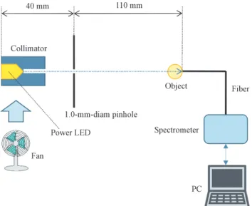
Figure 1 shows the experimental setup for measuring penetrating-photon spectra through three objects of a 3-mm-thickness chicken breast, a human finger and a glass vial filled with 10-mg/ml indigo-carmine solution.
The white line beam is formed using a 1.0-mm-diameter 10-mm-length graphite collimator and a 1.0-mm- diameter copper pinhole, and irradiated to the object. The penetrating photons are detected using a spectrometer (Hamamatsu, C10083CA) through a 1.5-m-length light fiber. The measured spectra are observed on the monitor and recorded by the personal computer (PC). The wavelength is calibrated using two LEDs of 567-nm yellow green (Sanyo, SLP-3118C-5) and 630-nm red (Sanyo, SLP-9118B-51-T1). The power LED with an aluminum heat-dissipation substrate is cooled using an electric fan. To measure two line spectra for the calibration, the same spectrometer was used, and the fiber tip was set to 10 cm from the LED without a collimator and a pinhole.
The electric circuit for driving the LED is shown in Fig. 2. The LED is connected directly to a DC power supply with a voltmeter and an ammeter, and the DC forward current is regulated by the applied forward voltage. The maximum values of the DC forward current, forward voltage and electric power are 1,500 mA, 3.25 V and 4.9 W, respectively (Table 1).
Fig. 1. Experimental setup for measuring spectra penetrating through the object using the white power LED. The line beam is formed using a graphite collimator and a copper pinhole, and irradiated to the object just in front of the light fiber.
Fig. 2. Electric circuit for driving the LEDs. The DC power supply is connected directly to the LED, and the forward voltage and current were measured using the voltmeter and ammeter in the power supply, respectively.
Characteristics Maximum value DC forward current (mA) 1,500
Forward voltage (V) 3.25 Electric power (W) 4.9
Table 1. Major characteristics of the power LED.
− 3 −
Measurement of penetrating-photon spectra using a white power light-emitting diode 11 3. Results
The VR spectra irradiated from the two LEDs for calibrating wavelength are shown in Fig. 3. The line spectra were sharp, and the peak wavelengths of the yellow green and red LEDs were 567 and 630 nm, respectively. These values corresponded well to those in the data table (Table 2), and we did not carry out any calculations for the calibration. Figure 4 shows the spectra from the white power LED. The spectra had two peak wavelengths, and the first and second peaks were 444 and 565 nm, respectively.
The spectra penetrating through the three objects are shown in Fig. 5 and Table 3. The spectra through the 3-mm-thickness chicken breast also had two peaks of 445 and 570 nm, and the first peak was lower than the second [Fig. 5(a)]. The spectra from the finger in the human-body window (HBW) had a peak of 616 nm [Fig.
5(b)], and the peak from the vial filled with indigo-carmine solution was 787 nm [Fig. 5(c)].
Object Penetrating peak wavelength (nm)
10-mg-ml indigo carmine 787
3-mm-thick chicken breast 570
Human finger 616
Table 3. Penetrating peak wavelength of the three objects.
Model number Peak-wavelength
data (nm) Measured peak wavelength (nm)
SLP-3118C-5 567 567
SLP-9118B-51-T1 630 630
Table 2. Peak wavelength of the yellow-green and red LEDs.
Fig. 3. VR spectra from the yellow-green and red LEDs for calibrating the wavelength.
Fig. 4. VR and NIR spectra from the power LED.
Fig. 5. Penetrating spectra through the three objects. (a) Spectra penetrating through the 3-mm- thickness chicken breast, (b) the human finger, and (c) the vial filled with 10-mg/ml indigo- carmine solution.
− 4 − Eiichi SATO et al.
12
4. Discussion
The white power LED is useful for measuring the spectra penetrating through the objects, and a wide variety of objects can be imaged using the CT scanner with the power LED. In the measurement of spectra, short-wavelength photons were absorbed by the chicken breast.
The peak wavelength of the HBW spectra is very important to perform CT for small animals. The first- peak photons of the power LED were absorbed by the muscle and blood, and the second LED peak shifted to long wavelength by the absorption.
Short-wavelength photons were absorbed effectively by the indigo-carmine solution, and the peak wavelength was in the first living-body window range of 700-900 nm determined by the absorption coefficients of water and hemoglobin. In addition, this solution may be used as an infrared filter, since the VR photons are absorbed.
In this experiment, we measured penetrating spectra including scattering photons, because the photon number substantially decreased using a second pinhole for reducing scattering photons from the objects.
5. Conclusion
We measured the spectra penetrating through three objects using a white power LED, and the peak wavelength varied substantially depending on the kind of object. Therefore, the VR-CT using the white power LED might be useful for imaging biomedical objects.
Acknowledgments
This work was supported by Grants from Promotion and Mutual Aid Corporation for Private Schools of Japan, Japan Science and Technology Agency (JST), and JSPS KAKENHI (17K10371, 17K09068, 17K01424, 17H00607). This was also supported by a Grant-in-Aid for Strategic Medical Science Research (S1491001, 2014–2018) from the Ministry of Education, Culture, Sports, Science and Technology of Japan.
References
[1] Matsukiyo, H., Sato, E., Oda, Y., Ishii, T., Yamaguchi, S., Sato, Y., Hagiwara, O., Enomoto, T., Watanabe, M., Kusachi, S., “Investigation of quad-energy photon counting for X-ray computed tomography using a cadmium telluride detector,” Appl. Radiat. Isot. 130, 54-59 (2017).
[2] Sato, E., Sato, T., Oda, Y., Sato, Y., Yoshida, S., Yamaguchi, S., Hagiwara, O., Matsukiyo, H., Enomoto, T., Watanabe, M., Kusachi, S., “Triple-energy high-count-rate X-ray computed tomography scanner using a cadmium telluride detector,” Health Technol. 8, 197-203 (2018).
[3] Moriyama, H., Watanabe, M., Kusachi, S., Oda Y., Sato, E., “Low-dose low-scattering X-ray computed tomography with high-spatial-energy resolutions using a cooled cadmium telluride detector,”
Ultramicroscopy 199, 62-69 (2019).
[4] Sato, E., Oda, Y., Sato, Y., Yamaguchi, S., Ishii, T., Hagiwara, O., Matsukiyo, H., Watanabe, M., Kusachi, S.,
“Investigation of a near-infrared-ray computed tomography scanner,” Proc. SPIE 9969, 99690I-1-6 (2016).
[5] Sato, Y., Takaoka, A., Sato, T., Sato, E., Oda, Y., Yoshida, S., Moriyama, H., Hagiwara, O., Matsukiyo, H., Enomoto, T., Watanabe, M., Kusachi, S., “850-nm-peak high-sensitivity near-infrared-ray computed tomography scanner in the living-body window,” Health Technol. 8, 205-210 (2018).
[6] Sato, E., Oda, Y., Sato, Y., Yoshida, S., Moriyama, H., Watanabe, M., “850-nm near-infrared-ray computed tomography with high spatial resolutions,” Proc. SPIE 11073, 110730N-1-6 (2019).