Mechanism of Manganese Uptake by a Green Alga,
Dunaliella tertiolecta Butcher
著者
NORO Tadahide
journal or
publication title
鹿児島大学水産学部紀要=Memoirs of Faculty of
Fisheries Kagoshima University
volume
34
number
2
page range
183-244
別言語のタイトル
緑藻Dunaliella tertiolecta Butcherにおけるマン
ガンの吸収機構
Vol. 34, No. 2, pp. 183-244 (1985)
Mechanism of Manganese Uptake by a Green Alga,
Dunaliella tertiolecta Butcher *]
Tadahide Noro *2
Abstract
Although trace metals are present at low concentrations in seawater, algae are capable of accumulating such metals in high concentrations against a gradient. In order to clarify the uptake mechanism of trace metals, manganese (Mn) uptake from a culture medium by a euryhaline alga, Dunaliella tertiolecta BUTCHER was studied.
D. tertiolecta was affected physiologically by Mn in the medium. Higher growth rates and
evident elongation in cell length were observed from 0.1 to 1.0 ppm Mn in contrast to other concentrations tested. Crude protein and carbohydrate increased with increasing Mn concentra tions with an optimum value at 0.05 to 0.1 ppm Mn. At 1.0 to 10 ppm Mn, protein and carbohydrate synthesis was inhibited. Chlorophyll-a content did not diminish in an Mn deficient medium, but apparently decreased when both Mn and Fe are deficient.
Live D. tertiolecta cells accumulated Mn from the culture medium subject to environmental factors. Maximum uptake of Mn occured at pH 9. 0, 10,000 lux and 20*C temperature. Dead cells are unable to take up Mn from the medium and, instead released accumulated Mn to the cell exterior. Mn uptake was activated during early exponential growth phase of the batch culture. It also synchronized with cell growth, with Mn content reduced at cell division and increased with cell growth. Mn levels inside the cell elevated sharply with increasing levels of exogenous Mn from 0. 0 to 0.1 ppm. The absorption systems was apparently saturated at 0.1 ppm Mn since further increase up to 1.0 ppm external Mn resulted in virtually no further
increase in the Mn content of D. tertiolecta. This saturation kinetics followed Michaelis Menten equation. Mn concentration factor in D. tertiolecta was 10-fold at 0. 3 to 5. 0 ppm and 100-fold at 0.1 ppm Mn in the medium. Mn uptake was inhibited by DCMU, TPAC, and KCN, and stimulated by respiratory substances, glucose. Zn and Cu ions in the medium also inhibited accumulation, while Na, K, Mg, Ca, P and Fe accelerated uptake of Mn. In D. tertiolecta, 72. 8% of accumulated Mn was found in the tonoplast, 25.1% in protoplast as free ion or low molecular weight compound, and only 2.1 % as firmly bounded to organelles or protoplast.
The presence of Mn-stimulated ATPase was confirmed in whole cell homogena-tes. Mn-ATPase activity was one-tenth lower than that in Na, K-ATPase, and was strongly temperature dependent with an optimum at 25*C. Optimum pH for the activity was pH 8. 0. With
* 1 Dissertation for the degree of Doctor of Fisheries Science at the Graduate School of Hokkaido
University (1985).
*2 Laboratory of Marine Botany, Faculty of Fisheries, Kagoshima University, 50—20 Shimoarata 4, Kagoshima City, 890 Japan.
184 Mem. Fac. Fish., Kagoshima Univ. Vol. 34, No. 2 (1985)
increasing Mn concentration, Mn-ATPase activity rose and reacheda limiting value at 1.0 mM
Mn (=55 ppm Mn). At higher concentrations, activity slowly diminished and eventually vanished at 8.0 mM Mn (=440 ppm Mn). Mn-ATPase was inhibited strongly by oligomycin and weakly by ouabine. The activity of Mn-ATPase was detected on cell plasma membranes.
A highly significant correlation was noted between Mn-ATPase activity and Mn influx at concentrations of 0.0 to 5.0 ppm Mn (P<0.025 ; r=0.896).
Form these results, Mn uptake mechanism of D. tertiolecta outlines an active transport
process. Channels or pumps in the cell membrane are activated by energy from Mn-ATPase,
which then takes up Mn from outside into the cell. Accumulated Mn are stored mostly in the
form of ions or low molecular weight compounds near the cell membrane, while minimum amounts are transported to organelles i. e., chloroplast. Trace metal accumulation in other algae are likely to follow a similar mechanism found in D. tertiolecta.
Table of contents
I. Introduction 185
II. State of manganese, taken up by Dunaliella tertiolecta Butcher 186
II -1. Introduction
II -2. Matetials and methods II -3. Results and discussion
HI. Structure of D. tertiolecta BUTCHER in relation to transport of manganese 189
HI—1. Introduction
III-2. Materials and methods ID-3. Results and discussion
IV. Effect of manganese on the growth and the metabolism in D. tertiolecta BUTCHER 195
Bf-1. Introduction
IV-2. Materials and methods IV-3. Results
(1) Growth rate
(2) Protein and carbohydrate content (3) Chlorophyll content
IV-4. Discussion
V. Manganese uptake and release in D. tertiolecta BUTCHER 200
V-l. Introduction
V-2. Materials and methods
V-3. Results
(1) Mn uptake as a function of time (2) Mn uptake as a function of cell density (3) Mn uptake in synchronous culture (4) Mn uptake in batch culture
(5) Mn uptake as a function of Mn concentration in the medium (6) Effect of environmental factors on Mn uptake
(7) Effect of glucose on Mn uptake
(8) Effect of metabolic inhibitors on Mn uptake and release (9) Effect of other ions on Mn uptake
(10) Effect of EDTA on Mn uptake
V~4. Discussion
VI. Distribution of Mn in the cell of D. tertiolecta BUTCHER 219 VI-1. Introduction
VI-2. Materials and methods VI-3. Results and discussion
1. Manganese sensitivity of ATPase in D. tertiolecta BUTCHER 221
W-l. Introduction
VI-2. Materials and methods 1-3. Results
(1) ATPase activity in whole homogenate of D. tertiolecta (2) Mn-ATPase activity as a function of time
(3) Effect of temperature on the activity of Mn-ATPase (4) Effect of pH on the activity of Mn-ATPase
(5) Effect of Mn concentration on the activity of Mn-ATPase (6) Effect of cations on the activity of Mn-ATPase
(7) Effect of metabolic inhibitor on the activity of Mn-ATPase
(8) Localization of Mn-ATPase in the cell of D. tertiolecta
MI-4. Discussion
VII. General discussion : Manganese uptake mechanism by D. tertiolecta BUTCHER 232
Acknowledgements 236
References 236
I. Introduction
In general, higher green plants take nutrients by their root. However, algae, whether marine or freshwater organisms, benthic or planktonic organisms, absorb many kinds of elements dissolved in the aquatic environment mainly through the surface of plants. This characteristic feature makes the algae favorable material for experiments of the nutrient utilization.
These nutrient elements are usually divided into macro-and micro-nutrients ( or trace metals ), though there is no clear differentiation between two definitions of nutrients. Most macro-nutrients (e. g. P, N, and C) are needed for the medium of algal culture at concentrations over 1 ppm, while micro-nutrients ( e. g. Fe, Al, Zn, Mn and Co ) are
enough at concentrations less than the above value. The concentration of micro-nutrients or
trace metals are quite low in the sea, but algae can highly accumulate them against a
gradient in concentration.
The existence of trace element in various forms in the natural water is recently becoming better known by examing nutrient elements which phytoplankton selectively utilized at
186 Mem. Fac. Fish., Kagoshima Univ. Vol. 34, No.2 (1985)
certain time of the year or by the presence of elements adsorbed by other colloidal particles. Although such elements may vary in concentration with depth and location, Fe, Al, Mo and
Zn are generally estimated to be in the order of 10 ppb, Cu, Mn, Ni and Ti in the order of 1 ppb and Co and Cr in the order of 0.1 ppb. Bowen (1966) reviewed the data published so far
on the mean values of trace elements concentrated by green, brown and red marine algae and showed that many algae could accumulate the elements at 103 to 104times or more as much as the sea contained, though their content was quite scanty in the environment.
Of these micro-nutrients, manganese is one of the most interesting elements. Manganese plays the roles of catalytic, activating or diminishing reaction for some enzyme in the following various ways : (1) in relation to the process of respiration, oxidative phosphoryla
tion, decarboxylaton, hydrolysis, citric-acid cycle, glycolysis and the metabolism of
carbohydrates in general; (2) in relation to the process of photosynthesis, chlorophyll synthesis, water photolysis and oxygen liberation ; (3) the reduction of nitrate to ammonia, and other reactions particularly in nitrogen assimilation; (4) plant hormone metabolism through the activation of oxidase ; (5) synthesis of aminoacid, peptide and protein, and metabolism of nitrogen and phosphorus; and (6) many other physiological functions
(Mulder & Gerretsen, 1952 ; Possingham & Spencer, 1962 ; Heath & Hind, 1969 ; Cheng & Ouellette, 1971 ; Amberger, 1973).
At the level of concentration in the sea, manganese is apparently not toxic to algae, but
essentially necessary for these organisms as their nutrient in the first part of marine food chain. In fact, the presence of Mn is ecologically significant in the sea with respect to affecting the growth of marine phytoplankton and finally causing the bloom of the red tide organisms such as Chattonella and Exuviaella (Iwasaki, 1978, 1979).
The nuclear power industry is developing to meet future energy needs, but in the process of treatment of the resultant by-products, radio-active wastes have leaked into the
environment. One of these radio-active substances possibly released into aquatic systems is
Mn54 which is an activation product formed from stable Mn55, and Fe54 in the presence of high energy neutrons (Neilson & Yakabe, 1966). But little is known about the distribution of radioactive manganese (Mn54) in aquatic systems and the uptake of this radioisotope by
marine organism.
In addition to except references quoted above, various biologically active roles of Mn in algae and terrestrial plants have been published. However, almost nothing is known of the forms of Mn taken up by algae or the process of incorporation in connection with passive or active uptake. Furthermore, few information is available in relation to other nutrients
present in the medium and Mn uptake.
This research was carried out to know what forms of Mn are taken up by algae and whether
transport of Mn is active or passive in euryhaline green alga Dunaliella tertiolecta Butcher.
I. State of manganese taken up by Dunaliella tertiolecta Butcher
1-1. Introduction
Manganese is commonly contained in igneous, sedimentary or metamorphic rocks on the earth. Interest in Mn has been aroused by the occurrence of Mn nodules which are widely
distributed as a certain form of pelagic sediments, particularly in the Pacific basin. Mason (1966) estimated Mn concentration in the crust of the earth to be 0.1% (1,000 ppm).
The concentration of manganese dissolved in the sea water generally lies in the range between 0.1 and 3 /*g//(=ppb) (Bender etal, 1977). The element seems to be present in the divalent state, and distributes throughout the water column in the sea as fine particles which are less than 0. 5 /im in size but more bigger in surface water (Riley & Roth, 1971 ; Kremling & Petersen, 1978). According to Hood (1966), a chemical form of Mn in the sea water is substantially an organic complex.
On the other hand, concentration gradients and other variations in geochemical conditions are seen in estuaries. This phenomenon has suggested that estuaries are just a transfering site at two states of Manganese : insoluble in the river sediment and dissolved in the sea (Murata, 1939 ; Morris & Bale, 1979).
H-2. Materials and methods
Chemicals
All chemicals used in this experiment were of guaranteed grade ( Wako Chemicals, Ltd. ) and reagents were prepared after dissolving in re-distilled water. The Mn solutions used for the experiments were prepared by the following procedure. Onegram of Mn metal (99. 99% )
was dissolved in 10 m/of 12 N HC1 and diluted to 1,000 m/with H2O to prepare the Mn stock
solution (1,000 ppm Mn2+/0.1 N HC1). The Mn54 tracer solution was obtained from Japanese Radioisotope Center, Tokyo, as MnCh> dissolved in 0.1 N HC1. This radiochemical was diluted to give a concentration of 1.0 ^Ci/1.0 m/ HC1 and stored at 4°C.
Determination of physical state of Mn in the medium
In order to elucidate the state of the particulate Mn in the medium, 3. 0 fi Ci of Mn54 were added to 20. 0 ml culture medium ( Table IV-1) in flasks to give a total Mn concentration of 0.0, 0.1 and 1.0 ppm. These flasks were autoclaved and shaken using the shaker under
continuous illumination of 3,000 lux and at 20°C. One m/aliquots were removed and filtered
through 0. 2 jum pore-sized membrane fillters and the filters were washed with 5.0 m/of
Mn-free culture medium. The radioactivities of the filters were measured as described
below. To know whether the filterable Mn was ionic or soluble Mn, 1 m/of the solution was removed periodically from the medium, and passed through a column of cationic exchange
resin ( Dowex 50). The ionic Mn was eluted with 6 N HC1 solution and its radioactivity was determined.
Radioactivity measurement of Mn54
The radioactivity measurements were carried out in a welltype gamma ray Nal scintillation counter at room temperature ( Ca. 10° to 20°C ). Either the time to total 10, 000
counts or the counts per 5 minutes were recorded.
1-3. Results and discussion
Amount of Mn particles retained on 0. 2 jum membrane filter after filtration was very low
in percentage ( Table II —1). On analysis, it becomes apparent that Mn dissolved in the medium is present in the remarkably high ratio of 97%, and 99% of the soluble Mn is ionic
188
Mem. Fac. Fish., Kagoshima Univ. Vol. 34, No. 2 (1985)
Table II - 1. Percentage of Mn retained on 0. 2 fim membrane filter as particles from medium containing different amount of nonactive Mn. Three /u Ci of Mn54 were added to 20 ml culture medium in flasks to give a total Mn concentration of 0.0, 0.1 and 1.0 ppm. Test flasks were shaken using the shaker at 20°C. One m/of aliquots were removed and filtered through
0. 2 fi m pore-size membrane filters. After washing, radioactivities of the filter were measured. Less than 4 % of the Mn added to the medium was present as particulate Mn. So that, the manganese taken up by cell was dissolved in the medium as Mn2+ ion.
Added Mn (ppm) Particulate Mn Total Mn (%) 0.5 24 48 72 hrs* 0.0 0.1 1.0 0.9 0.8 2.8 1.5 0.5 3.3 0.4 1.9 2.9 0.9 1.9 2.4
* Hours after the addition of Mn.
Table II - 2. Relationship between pH and ionc Mn2+ in the medium. To know whether the filterable Mn was ionic or soluble Mn, 1 ml of the solution
was removed from the medium described in Table E - 1 , and passed
through a column of cationic exchange resin ( Dowex 50). The ionic Mn
was eluted with 6 N HC1 solution. Percentage of Mn2+ was calculated from Mn54 absorbed by anion exchange resin. More than 99 % of the manganese was ionic in wide range of pH. This result shows the man ganese absorbed by D. tertiolecta was Mn2+ ion.
Mn2 + (%) PH Total Mn 0.5 4 24 hrs* pH 6.0 8.0 10.0 99.8 99.2 99.9 100.0 99.5 99.2 99.5 99.3 99.9
* Hours after the addition of Mn.
Mn2+ (Table II-2).
According to Ichikuni (1972), several forms of Mn may be present in aqueous solutions
depending upon pH value, Mn concentration, reduction-oxidation potential, ionic strengthand pH-dependent forms of Mn can be described.
In general, chemical form of trace element in sea water is classified as follows : (1) soluble
( = ionic ) ;(2) colloidal; and (3) particulate form. But it is difficult to distinguish soluble
form from colloidal form, because colloidal trace elements can pass through membrane
filter, for example. Ichikuni(1972) showed the physical composition of Mn dissolved in aqueous solution and considered that Mn seemed to be present in the sea mostly as Mn2+,Mn02 and Mn(0H)2. It has been known that in the cultural experiments of marine
planktonic algae metals are utilized for their growth not only in ionic form but also in
particulated form, e. g. Asterionella japonicatakes upparticulate or colloidal iron (Goldberg,
1952) and Nitzschia cksteriwn, the particulate cesium (Rice & Willis, 1959). However, as shown in Tables II -1 and II -2, the results obtained in the present experiment show that the manganese added to the medium is present as Mn2+ ion.II. Structure of Dunaliella tertiolecta Butcher in relation to transport of manganese 11~1. Introduction
The genus Dunaliella Teodoresco is a naked, unicellular flagellate alga in which 12 species are enumerated (Butcher, 1959) and is a member of the family Polyblepharidaceae
belonging to the division Chlorophycophyta (Bold & Wynne, 1978). This genus is widely
distributed in the sea and often found in hypersaline environments and/or in environments subjected periodically or frequently to drastic salinity fluctuation.Dunaliella tertiolecta Butcher is an oceanic euryhaline species and used in the present research by the reason why it is not considered typical of oceanic phytoplankton but profitable in (1) the ease in handling and (2) the tolerance to highly concentrated toxic heavy metals than most of other marine planktonic algae ( Davies, 1976). The properties mentioned above made this alga more useful test organism than other marine phytoplankton with respect to the investigation of the heavy metals uptake ( Overnell, 1975 ; Saraiva &
Fraizier, 1975 ; Davies, 1976 ; Betz, 1977 ; Jennings, 1979 ; Jennings & Rainbow, 1979 ; Wheeler et al, 1982).
Prior to studying the Mn uptake mechanisms in D. tertiolecta, the ultrastructure of the organism was described in this chapter.
11-2. Materials and methods
Organism and culture conditions
The culture of Dunaliella tertiolecta * was obtained through the kind offers of Dr. Y.
Nishihama, Hokkaido Institute of Mariculture. The culture was cleaned of contaminated
bacteria using capillary and was grown axenically. The culture medium used was modified from Johnson etal (1968) by Noro (1978) as shown in Table IV-1. Cells were added to 5 m/ of medium in a 15 m/ test tube with screw and incubated at 20°C under a light intensity of approximately 3,000 lux from fluorescent lamps, 12:12 h LD.
Electron microscopy
For electron microscopy, the cells were harvested at a stationary phase by centrifugation and fixed with 2. 5% (V/V) glutaraldehyde in 0.1 M 7 -collidine buffer, pH 7. 4, for 1. 5 hr
at 4°C. The osmolarity of the fixative was adjusted by the addition of 0. 25 M sucrose.
Samples were subsequently embedded in 2% noble agar and washed 3 times with 0.1 M 7-collidine buffer at 5 min interval at 4°C. Small agar blocks ( Ca. 1mm3) containing cells were
190 Mem. Fac. Fish., Kagoshima Univ. Vol. 34, No. 2 (1985)
then postfixed in 1. 0% Os04 buffered with 0. 2 M 7 -collidine for 30 min at 4°C. The blocks
were washed, dehydrated in an increasing ethanol series (25—100% ), placed in propylene oxide ( QY—1) and finally embedded in pure EPON. The EPON embedded cells were
polymerized at 65°C. Blocks were sectioned on an MT-1 ultramicrotome and sections were
post-stained with saturated aqueous uranyl acetate for 30 min followed by Reynold's lead citrate for 5 min. Grides were examined under Hitachi H-300 electron microscope.
II ~3. Results and discussion
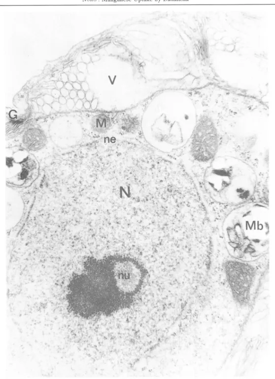
Fig m-1. is a longitudinal section of the organism showing profiles of the organelles. The cell is found only by a cytoplasmic membrane, and lacks a cell wall or scales. Two flagella extrude through the apex. The axonemes appear to have the typical 9+ 2 microtubular arrangement. Below the flagellar bases lies the Golgibody, consisting of one or more dictyosomes. In the centre of the anterior portion of the cell lies the nucleus with a porous nuclear envelope. The nucleolus is nearly central, surrounded by clumped heterochromatin. Although typical of both logarithmic and stationary phase cells, vacuoles and vacuolar inclusions are somewhat more abundant in the latter ( Figs. IE-3, & III-4). Vacuoles contain portions of membrane and vesicles. The campanulate chloroplast occupies the entire posterior portion of the cell and extends anterioly along the sides, almost completely enclosing the central region of cytoplasmic matrix and nucleus ( Fig. EI-2). The lamellae runs approximately parallell to the longitudinal axis of the chloroplasts, each band consisting of 1—4 tylakoids. The chloroplast stroma contains small electron dense materials which are assumed to be lipid droplets. The pyrenoid is immersed in chloroplast matrix located at the posterior end of the cell and is usually penetrated by lamellae on all sides. These lamellae normally consist of pairs of thylakoids and they terminate before reaching the centre of the pyrenoid matrix. Between the pyrenoid matrix and chloroplast proper is found the starch sheath which may either consist of a few large curved plates or a
number of smaller discoidal grains.
The use of giant algal cells has been very important in recent-advances in our understanding of mechanism of solute and water transport in plants. On the other hand, studies with unicellular micro algae have contributed to our knowledge of Na+/K+
regulation ( Barber, 1986) and the transport of metabolized nutrients such as phosphate,
nitrate and sugars (Nissen, 1974).In these micro algae, Z). tertiolecta is one of the most useful alga for nutritional study. This species grows actively in the artificial medium and is a widely used organism in the study of the effect of nutrient on the growth of algae. Nutritional study of heavy metals also studied in D. tertiolecta. But, uptake mechanisms of heavy metals is not clear.
In the analysis of the uptake mechanism of D. tertiolecta, adsorption by cell wall is neglected ( omitted ) because this flagellate alga is a wall-less or naked cell organism. The
present study of D. tertiolecta was almost the same as that of Hoshaw & Maluf (1981) of D.
tertiolecta. The relatively large fraction of the cytoplasm occupied by the chloroplast and vacuoles, makes it important in considering the distribution of solutes and the significance of electrical measurements in the cytoplasm. Quantitative electron microscopy shows that
„G
iPm
Fig. ffl-1. Electron micrograph of D. tertiolecta. Longitudinal section showing flagella ( F ), nucleus ( N ) , nucleous ( nu ) , chloroplasts ( ch ) , vacuoles ( V ) and Golgi bodies ( G ). The plasmalemma ( Pm )surrounds the cell. Starch ( St ) surrounds pyrenooid ( py ). Scale = l,«m.
192
.
Mem. Fac. Fish., Kagoshima Univ. Vol. 34, No. 2 (1985)
Pm
n e-2. Electron micrograph of D. tertiolecta. Numerous chloroplast lamellae penetrate
fvf
n eN
-\ V ^ .V. •„ i: •:i:.:;-:;?;Mn
Fig. ID -3. Electron micrograph of D. tertiolecta. The nuclear envelop ( ne ) is characterized
by many nuclear pores. Heterochromatin scatters within the nucleus. Mitochondria
194 Mem. Fac. Fish., Kagoshima Univ. Vol. 34, No. 2 (1985)
/
Pm
^fifej
Fig. HI -4. Electron micrograph of D. tertiolecta. Vacuoles ( V ) are seen near Golgi bodies ( G ). Scale = l /j m.
D. tertiolecta has about 50% of their cell volume occupied by vacuoles. Chloroplast, nucleus, starch and pyrenoid occupied about 20, 15, 10 and 5% of the cell, respectively. This chapter showed how D. tertiolecta can make good experimental material for a number
of transport problems as well as for the biophysical approaches to which giant algal cells are
so well suited.
W. Effect of manganese on the growth and
the metabolism in Dunaliella tertiolecta Butcher
F ~ 1. Introduction
The recent realisation of the significant effects of dissolved heavy metals on the growth of marine phytoplanktons has been reflected in several literature ( Mandelli, 1969 ; Steemann Nielsen & Wium-Anderson, 1970;Hannan & Patouillet, 1972; Jensen et al, 1974, 1976; Overnell, 1975 ; Braek et al, 1976 ; Berland et al., 1977). The position of the phytoplankton at the base of many marine food chains means that any inhibition or promotion of their growth may cause changes in the population levels of herbivores and carnivores of the food chain. Additionally, many phytoplankton species accumulate heavy metals, ( Saraiva & Fraizier, 1975 ; Cossa, 1976 ; Schulz-Baldes & Lewin, 1976 ; Styron etal, 1976 ; Gerhards & Weller, 1977 ; Jennings & Rainbow, 1979) and such heavy metals
are further accumulated at higher food chain levels.
The initial report on the manganese requirements for algae was made by Hopkins (1930 a, b). Harvey (1947) reported the manganese starvation in natural inshore waters. Barber et al (1973) also demonstrated that poor growth of phytoplankton in fresh upwelling seawater was reversed by addition of Fe or Mn. In these situations, it is apparent that the growth of
phytoplankton is limited when manganese is at low concentration. The effects and functions
of manganese on algal metabolism has been reviewed by many phycologists including
Bowen (1966), Iwasaki (1967), O'kelley (1974) and Sanders (1978). The most studied
effect of manganese on algal metabolism is the role of Mn in the Ch-evolving system of photosynthesis. This micronutrient activates a number of phosphate transferases and decarboxylases, notably those used in the Krebs cycle.
The marine alga, D. tertiolecta, has been used in the physiological experiments of nutrients. Harvey (1947) investigated the effects of added Mn on the growth of D. tertiolecta (as Chlamydomonas sp. ). In the report of Pirson & Bergmann (1955) and Reisner & Thompson (1956), Mn-requirement for trophical growth and no growth carried out by Mn deficiency in Chlorella under heterotrophic carbon nutrition was conspicuous. Manganese concentration of D. tertiolecta in culture was analyzed by Riley & Roth (1971). However,
the function of Mn on the physical condition of D. tertiolecta is obscure.
This chapter treated with the growth rate and the variation of three cell contents, protein,
carbohydrate and chlorophylls, at various level of Mn concentration in the medium.
W~2. Materials and methods
196 Mem. Fac. Fish., Kagoshima Univ. Vol. 34, No.2 (1985)
Table IV- 1. Compositionof culture medium*)
NaCl 2.4 g MgCl2.6H20 1.5 g MgS04. 7H20 0.5 g KC1 0.2 g CaCl2 0.2 g KN03 1.0 g NaHCOs 0.043 g TRIS 2.45 g**) K2HP04 0.045 g Fe-EDTA 3.64 mg EDTA-2Na 1.89 mg ZnS04.7H20 0.087mg H3BO3 0.61 mg CoCl2. 6H20 0.015mg CuS04. 5H20 0.06 mg MnCl2 0.23 mg***} (NH4)6Mo7024. 4H20 0.38 mg H20 1,000 ml
* ) Modified from medium used by JOHNSON etal. (1968). * * ) Adjusted to pH 8.0 with HC1.
***) Stock manganese solution:dissolve 1.000 g of manganese metal (99. 99%) in the minimum volume of HNO3 (40% ). After evaporating to dryness on a water bath, again dissolve in 10 ml of cone. HC1 and dilute
to 1,000 m/with H2O.
i
modified Johnson's medium ( Table IV-1) at 20*C under 12 :12 hrs LD cycle ( cool white fluorescent lamps giving an illumination of ca. 5,000 lux). Manganese solution was
prepared by dissolving metal Mn (p. 187). Before the experiment, D. tertiolecta cells were
Mn-starved in Mn-free medium with aeration.
The number of cells of D. tertiolecta was determined using a haemocytometer. Chlo
rophyll content was determined according to the procedure of Jeffrey & Humphrey (1975).
Crude protein extraction using 1. 0 N NaOH took 3 hrs at roomtemperature, was determinedby the modified Lowry'S method (Hartree, 1972). The protein value was converted to
albumine. Crude hydrocarbon analyzed by the method of Dubois etal (1956) was convertedto glucose value.
F-3. Results
(1) The relationship between the growth and the manganese content of the medium
The growth in cell number responses differetly in proportion to the concentration of Mn in the medium as illustrated in Fig. IV-1. Fig. IV-2 and Table IV-2 also show the cell density on the 10th day after inoculation and the growth rate constant (K) in logarithmic phase,
respectively. At the lowest concentration (0.05 ppm Mn), the growth continued
exponentially after the addition of Mn, though the growth ceased up at 16 ppm Mn concentrations. Higher growth rate and a slight enlargement of cell length was observed in10 -C EL <t> or-(J
° o-#::?:r::^fi 0.5 ppm Mn
1.0 —100 ppm Mn A 8 12 16 20 24 25 Days 001 0.1 1 10 Mn concentration(ppm)Fig. IV-1. Effect of Mn at various concentrations on the growth of Dunaliella tertiolecta in batch culture. Growth curve is expressed as a function of time ( A ) and shown on the end of 10 days culture ( B ). The results shown in ( A ) and ( B ) come from different experiments, so that the cell density attaiend is different between ( A )
and ( B ). Cells were incubated at 20°C, 12:12 hrs LD, 5,000 lux.
, }H"V
0.01 0.1 1 10
Mn concentration (ppm)
Fig. IV -2. Effect of Mn at various concentration on the cell enlargement of Dunaliella ter
tiolecta maintained in batch culture for ten days. Cells were incubated at 20°C ,
12:12 hrs LD, 5,000 lux. Slight enlargement of cell length was observed in Mn concentration from 0.1 to 1. 0 ppm.
198 Mem. Fac. Fish., Kagoshima Univ. Vol. 34, No. 2 (1985)
Table IV - 2. Mn effects on the growth rate constant ( K at 7 concentrations from 0 to 16 ppm Mn at 20*C calculated using the equation:
of Dunaliella tertiolecta incubated
1212 hrs LD, 5,000 lux. K is
T2-T1
where Ni and N2 are the cell number at 3-day ( Ti ) and 5-day ( T2 ) growth respec tively. Higher growth rate constant ( K ) in logarithmic phase is observed from 0.1 to 0. 5ppm Mn concentration. Concentration of Mn in medium (ppm) 0.00 0.01 0.10 0.50 1.0 10.0 16.0 K 0.08 0.22 0.38 0.38 0.30 0.26 0.00 100(-
--O- Protein in 1ml of culture -••--Protein in 10^cells
/£\
/>' V 6/ •iT*"*"" ^ 1^....,<f" ~~*-"^L
•/ _ + cr \l J 1 1X 1.0 3If-8< .£0. 5 3
I
0.01 0.1 1 10 Mn concentration (ppm) c i= 2 E 5 5 o ^ 0.5Fig. IV-3. Protein content of Dunaliella tertiolecta grown at various levels of Mn in batch cul ture for ten days. Cells were incubated at 20*C, 12:12 hrs LD, 5,000 lux. Protein was analysed using the method of HARTREE ( 1972 ). Protein content attained a
maximum value when 0.05-0. lppm Mn was added to medium.
0.0? 0.1 1 10
Mn concentration (ppm)
Fig. IV -4. Carbohydrate content of Dunaliella tertiolecta grown at various levels of Mn in batch culture for ten days. Cells were incubated at 20*0, 12:12 hrs LD, 5,000 lux. Carbohydrate was analysed using the method of DUBOIS et al. ( 1956 ). Carbo hydrate content, as well as protein content, attained a maximum value when 0. 05-0. lppm Mn was added to medium.
Chl.a in 1ml ot culture
--0-- Chl.a in 10* cells
0.01 0.1 1
Mn concentration (ppm)
Fig. IV -5. Chlorophyll-a content of Dunaliella tertiolecta grown at various levels of Mn in batch culture for ten days. Cells were incubated at 20°C, 12:12 hrs LD, 5,000 lux. Chlorophyll-a was analysed using the method of JEFFREY et al. ( 1975 ).
Chlorophyll-a in cells did not decrease in Mn deficient medium. 0.001*
0.002
u
o
Table IV - 3. Chlorophyll-a and -b of Dunaliella tertiolecta grown in batch culture system with and without Mn and Fe for ten days. Cells were incubated at 20°C , 12:12 hrs LD, 5,0001ux. JOHNSON'S medium ( JM ) was shown in Table HI — 1 . JM-Mn:Mn
deficient JOHNSON'S medium, JM-Fe:Fe deficient JOHNSON'S medium, JM-Mn,
Fe:Mn and Fe deficient JOHNSON'S medium. Chlorophyll-a in cells did not de crease so much in Mn deficient medium but decreased remarkably under Mn and Fe
deficient condition. Medium (1) Cell No/m/ (xl04cells/m/) (2) Chl.a/m/ (/ig/m/j (3) Chla /cell (xlO-Wl04cells) (4) Chl.b/m/ {fig/ml) (5) Chl.b/cell (xl0-3/ig/104cells) (6) Chl.a/Chl.b JM 452 ±63 0.63+0.08 1.39 0.15±0.02 0.33 4.21 JM-Mn 337 ±26 0.49±0.03 1.45 0.13±0.01 0.39 3.71 JM-Fe 349±35 0.37±0.03 1.06 0.09±0.01 0.26 4.08 JM-Mn, Fe 353±27 0.30 ±0.02 0.85 0.07±0.01 0.20 4.25 (3) = (2)/(l), (5) = (4)/(l), (6) = (3)/(5), ±S. D. Mn concentration from 0.1 to 1.0 ppm.
(2) Effect of manganese on protein and carbohydrate contents
Crude protein and carbohydrate composition of D. tertiolecta are shown in Figs. IV-3 and IV_4, respectively. As expected from the growth data shown above, some relationship
exists between the protein and carbohydrate content of cells and Mn concentrations in the medium. These cell components increased with the increase in Mn concentration in medium and attained a maximum value when 0. 05—0.1 ppm Mn was added to medium. From 0.1 to 10 ppm Mn, protein and carbohydrate contents were constant but decreased above 10 ppm
Mn.
(3) Effect of manganese on chlorophyll contents
The variations of chlorophyll-a content per cell in each culture at increasing concentra
tion of Mn were little different from those of protein and carbohydrate (Fig. IV~5). Chlorophyll-a in cells did not decrease so much in Mn deficient medium (Fig. IV_5 & Table
200 Mem. Fac. Fish., Kagoshima Univ. Vol. 34, No. 2 (1985)
IV-3), but decreased remarkably under Mn and Fe deficient condition ( Table IV-3, see column (3)). This chlorosis was apparent in deficiency of Mn and Fe, but not obvious in Fe
free culture.
IV~4. Discussion
As mentioned above, it has been well known that trace amount of Mn is essential for algal
growth. In Dunaliella tertiolecta, Harvey (1947) reported that the growth was activated by the addition of 0. 5—2. 0 mg Mn/m3(ppb). However, in the present batch culture experiment the organism in problem grew actively in the presence of 0.1 ppm Mn and this fact was supported by the results that the growth rate and cell components became to the highest
value at the level of Mn in the medium. This value of Mn concentration is almost the same level as Mn component in several artificial sea water culture medium for algal growth.
There are many reports on the effect of culture condition on the chemical composition of
algae. However, the effects of Mn on the metabolism of protein and carbohydrate was obscure. In the present study, Mn sensitivity and relationship with metabolism are
re-proved.
According to Constantopoulos (1970), the presence of Mn affects the growth of Euglena gracilis but do not affect chlorophyll contents. This phenomenon was observed also in D. tertiolecta. Algae without hydrogenase particularly show chlorosis if deprived of Mn, but the chlorophyll content of hydrogenase-containing algae is more stable (Kessler, 1968). Iron is known as an activator of hydrogenase (Waring & Werkman, 1944). It is interesting to
know that D. tertiolecta has become chlorosis in the present study when cultivated in the Fe and Mn deficient medium.
V. Manganese uptake and release in Dunaliella tertiolecta Butcher
V-l. Introduction
Algae, like other organisms, accumulate a number of trace metals and require these elements for growth. The accumulation of trace metals and their radioisotopes by algae in culture has been the object of numerous studies. In the case of essential trace metals, Jensen etal (1976) and Sakaguchi etal (1977) have investigated the uptake of Cu by three diatoms and Chbrella, and Bernhard & Zattera (1969) and Parry & Hayward (1973) have studied the uptake of Zn by Phaeodactylum tricornutum and D. tertiolecta, respectively. Some emphasis has also been placed on the uptake of non-essential metals such as Cd (Saraiva & Fraizier, 1975 ; Cossa, 1976 ; Gerhards & Weller, 1977 ; Jennings & Rainbow 1979), Pb (Schulz-Baldes & Lewin, 1976), Hg (Gerhards & Weller, 1977) and Ni (Gerhards &
Weller, 1977 ; Skaar et al, 1974).
Previous studies on Mn uptake by algae have been conducted by Tsukidate (1974), who
found that the amount of Mn absorbed by Porphyra in cultures varied with the illumination and temperature.
A number of workers have studied the effects of trace metals on algal growth, but the
(1975) concluded that Zn and Hg uptake by Viva lactuca and Synedra ulna was a passive
process since it was affected neither by metabolic inhibitors nor by anaerobic conditions.
However, Paton & Budd (1972) have concluded that Zn uptake by Neocosmosphora vasinfectaoccurs both by a rapid passive process or iox-exchange and also by a slower, metabolically
dependent process.
Rothstein etal (1958) and Jennings etal (1958) showed that Mn uptake by yeast cell was
stimulated by phosphate and they concluded that the uptake was a metabolically active
process. While, Parry & Haywars (1973) demonstrated that the uptake of Zn by D.
tertiolecta was temperature and pH-dependent but not directly linked with metabolism. The
relationship between iron uptake and various environmental factors (e.g. light, pH and
temperature) which influence cell metabolism has been studies also in Porphyra (Tsukidate,
1974). However, little is known as to how such factors affect the Mn uptake by D. tertiolecta. In this chapter the existence of an energy-coupled transport system of Mn in D.
tertiolecta was proved.
Table V- 1. Capture of Dunaliella tertiolecta by filtration ( Whattman GF/C ) and centrifugation ( 2,000 rpm,2 min. ). Stationary growing D. tertiolecta was captured by above both methods and the biomass was evaluated by chlor ophyll-a concentration in one mlof culture. The cells captured by filtra tion is as by centrifugation. The destruction of cells by filtration is neg ligible. Chlorophyll-a (xl0_3mg Chi. a/m/-culture) Centrifugation Filtration
fc
Cpm
g 2,000
c o c LO •D <b 8 < 1,000 6.80±0.37 7.06 ±0.28 ±95% C. I. 0 20 40 mlVolume of washing solution
Fig. V -1. Residue of adsorded Mn54 on filter paper after repeated washing. Mn deprived culture medium was used for washing.
202 Mem. Fac. Fish., Kagoshima Univ. Vol. 34, No.2 (1985)
V~2. Materials and methods Organism and culture
The experimental techniquesemployed for culturingD. tertiolecta werebasically identical as described in chapters IE and IV(see p. 189, 196).
Manganese uptake studies
After the medium was inoculated with D. tertiolecta (1—3X10⁴ cells/m/)and the culture was grown to an appropriate number of cells (200—400X104 cells/m/), 0. 05 ju Ci of Mn54 was added to the 5. 0 ml of the culture suspension, giving a total Mn concentration of 0.1 ppm. Before the experiment, D. tertiolecta was Mn-starved in Mn-free medium with aeration. After shaking the culture by means of mini-shaker ( Shaker Mini SS-80, Tokyo
Rikakikai Co. Ltd. ) under the light, a sample of the culture was periodically filtered
through Whattman GF/C glass fiber filter to separate the cells. Filtration was carried out using vacuum pump. Controls were also prepared with the same amount of radio-active Mn54, but containing no cells. Similarly, the controls were filtered. The filtrate were terminated by rinsing with 30 m/each in cold Mn-free culture medium ( Table V-l, Fig. V -1). The filter plus cells were dried under a lamp. The radioactivities of filtrate were measured by a well-type gamma ray Nal scintillation counter. Further procedures are described in detail in result of respective part.V-3. Results
(1) Mn uptake by living and dead cells as a function of time
% u o Ui f o c 2 •a. Living cell (Light) Living cell (Dark) Dead cell Time (hrs)
Fig. V -2. Uptake of Mn by living and dead cells of D. tertiolecta. Cells were incubated in 50 ml flasks using a shaker at 20*0, 5,000 lux for 4 hrs. Living cell showed a rapid rate of uptake during the first hour, followed by a slower uptake. The amount of Mn accumulated by D. tertiolecta was stimulated by light. Dead cells initially took up slight amount of Mn but this uptake ceased within 10 min.
One method of trying to understand howcells concentrate Mn is to compare the uptake by
living and dead cells. The uptake of Mn54 by living cells and by cells killed with
formaldehyde was compared (Fig. V~2). Cells, preincubated for 12hrs in Mn-free medium in darkness, were incubated in 50 ml flasks using a shaker at about 20°C, 5, 000 lux for 4 hrs. Living cells showed a rapid rate of uptake during the first hour, followed by a slower uptake. The amount of Mn accumulated by the D. tertiolecta reached a maximum value after incubation period of 2—4 hours. Fig. V-2 also shows that in this experiment Mn54 uptake was stimulated more in the light than in the dark. Dead cells initially took up slight amount of Mn54 but this uptake ceased within 10min. Mn54 was also rapidly bound on the living cellswithin the initial 10 min after the addition of Mn, even in the absence of an energy source.
This Mn adhesive phenomenon was also observed in dead cells. This kind of uptake of Mn54 seems to be due to the physical process of adsorption to the cell surface.
Cells of D. tertiolecta, which had been exposed to Mn54 for 3 hrs, were transferred to Mn-free medium and killed by heating. So that, Mn54 was lost from the dead cells, rapidly at first but then more slowly. Living cells transferred to Mn54-free medium did not appear to lose Mn to the medium but little for the duration of the experiment (Fig. V-3). (2) Mn uptake as a function of cell density
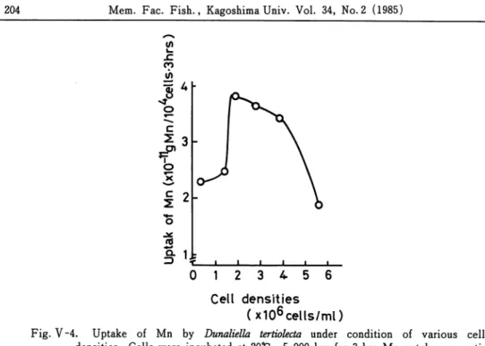
The amount of manganese taken up by D. tertiolecta was measured at the different population sizes. In this experiment, Mn-starved cells harvested in logarithmetic phase were incubated for 3 hrs after the cell density in the culture medium was regulated from 0. 5 to 6. 0 x 106 cells/mZ. As shown in Fig. V~4, Mn uptake was activated under the condition
Living cell o c 2 3 , t-O* o * 2 %
.c1
(
c\
- • Dead cell - • 0 1 2 3 Time (hrs)Fig. V -3. Retention of Mn by living cells and loss of Mn from dead cells of D. tertiolecta. The measurement was done after cells was labelled with Mn54 and suspended in Mn54 free medium.
204 Mem. Fac. Fish., Kagoshima Univ. Vol. 34, No. 2 (1985) JZ CO J)
J'
o c£3
c 2-rd a i -3 t 0 1 2 3 4 5 6 Cell densities(x106 cells/ml)
Fig. V-4. Uptake of Mn by Dunaliella tertiolecta under condition of various cell densities. Cells were incubated at 20*C, 5,000 lux for 3 hrs. Mn uptake was acti
vated under the density of 2-4 x 10⁶ cells/m/. The depression of Mn accumulation
occurred under low and high cell desities.
3 9
o'clock
Light and dark cycle
Fig. V -5. Changes in Mn content and numbers during the synchronized cycle of Dunaliella
tertiolecta. Synchronization was tried under the conditions of 3,000 lux, 12:12 hrs
LD and 20*C with aeration. The cell division mostly occurred in the early part of the dark period. And Mn content was reduced by the divisions and increased with the cell growth.
of 2—4 x 10⁶ cells/m/. The depression of Mn accumulation occurred under high cell
density.
(3) Mn uptake in synchronous culture
Synchronization of D. tertiolecta was tried under the conditions of 3,000 lux, 12 :12 hrs
LD. and 20°C with aeration (Eppley & Coatsworth, 1966 ; Wegmann & Metzner, 1971) and was successfully established. The degree of synchrony was rather high up to 3 x 10⁶ cells/m/. For uptake experiment in the synchronized culture, Mn starved cells werepreincubated with aeration. When the cell density reached 1 x 10⁶ cells/m/, Mn54 and
non-active Mn were added to the culture medium, giving to the concentration of 0.1 ppm Mn, and the number of cell and uptake of Mn54 was determined every 3 hrs.
Microscopic controls showed that the cell division mostly occurred in the early part of the
darkperiod. Fig. V-5 gives the Mncontent and cell number during the synchronized cycle.
The data shows that Mn content was reduced by the cell divisions and increased with the cellgrowth.
(4) Mn uptake in batch culture
Mn starved cells were inoculated in 100 m/of Johnson's medium ( 5 //CiMn54, 0.1 ppm non-active Mn ). The culture was aerated and maintained at about 20°C, 5,000 lux and 12 : 12hrs LD. Every 3 : 00p. m., 5 m/of aliquotswere removed and Mn54 concentrationin cells and number of cells were determined.
The time course of Mn uptake by cells of Dunaliella tertiolecta in batch culture for 10 days is
5 10
Time (Days)
Fig. V -6. Changes in Mn content and cell numbers in the batch culture of Dunaliella ter tiolecta, maintained at 20°C, 5,00Q lux. During the first 2 days after the incubation, Mn was taken up rapidly.
206 Mem. Fac. Fish., Kagoshima Univ. Vol. 34, No.2 (1985)
shown in Fig. V-6. During the first 2days after incubation, Mn was taken up rapidly. The
actual quantity of Mn per cell continued to be diluted. Sixth day after the initiation of
culture, the Mn concentration in the cells remained constant.
It is well known that physiological activities of phytoplankton, e. g. photosynthetic rate,
growth rate etc., are high at early exponential phase ( Daley & Brown, 1973 ). Similarphenomenon was seen in this result.
(5) Mn uptake as a function of Mn concentration in the medium
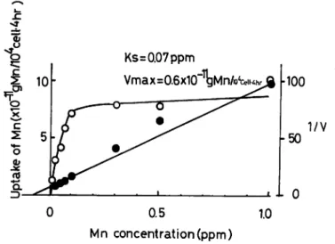
The uptake of Mn by D. tertiolecta was measured as a function of the external Mn
concentrations ( Fig. V-7). D. tertiolecta was incubated in Johnson's medium containing
different concentrations of Mn from 0.0 to 0.1 ppm for 4 hrs. The Mn was labelled with
Mn54.The Mn levels in D. tertiolecta sharply increased as exgenous Mn increse from 0. 0 to 1. 0
ppm. The absorption system was apparently saturated at 0.1 ppm since further increases in
external concentrations up to 1.0 ppm accompanied with no additional increases in the Mncontent ofD. tertiolecta. Such saturation kinetics follow the Michaelis-Menten equation for
enzymic catalysis. This saturation of the absorption mechanism of an ion has been taken as
evidence for active ion transport ( Epstein, 1973). The graphical methods of Lineweaver
and Burk was used for the estimation of the kinetic constants Ks of 0.07 ppm and Vmax of
0.6 x 10⁻¹¹ g Mn/104 cells-4 hr for the absorption process.Fig. V-8 is the effect of Mn concentration on the concentration factor ( as wetweight ).
x:
i
c Ks=0.07 ppmVmax=0.6x10"l^Mn/o4ceii
100 1/V -50 i l 0 0 0.5 1.0 Mn concentration (ppm)Fig. V -7. Uptake of Mn by Dunaliella tertiolecta incubated for 4 hrs in JOHNSON'S medium containing various amounts of available Mn ( —O— ). The line is a plot of the Michaelis-Menten equation;
VS v Ks+S
( v: uptake rate of Mn, S: concentration of Mn in medium, V: rate of absorption, Vmax: maximum rate of absorption, Ks: Michaelis constant ). The graphical
methods of Lineweaver and Burk (—•— ) was used for the estimation of kinetic
c o
"E 1
(U u c 0.5 Added Mn (ppm)\
JL*J / / -1.0 5.0Fig. V -8. Effect of added Mn on the Mn concentration factor of Dunaliella tertiolecta. Con centration factor is logarithmically expressed. The date used is the same as those in
Fig. V~7. Manganese concentration factor ( as wet weight ) is the order of 10-fold in 0. 3-0.5ppm Mn, but 100-fold in <0.1 ppm Mn. 6h 0) o en
12
=> 0 5 10 20 30 Temperature(°C)Fig. V-9. Effect of temperature on Mn uptake by Dunaliella tertiolecta. Manganese uptake was observed in JOHNSON'S medium at different temperature from 5*C to 30°C under
a light intensity of 3,000 lux for 3 hrs.
The data used in Fig. V-8 is the same as those in Fig. V-7. Manganese concentration factor is the order of 10- fold in 0.3-5.0 ppm added Mn, but 102-fold in 0.1 ppm Mn. (6) Effect of environmental factors on Mn uptake
Temperature: The manganese uptake by D. tertiolecta was observed at differenat tempera
tures from 5°C to 30°C under a light intensity of 3,000 lux for 3 hrs. As shown in Fig. V-9,
Mn uptake was strongly temperature-dependent. Maximum Mnwas taken up at about 20°C.
208 Mem. Fac. Fish., Kagoshima Univ. Vol. 34, No. 2 (1985)
These data indicate the mediation of metabolism in the absorption process.
Light intensity: The accumulation of Mn by D. tertiolecta was observed at different light
intensities from 0 to 60,000 lux at 20°C for 4 hrs. The intensity of illuminations were
obtained to regulate the natural light using tracing papers. The action of light was to
stimulatethe influx of Mn and this sensitivity is clearly shown in Fig. V-10. The cells used werecarbonstarved by being kept in the darkfor 12hrs before the experiment. It can be seenthat this process saturated at about 8,000 lux and inhibited at high light intensity. Reyther
(1956) showed photosynthesis-light curve of Dunaliella. And the present result concerning
Mn uptake-light curve was also of the same pattern.
1 2 3 4 5 6
Light intensity (xioAiux)
Fig. V -10. Effect of light intensities on Mn uptake by Dunaliella tertiolecta. Mn uptake was observed in JOHNSON'S medium at different light intensities from 0 to 6,000 lux at
20°C, 3,000 lux for 4 hrs.
Living cell
Fig. V-ll. Effect of pH on Mn uptake by living and dead cells of Dunaliella tertiolecta. Mn
uptake was observed in JOHNSON'S medium at different pH from pH 6. 0 to pH 9. 0 at20t;, 3,000 lux for 3 hrs.
pH: Mn absorption was investigated in a series of pH value from 6. 0 to 9. 0 using living
and dead Z). tertiolecta. In this series the media were all buffered with TRIS at a concentration of 2. 45 g//, then adjusted to the desired pH with HC1. Fig. V-ll shows the
uptake of Mn by living and dead D. tertiolecta at different pH's. A pronounced pH
dependency of Mn absorption by living cell is indicated. Absorption of Mn by living cell
was 102-fold as high as formaldehyde treated cell. The amount of Mn found in living cell
increased with the increase of pH. At pH values greater than 8. 0, Mn uptake was markedly increased. While D. tertiolecta could not grow actively at pH 9. 0, the organism took up moreMn at the same pH.
(7) Effect of glucose on Mn uptake
A respiratory substrate, glucose, was added to observe the evidence of coupling of
oxidative phosphorylation in relation to manganese uptake. As shown in Fig. V-12,
accumulation of Mn was stimulated 2 times by glucose. With increasing glucose concentration the amount of Mn accumulated by D. tertiolecta rose and reached a limiting value at concentration of 0. 5 g glucose//. Therefore, the energy for Mn transport, can be provided from glycolysis.1 ^
=>0 j _
0.5 1.0
Glucose(g/D
Fig. V-12. Effect of glucose on uptake by Dunaliella tertiolecta. D. tertiolecta was incubated in JOHNSON'S medium with various concentrations of glucose at 20*0, 3,000 lux. After 4 hrs, D. tertiolecta was harvested by filter paper to analyze Mn54 in the cells.
(8) Effect of nutrients on Mn uptake
Figs. V-13 to V-16 illustrate the effect of components in the culture medium on Mn54
uptake by D. tertiolecta. D. tertiolecta was harvested by centrifugation, then washed twice with a sodium chloride solution at the same concentration as that of the growth medium. Na, K: D. tertiolecta was incubated in Johnson's medium containing different concentration
of NaCl from 10 to 160 mM ( =0. 58-9. 3 % ) for 3 hrs to study the effect of NaCl on uptake of Mn54. Manganese accumulation is reduced in 25 mM NaCl and below and reached the
210 Mem. Fac. Fish., Kagoshima Univ. Vol. 34, No. 2 (1985) 50 100 NaCl(mM) 1 ' — / / ' / / ' 10 20 25 50 100 KCKmM) 10 20 MgCl2(mM) 25 50 100
Fig. V -13. Effect of Na, K, and Mg on Mn uptake by Dunaliella tertiolecta. Exponential phase of D. tertiolecta was harvested and washed with NaCl solution or Tris-HCl
buffer. After that cell was incubated in each nutrient solution with Mn54 for 3 hrs
to analyze Mn which is taken up by cell.
mM NaCl.
Johnson et al (1968) studied the effect of NaCl on the growth and enzymatic activity of
halophilic Dunaliella viridis. The results obtained in the present study were similar to thosein D. viridis of Johnson et al
KCl was added to the culture medium at differentconcentration from 0 to 100mM to study the effect on Mn uptake. Mn uptake was stimulated between 0 and 2 mM KCl, although at the concentrations above 5 mM KCl the Mn uptake was reduced ( Fig. V-13).
N, P, C: Nitrogen, phosphorus and carbon are important constituents of a variety of organic compounds which are essential to metabolism and the following construction of structure for plants. The effects of N, P and C on Mn absorption were studied using labelled Mn at 0.1 ppm, as same as other experiments. Mn uptake was slightly stimulated in 6-8 x 10"4 M K2HPO4 and 1-5 x 10⁻⁴ M NaHCOs while there was no stimulation of Mn uptake by the addition of NaNOs ( Figs. V-14 & V-16).
These elements were less effective than micronutrients as discussed below. D. tertiolecta
?0
10 20 CaCl2(mM) 10 20NaN03(mM)
0 10 20 NaHCO3(xi0"AM)Fig. V-14. Effect of Ca, NO2-N and HCO3-C on Mn uptake by Dunaliella tertiolecta. Detail
method was described previously in Fig. V-13.
confirm distinctive effects on Mn uptake in short expeiment period.
Mg, Ca: Magnesium and calcium ions have a function as activators for a number of enzymes in nature. Calcium salts of phosphatidic acid occur in cell membranes and are essential to the maintenance of their structure. And Mn transport process was expected to be affected by Mg and Ca in the medium while low concentration of these divalent cations
slightly accelerated the Mn uptake by D. tertiolecta ( Figs. V-13 & V-14). The uptakes of
Ca and Zn byexcised rice roots areknown to beinhibited by Mn ( Ramani & Kannan, 1976).
Co, Cu, Mo: Cobalt is a component of vitamin B12. Copperis also the component of several metalloenzyme including ascorbic acid oxidase, phenolase and cytochrome oxidase. Molybdenium is essential for algal growth.
Mn uptake was a little increased in low concentration of these metals while Mn absorbtion was extremly inhibited in the order of 1 x 10⁻⁷ M of C0CI2, 1 x 10⁻⁶ M of CuCh and
(NH4)6Mo7024. Sunda etal (1981) showed that Cu competes with Mn for metabolically
active site in Chaetoceros socialis ( Fig. V-15).
Fe: Iron occurs in the prosthetic group of certain proteins, notably the cytochromes which function in electron transport, and in the enzymes, peroxidase and dehydrogenases. It has a specific role in chlorophyll synthesis.
Unlike other nutrients, Fe ( as Fe-EDTA ) helped the uptake of Mn at the concentration
30 40
212 Mem. Fac. Fish., Kagoshima Univ. Vol. 34, No. 2 (1985) Q. 0 10'8 107 10"6 105 H3BO3 (M) 10 20 CoCl2(xio~8M) 10 20 CuS04(x10~7M) 10"3 Co 30 Mo l l 0 10 20 50 100 (NHOeMOTOa^xIC^M)
Fig. V-15. Effect of BO3, Co, and Mo on Mn uptake by Dunaliella tertiolecta. Detail method was described previously in Fig; V-13.
of 1-20x 10~5 M Fe, especially in 5 x 10"5 M Fe. As described before ( p. 199) it is known
that Mn physiologically cooperates with Fe. The data obtained here confirm the above Fe— Mn relationship ( Fig. V-16).Zn: Zinc involved in several enzymes as either a constituent or an activator ( Epstein, 1973). In this experiment, the absorption of Mn is reduced at all Zn concentration used. Zinc is different from other heavy metals in this respect (Fig. V-16).
B: The role of boron in the translocation of sugars has been much discussed ( Epstein,
1973). Boron ( as H3BO3 ) is the only trivalent ion used in this experiment. As shown in
Fig. V-15, there is no effect on absorption of Mn by BOa3 addition ( Fig. V-15).
(9) Effect of metabolic inhibitors on Mn uptake and release
The experiments already described in articles from (l) to (8) are based on physical and
chemical conditions andthe data obtained show that the mechanism of the Mn uptake by D.
tertiolecta is dependent on metabolism. To confirm further above results, the effects of several metabolic inhibitors at the concentrations over 0.1 ppm upon the absorption of Mn10 PO4 c M- 0 o
w \
10 K2HPOa(10'*m) 10 20 50 100 Fe-EDTA(xiO~5M) ZnSOA(xio;8M)Fig. V -16. Effect of PO4-P, Fe and Zn on Mn uptake by Dunaliella tertiolecta. Detail method was described previously in Fig. V-13.
were studied.
D. tertiolecta was cultured in Johnson's medium and harvested in exponential phase as described before ( see p. 195). On the day of the exponential growth, triplicate samples of D. tertiolecta were incubated in the medium containing KCN (0-50 mM), TPAC (0-10 mM)
and DCMU ( 0-10"5 M ) at 20°C, 5,000 lux for 3 hrs ( Table V-2).
Fig. V-17 shows the effect of the respiration inhibitor, KCN, on the uptake of Mn byD. tertiolecta. It is clear that the uptake of Mn is strongly inhibited by 40 mM KCN. This involves respiratory chain providing energy for Mn transport in the cells.
As described later, Mn absorption process may need the electronpotential of membrane
( p. 217). Theoretically, cell membrane must keep -50 to -70 mV of electronpotential to take up divarent anion ( Clarkson, 1974, see Fig. 3.1). Asano (1974) used tetraphenylarso-nium chloride ( TPAC ) as a metabolic inhibitor of electronpotential process in cell
membrane. To prove the role of electronpotential in Mn uptake mechanisms, the TPAC
was used.
214 Mem. Fac. Fish.., Kagoshima Univ. Vol. 34, No.2 (1985)
Table V- 2 . The inhibitors used, their concentration, and metabolic effect.
Inhibitor Dichloropheny1-1,1-dimethylurea (DCMU) Tetraphenylarsonium chloride (TPAC) KCN Effective Metabolic cones. (M) effect
Inhibits the
photo-lxlO"5 synthetic evolution
without effect on
absorption of K
Inhibits the
lxlO"2 electropotential
of membrane
5xlO-2 Forms complexes
with metaloenzymes
and attacks the
cytochrome oxidases of phosphorylation
10 30 50
KCN(mM)
Fig. V-17. Effect of the respiration inhibitor, KCN, on Mn uptake by Dunaliella tertiolecta. D. tertiolecta was inoculated at 20°C, 3,000 lux in the modified JOHNSON'S medium
which contain Mn54 and various amount of KCN. After 3 hrs, cell was filtrated to
analyzed Mn. The uptake of Mn was inhibited at the concentration of 10-50 mM KCN.
TPAC(mM)
5 10
TPAC(mM)
Fig. V-18. Effect of TPAC on the uptake and retention of Mn by Dunaliella tertiolecta. (A) :
D. tertiolecta was incubated in JOHNSON'S medium with TPAC and Mn54. Mn up
take by cell was inhibited with 1 mM TPAC. ( B ) : D. tertiolecta, which was in
cubated with Mn54, was transfered to TPAC-added JOHNSON'S medium. The cell
discharged Mn to the culture medium.
o
io9io8 id7io"6io"5
DCMU(M)
Fig. V -19. Effect of the photosynthesis inhibitor, DCMU, on Mn uptake by Dunaliella ter tiolecta which is incubated in DCMU-added medium, at 20*C , 3, 000 lux, for 4 hrs.
hand, Mn ion in cell was discharged to outside by incubating cells with TPAC ( Fig.
V-18(B)).
Fig. V-18 shows the effect of photosynthesis inhibitor, 3-( 3, 4-dichlorophenyl )-l, 1-dimethylurea ( DCMU ) on the light-dependent uptake of Mn. As Orr et al (1976)
216 Mem. Fac. Fish., Kagoshima Univ. Vol. 34, No.2 (1985)
suggested DCMU was an inhibitor of noncyclic photophosphorylation. Mn uptake is slightly reduced from 10"9 to 10"7 M DCMU and strongly in 10"5 M DCMU. This result means that the metabolic relationship between photosynthesis and Mn absorption exists. Similar inhibition was reported in Zn accumulation by Dunaliella ( Parry & Hayward, 1973). (10) Effect of EDTA on Mn uptake
Ethylenediaminetetraacetic acid ( EDTA ) is most widely used as chelator in marine medium, and is not readily metabolized by microbes ( Johnston, 1964).
To study the effect of chelator on Mn uptake, Dunaliella tertiolecta was suspended in the modified Johnson's medium containing different concentrations of EDTA from 0 to 20 x 10"6
M. After 0.1 ppm Mn was added, the cultures were incubated at 20°C, 3,000 lux for 3 hrs. Then the amount of the Mn taken up by cell was measured by Nal scintilation counter.
Manganese uptake was markedly stimulated by the addition of EDTA ( Fig. V-20). It was evident that D. tertiolecta was capable of accumulating Mn from pure and chelated forms. Kannan & Joseph (1975) has also reported the occurrence absorption of Mn with
EDTA by germinating sorghum. Theoretically, a chelate should combine with a metal in a 1:1 molar ratio. The data shown in Fig. V-20 exhibited the same conclusion. However, it is
not known whether the molecule of EDTA is transported to inside the cell with manganese ion.
If Mn uptake were stimulated by the addition of chelate substances, the organic substances which were released from algae would promote the Mn uptake process. According to Huntsman & Barber (1975) and Murphy et al (1976), algae released ion-selective chelators to the culture solution. So, used culture medium were added to Mn absorption experimental culture. As expected, Mn accumulation was stimulated by this
c 10 c 3 (Mn:EDTA=1:1)
j
10 EDTA-2Na(xKT6M) 20Fig. V -20. Effect of EDTA on Mn uptake by Dunaliella tertiolecta. After D. tertiolecta was suspended in modified JOHNSON'S medium containing different concentration of
EDTA. The culture were incubated at 20*0, 3,000 lux for 4 hrs. Then the amount
Table V - 3. Effect of used medium and EDTA on Mn uptake by Dunaliella ter
tiolecta. Culture medium which was separated from stationary phase of
culture by filtration was added to JOHNSON'S medium as used medium.
After D. tertiolecta was cultured at 20°C, 3,000 lux for 3 hrs, Mn54 absor
bed cell was analyzed.
Medium added Mn uptake
(xl0-ngMn/104cells. 4hrs)
J'M 4.5 + 1.3
J'M + O.lm/ of used medium 6.3±0.4 J'M + 0.5m/of used medium 7.0±1.3
J'M + IO"5 M EDTA 11.2±2.6
J'M : Johnson's medium, ±95% C. I.
used medium and it means metabolic organic substances are able to act as metal chelators
(Table V-3. ).
V~4. Discussion
Metabolic aspects of manganese uptake
Data obtained support the hypothesis that Mn transport in D. tertiolecta is
energy-dependent. Mn transport is dependent on endogenous sources of energy, glucose, but is
inhibited by DCMU, KCN and TPAC. These energy-dependent transport of Mn is known
in yeast cell ( Okorokov etal, 1977). As with the present study on D. tertiolecta the previous investigations generally demonstrated the two distinct processes in accumulation of the cation. First, there is a rapid but limited, energy- and temperature-independent system binding to the cell surface. The amount of such binding system was low in comparison withvalues reported in other similar studies ( Failla etal, 1976). These authors have shown
that cations compete for non-specific anionic sites on the cell surface.The second process is the slower, but sustained, energy- and temperature-dependent translocation across the cell membrane. Energy-dependent Mn uptake is a highly specific process that exhibits saturation kinetics. In D. tertiolecta, Mn was mostly accumulated by this second process. According to Gutknecht (1961), physical process of adsorption of cation exchange in Viva lactuca is primarily responsible for Zn uptake. Results of the present experiments indicate that Mn uptake is affected by several metabolic effects.
If it is assumed that 80 % of each cell content consists of water and that all of the intracellular Mn exists as the free cation, the uptake of Mn from the solution under 0.1 ppm
Mn represents the order of 1 x 10² of concentration factor. Further, when the electrical
potential of biological membrane is -40mV ( Barber, 1968), approximately 10 to 100 of the
concentration factor is necessary for active transport ( Nernst potential equation ; Clarkson, 1974,p. 59).As a conclusion, Mn is transported actively in the solution at the concentration less than 1 ppm ( Fig. V-8). While the cation may be exhausted actively in the solution at the concentration larger than 5 ppm. This result means algae/phytoplankton can accumulate
218 Mem. Fac. Fish., Kagoshima Univ. Vol. 34, No.2 (1985)
The fact which Mn uptake is inhibited by KCN, DCMU and TPAC also proves the active transport in D. tertiolecta.
Kinetic approach
In conclusion, it has been shown that D. tertiolecta has a high ability to concentrate Mn from a medium. The uptake of Mn appears to be proportional to the amount of the metal in the solution to a certain degree but in the concentration more than about 0.1 ppm no further
accumulation occurs.
D. tertiolecta incubated in the medium containing 0.1 ppm Mn, absorbed 7 x 10~u
gMn/104 cell ( Fig. V-7). If the cell was regarded as an oval ( 10 pm x 6 fim ), the volume of 104 cell would be:
4/3 x (5 x 10"4) x (3 x 10-4)2 x 104=1.9 x 10⁻⁶ ( m//104 cells) And the amount of Mn absorbed by this alga was calculated as follow:
7 x 10-11
—-—iH—=37 ppm Mn ( wet weight ). i.y x 10
Riley (1971) reported that D. tertiolecta accumulated 3. 8 ppm Mn as dry weight. Consider ing that the dry weight in this organism was about 10 %of the wet weight, these values were
considerably different.
In biological systems ions can be considered to be acted on by two physical forces: the
chemical potential gradient and the electropotential gradient. Together, they constitute the
electrochemical potential gradient: u=d(RT In. a)/dx+*F d^/dx.
In this equation, u is the elctron chemical potential gradient, R is the gas constant, T is the absolute temperature, a is the chemical activity, z is the algebraic valency, F is the Faraday constant, and 0 is the electrical potential.
If an ion moves "uphill", from a lower to a higher electrochmical potential during its transport through a membrane, the process requires an input of energy and this transport process is said to be active. When the tendency for an ion to move down its chemical potential gradient in the opposite direction, the equilibrium state is described as Nernst equation. The expression has the form:
En^
ZFln (-5?)
where En represents the Nernst potential and Cin and Com* the concentrations in inside and
outside the cell. The Nernst potential En=-74 mV was calculated from the Nernst equation
using Cln=3. 7 ppm, Coul=0.1 ppm, z=-2 and T=293 K. Although the membrane potential of D. tertiolecta was lacking, Barber (1968) measured the membrane potential of -40 mV in
an unicellular green alga, Chlorella pyrenoidosa. The membrane potential of D. tertiolecta
must be supposed to be not far from -40 mV. The difference between -40 mV and -74 mV means the system was not in equilibrium and energy must be expended to maintain the non-equilibrium state. ( i. e, it would require a metabolically driven active transport ).
It has been observed that the kinetics of Mn transport across membranes are similar to
those of enzymic catalysis ( Michaelis-Menten kinetics ), and this showed that reversible